Loading
PDBj
MenuPDBj@FacebookPDBj@X(formerly Twitter)PDBj@BlueSkyPDBj@YouTubewwPDB FoundationwwPDBDonate
RCSB PDBPDBeBMRBAdv. SearchSearch help

1O64
DownloadVisualize
BU of 1o64 by Molmil
Crystal structure of an ATP phosphoribosyltransferase
Descriptor: ATP phosphoribosyltransferase, PHOSPHATE ION
Authors:Structural GenomiX
Deposit date:2003-10-23
Release date:2003-11-11
Last modified:2024-10-30
Method:X-RAY DIFFRACTION (2.1 Å)
Cite:Structural analysis of a set of proteins resulting from a bacterial genomics project
Proteins, 60, 2005
1O69
DownloadVisualize
BU of 1o69 by Molmil
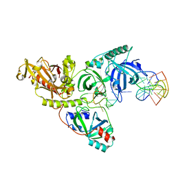
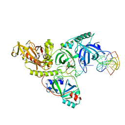
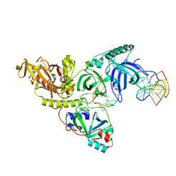
Crystal structure of a PLP-dependent enzyme
Descriptor: (2-AMINO-4-FORMYL-5-HYDROXY-6-METHYLPYRIDIN-3-YL)METHYL DIHYDROGEN PHOSPHATE, BETA-MERCAPTOETHANOL, aminotransferase
Authors:Structural GenomiX
Deposit date:2003-10-23
Release date:2003-11-11
Last modified:2023-12-27
Method:X-RAY DIFFRACTION (1.84 Å)
Cite:Structural analysis of a set of proteins resulting from a bacterial genomics project
Proteins, 60, 2005
5OR3
DownloadVisualize
BU of 5or3 by Molmil
Crystal structure of Aspergillus oryzae catechol oxidase in met/deoxy-form
Descriptor: 1,2-ETHANEDIOL, 2-acetamido-2-deoxy-beta-D-glucopyranose, 2-acetamido-2-deoxy-beta-D-glucopyranose-(1-4)-2-acetamido-2-deoxy-beta-D-glucopyranose, ...
Authors:Hakulinen, N, Penttinen, L, Rutanen, C, Rouvinen, J.
Deposit date:2017-08-15
Release date:2018-05-09
Last modified:2024-11-20
Method:X-RAY DIFFRACTION (1.795 Å)
Cite:A new crystal form of Aspergillus oryzae catechol oxidase and evaluation of copper site structures in coupled binuclear copper enzymes.
PLoS ONE, 13, 2018
1O62
DownloadVisualize
BU of 1o62 by Molmil
Crystal structure of the apo form of a PLP-dependent enzyme
Descriptor: ACETATE ION, BETA-MERCAPTOETHANOL, aminotransferase
Authors:Structural GenomiX
Deposit date:2003-10-23
Release date:2003-11-11
Last modified:2023-12-27
Method:X-RAY DIFFRACTION (2.1 Å)
Cite:Structural analysis of a set of proteins resulting from a bacterial genomics project.
Proteins, 60, 2005
1O65
DownloadVisualize
BU of 1o65 by Molmil
Crystal structure of an hypothetical protein
Descriptor: Hypothetical protein yiiM
Authors:Structural GenomiX
Deposit date:2003-10-23
Release date:2003-11-11
Last modified:2023-12-27
Method:X-RAY DIFFRACTION (2.33 Å)
Cite:Structural analysis of a set of proteins resulting from a bacterial genomics project
Proteins, 60, 2005
1O68
DownloadVisualize
BU of 1o68 by Molmil
Crystal structure of 3-methyl-2-oxobutanoate hydroxymethyltransferase
Descriptor: 3-METHYL-2-OXOBUTANOIC ACID, 3-methyl-2-oxobutanoate hydroxymethyltransferase, SODIUM ION
Authors:Structural GenomiX
Deposit date:2003-10-23
Release date:2003-11-11
Last modified:2024-10-30
Method:X-RAY DIFFRACTION (2.1 Å)
Cite:Structural analysis of a set of proteins resulting from a bacterial genomics project
Proteins, 60, 2005
5ZCI
DownloadVisualize
BU of 5zci by Molmil
Crystal structure of apo form of Xylose reductase from Debaryomyces nepalensis
Descriptor: Aldose reductase
Authors:Manoj, N.
Deposit date:2018-02-17
Release date:2018-11-07
Last modified:2023-11-22
Method:X-RAY DIFFRACTION (2 Å)
Cite:Crystal structure of yeast xylose reductase in complex with a novel NADP-DTT adduct provides insights into substrate recognition and catalysis.
FEBS J., 285, 2018
1R4V
DownloadVisualize
BU of 1r4v by Molmil
1.9A crystal structure of protein AQ328 from Aquifex aeolicus
Descriptor: CACODYLATE ION, Hypothetical protein AQ_328, ZINC ION
Authors:Qiu, Y, Tereshko, V, Kim, Y, Zhang, R, Collart, F, Joachimiak, A, Kossiakoff, A, Midwest Center for Structural Genomics (MCSG)
Deposit date:2003-10-08
Release date:2004-03-30
Last modified:2024-10-30
Method:X-RAY DIFFRACTION (1.9 Å)
Cite:The crystal structure of Aq_328 from the hyperthermophilic bacteria Aquifex aeolicus shows an ancestral histone fold.
Proteins, 62, 2006
5OR4
DownloadVisualize
BU of 5or4 by Molmil
Crystal structure of Aspergillus oryzae catechol oxidase in deoxy-form
Descriptor: 2-acetamido-2-deoxy-beta-D-glucopyranose, 2-acetamido-2-deoxy-beta-D-glucopyranose-(1-4)-2-acetamido-2-deoxy-beta-D-glucopyranose, COPPER (II) ION, ...
Authors:Hakulinen, N, Penttinen, L, Rutanen, C, Rouvinen, J.
Deposit date:2017-08-15
Release date:2018-05-09
Last modified:2024-10-09
Method:X-RAY DIFFRACTION (2.445 Å)
Cite:A new crystal form of Aspergillus oryzae catechol oxidase and evaluation of copper site structures in coupled binuclear copper enzymes.
PLoS ONE, 13, 2018
6BY0
DownloadVisualize
BU of 6by0 by Molmil
Crystal structure of catalase HPII from E. coli in space group P1
Descriptor: Catalase HPII, PROTOPORPHYRIN IX CONTAINING FE
Authors:Lisa, M.N, Buschiazzo, A.
Deposit date:2017-12-19
Release date:2018-01-24
Last modified:2024-11-06
Method:X-RAY DIFFRACTION (2.93 Å)
Cite:SIMBAD: a sequence-independent molecular-replacement pipeline.
Acta Crystallogr D Struct Biol, 74, 2018
1TEW
DownloadVisualize
BU of 1tew by Molmil
STRUCTURE OF HEXAGONAL TURKEY EGG WHITE LYSOZYME AT 1.65 ANGSTROMS RESOLUTION
Descriptor: THIOCYANATE ION, TURKEY EGG WHITE LYSOZYME
Authors:Howell, P.L.
Deposit date:1994-11-17
Release date:1995-01-26
Last modified:2024-10-16
Method:X-RAY DIFFRACTION (1.65 Å)
Cite:Structure of hexagonal turkey egg-white lysozyme at 1.65A resolution.
Acta Crystallogr.,Sect.D, 51, 1995
1O6B
DownloadVisualize
BU of 1o6b by Molmil
Crystal structure of phosphopantetheine adenylyltransferase with ADP
Descriptor: ADENOSINE-5'-DIPHOSPHATE, CHLORIDE ION, MAGNESIUM ION, ...
Authors:Structural GenomiX
Deposit date:2003-11-03
Release date:2003-11-25
Last modified:2024-11-13
Method:X-RAY DIFFRACTION (2.2 Å)
Cite:Structural analysis of a set of proteins resulting from a bacterial genomics project
Proteins, 60, 2005
6AJD
DownloadVisualize
BU of 6ajd by Molmil
Crystal structure of a monometallic dihydropyrimidinase from Pseudomonas aeruginosa PAO1 reveals no lysine carbamylation within the active site
Descriptor: D-hydantoinase/dihydropyrimidinase, ZINC ION
Authors:Huang, Y.H, Huang, C.Y.
Deposit date:2018-08-27
Release date:2018-10-24
Last modified:2023-11-22
Method:X-RAY DIFFRACTION (2.223 Å)
Cite:Crystal structures of monometallic dihydropyrimidinase and the human dihydroorotase domain K1556A mutant reveal no lysine carbamylation within the active site
Biochem. Biophys. Res. Commun., 505, 2018
1O66
DownloadVisualize
BU of 1o66 by Molmil
Crystal structure of 3-methyl-2-oxobutanoate hydroxymethyltransferase
Descriptor: 3-methyl-2-oxobutanoate hydroxymethyltransferase, GLYCEROL
Authors:Structural GenomiX
Deposit date:2003-10-23
Release date:2003-11-11
Last modified:2024-10-30
Method:X-RAY DIFFRACTION (1.75 Å)
Cite:Structural analysis of a set of proteins resulting from a bacterial genomics project
Proteins, 60, 2005
1XQI
DownloadVisualize
BU of 1xqi by Molmil
Crystal Structure Analysis of an NDP kinase from Pyrobaculum aerophilum
Descriptor: 2-AMINO-2-HYDROXYMETHYL-PROPANE-1,3-DIOL, Nucleoside diphosphate kinase, TRIETHYLENE GLYCOL
Authors:Pedelacq, J.D, Waldo, G.S, Cabantous, S, Liong, E.C, Berendzen, J, Terwilliger, T.C.
Deposit date:2004-10-12
Release date:2005-09-20
Last modified:2024-10-30
Method:X-RAY DIFFRACTION (2.5 Å)
Cite:Structural and functional features of an NDP kinase from the hyperthermophile crenarchaeon Pyrobaculum aerophilum
Protein Sci., 14, 2005
1TKQ
DownloadVisualize
BU of 1tkq by Molmil
SOLUTION STRUCTURE OF A LINKED UNSYMMETRIC GRAMICIDIN IN A MEMBRANE-ISOELECTRICAL SOLVENTS MIXTURE IN THE PRESENCE OF CsCl
Descriptor: GRAMICIDIN A, MINI-GRAMICIDIN A, SUCCINIC ACID
Authors:Xie, X, Al-Momani, L, Bockelmann, D, Griesinger, C, Koert, U.
Deposit date:2004-06-09
Release date:2004-07-13
Last modified:2025-03-26
Method:SOLUTION NMR
Cite:An Asymmetric Ion Channel Derived from Gramicidin A. Synthesis, Function and NMR Structure.
FEBS J., 272, 2005
1XFT
DownloadVisualize
BU of 1xft by Molmil
Synchrotron X-ray Powder Diffraction Study of Hexagonal Turkey Egg-white Lysozyme
Descriptor: Lysozyme C
Authors:Margiolaki, I, Wright, J.P.
Deposit date:2004-09-15
Release date:2005-04-05
Last modified:2024-11-20
Method:POWDER DIFFRACTION (3.35 Å)
Cite:Synchrotron X-ray powder diffraction study of hexagonal turkey egg-white lysozyme.
Acta Crystallogr.,Sect.D, 61, 2005
1TE7
DownloadVisualize
BU of 1te7 by Molmil
Solution NMR Structure of Protein yqfB from Escherichia coli. Northeast Structural Genomics Consortium Target ET99
Descriptor: Hypothetical UPF0267 protein yqfB
Authors:Atreya, H.S, Shen, Y, Yee, A, Arrowsmith, C, Szyperski, T, Northeast Structural Genomics Consortium (NESG)
Deposit date:2004-05-24
Release date:2005-01-04
Last modified:2024-05-22
Method:SOLUTION NMR
Cite:G-Matrix Fourier Transform NOESY-Based Protocol for High-Quality Protein Structure Determination
J.Am.Chem.Soc., 127, 2005
1Y4O
DownloadVisualize
BU of 1y4o by Molmil
Solution structure of a mouse cytoplasmic Roadblock/LC7 dynein light chain
Descriptor: Dynein light chain 2A, cytoplasmic
Authors:Song, J, Tyler, R.C, Lee, M.S, Tyler, E.M, Markley, J.L, Center for Eukaryotic Structural Genomics (CESG)
Deposit date:2004-12-01
Release date:2005-01-18
Last modified:2024-05-22
Method:SOLUTION NMR
Cite:Solution structure of isoform 1 of Roadblock/LC7, a light chain in the dynein complex.
J.Mol.Biol., 354, 2005
1PH2
DownloadVisualize
BU of 1ph2 by Molmil
CRYSTAL STRUCTURE OF THE OXYTRICHA NOVA TELOMERE END-BINDING PROTEIN COMPLEXED WITH NONCOGNATE SSDNA GGGGTTTTG
Descriptor: 5'-D(*GP*GP*GP*GP*TP*TP*TP*TP*G)-3', 5'-D(*GP*GP*GP*GP*TP*TP*TP*TP*GP*GP*GP*GP*T)-3', CHLORIDE ION, ...
Authors:Theobald, D.L, Schultz, S.C.
Deposit date:2003-05-29
Release date:2003-06-17
Last modified:2024-02-14
Method:X-RAY DIFFRACTION (3.1 Å)
Cite:Nucleotide Shuffling and ssDNA Recognition in Oxytricha Nova Telomere End-Binding Protein Complexes
Embo J., 22, 2003
1PH4
DownloadVisualize
BU of 1ph4 by Molmil
CRYSTAL STRUCTURE OF THE OXYTRICHA NOVA TELOMERE END-BINDING PROTEIN COMPLEXED WITH NONCOGNATE SSDNA GGGGTTTTGGCG
Descriptor: 5'-D(*GP*GP*GP*GP*TP*TP*TP*TP*GP*GP*CP*G)-3', 5'-D(*GP*GP*GP*GP*TP*TP*TP*TP*GP*GP*GP*GP*T)-3', CHLORIDE ION, ...
Authors:Theobald, D.L, Schultz, S.C.
Deposit date:2003-05-29
Release date:2003-06-17
Last modified:2024-02-14
Method:X-RAY DIFFRACTION (2.3 Å)
Cite:Nucleotide Shuffling and ssDNA Recognition in Oxytricha Nova Telomere End-Binding Protein Complexes
Embo J., 22, 2003
1TTO
DownloadVisualize
BU of 1tto by Molmil
Crystal structure of the Rnase T1 variant R2
Descriptor: 2-AMINO-2-HYDROXYMETHYL-PROPANE-1,3-DIOL, RNase T1
Authors:Hahn, U, Czaja, R, Perbandt, M, Betzel, C.
Deposit date:2004-06-23
Release date:2005-09-06
Last modified:2024-11-20
Method:X-RAY DIFFRACTION (2.1 Å)
Cite:Purine activity of RNase T1RV is further improved by substitution of Trp59 by tyrosine
Biochem.Biophys.Res.Commun., 336, 2005
1PH3
DownloadVisualize
BU of 1ph3 by Molmil
CRYSTAL STRUCTURE OF THE OXYTRICHA NOVA TELOMERE END-BINDING PROTEIN COMPLEXED WITH NONCOGNATE SSDNA GGGGTTTTGGTG
Descriptor: 5'-D(*GP*GP*GP*GP*TP*TP*TP*TP*GP*GP*GP*GP*T)-3', 5'-D(*GP*GP*GP*GP*TP*TP*TP*TP*GP*GP*TP*G)-3', SODIUM ION, ...
Authors:Theobald, D.L, Schultz, S.C.
Deposit date:2003-05-29
Release date:2003-06-17
Last modified:2024-02-14
Method:X-RAY DIFFRACTION (2.3 Å)
Cite:Nucleotide Shuffling and ssDNA Recognition in Oxytricha Nova Telomere End-Binding Protein Complexes
Embo J., 22, 2003
1PH5
DownloadVisualize
BU of 1ph5 by Molmil
CRYSTAL STRUCTURE OF THE OXYTRICHA NOVA TELOMERE END-BINDING PROTEIN COMPLEXED WITH NONCOGNATE SSDNA GGGGTTTTG(3DR)GG
Descriptor: 5'-D(*GP*GP*GP*GP*TP*TP*TP*TP*GP*(3DR)P*GP*G)-3', 5'-D(*GP*GP*GP*GP*TP*TP*TP*TP*GP*GP*GP*GP*T)-3', CHLORIDE ION, ...
Authors:Theobald, D.L, Schultz, S.C.
Deposit date:2003-05-29
Release date:2003-06-17
Last modified:2024-02-14
Method:X-RAY DIFFRACTION (2.3 Å)
Cite:Nucleotide Shuffling and ssDNA Recognition in Oxytricha Nova Telomere End-Binding Protein Complexes
Embo J., 22, 2003
5OQ1
DownloadVisualize
BU of 5oq1 by Molmil
Crystal structure of Serratia marcescens ChiX (used as MR model for superior PDB 5OPZ)
Descriptor: CHLORIDE ION, ChiX, ZINC ION
Authors:Owen, R.A, Fyfe, P.K, Lodge, A, Biboy, J, Vollmer, W, Hunter, W.N, Sargent, F.
Deposit date:2017-08-10
Release date:2018-01-17
Last modified:2024-05-08
Method:X-RAY DIFFRACTION (1.34 Å)
Cite:Structure and activity of ChiX: a peptidoglycan hydrolase required for chitinase secretion by Serratia marcescens.
Biochem. J., 475, 2018

245663

PDB entries from 2025-12-03

PDB statisticsPDBj update infoContact PDBjnumon