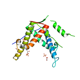
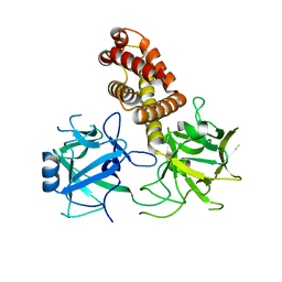
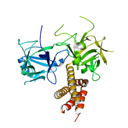
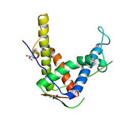
Loading
PDBj
メニューPDBj@FacebookPDBj@TwitterPDBj@YouTubewwPDB FoundationwwPDB
RCSB PDBPDBeBMRBAdv. SearchSearch help
PDB: 143 件

3FYQ
DownloadVisualize
BU of 3fyq by Molmil
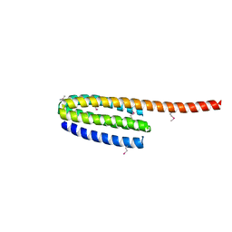
Structure of Drosophila melanogaster talin IBS2 domain (residues 1981-2168)
分子名称: CG6831-PA (Talin)
著者Cheung, T.Y.S, Fairchild, M.J, Zarivach, R, Tanentzapf, G, Van Petegem, F.
登録日2009-01-22
公開日2009-02-03
最終更新日2018-01-24
実験手法X-RAY DIFFRACTION (1.95 Å)
主引用文献Crystal structure of the talin integrin binding domain 2.
J.Mol.Biol., 387, 2009
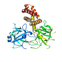
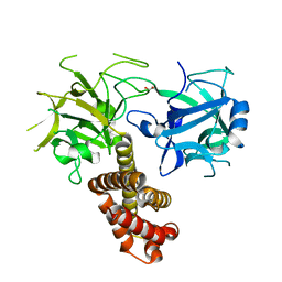
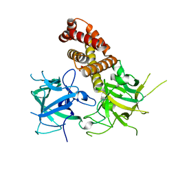
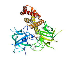
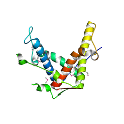
4DJC
DownloadVisualize
BU of 4djc by Molmil
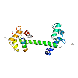
1.35 A crystal structure of the NaV1.5 DIII-IV-Ca/CaM complex
分子名称: CALCIUM ION, Calmodulin, ISOPROPYL ALCOHOL, ...
著者Sarhan, M.F, Tung, C.-C, Van Petegem, F, Ahern, C.A.
登録日2012-02-01
公開日2012-02-22
最終更新日2024-02-28
実験手法X-RAY DIFFRACTION (1.35 Å)
主引用文献Crystallographic basis for calcium regulation of sodium channels.
Proc.Natl.Acad.Sci.USA, 109, 2012
4ETT
DownloadVisualize
BU of 4ett by Molmil
Crystal structure of rabbit ryanodine receptor 1 mutant E2764K
分子名称: GLYCEROL, Ryanodine receptor 1
著者Yuchi, Z, Lau, K, Van Petegem, F.
登録日2012-04-24
公開日2012-06-13
最終更新日2023-09-13
実験手法X-RAY DIFFRACTION (2.195 Å)
主引用文献Disease mutations in the ryanodine receptor central region: crystal structures of a phosphorylation hot spot domain.
Structure, 20, 2012
4ESU
DownloadVisualize
BU of 4esu by Molmil
Crystal structure of rabbit ryanodine receptor 1 mutant S2776M
分子名称: GLYCEROL, Ryanodine receptor 1
著者Yuchi, Z, Lau, K, Van Petegem, F.
登録日2012-04-23
公開日2012-06-13
最終更新日2023-09-13
実験手法X-RAY DIFFRACTION (1.59 Å)
主引用文献Disease mutations in the ryanodine receptor central region: crystal structures of a phosphorylation hot spot domain.
Structure, 20, 2012
4ERV
DownloadVisualize
BU of 4erv by Molmil
Crystal structure of human ryanodine receptor 3 (2597-2800)
分子名称: GLYCEROL, Ryanodine receptor 3, SULFATE ION
著者Yuchi, Z, Lau, K, Van Petegem, F.
登録日2012-04-20
公開日2012-06-13
最終更新日2024-02-28
実験手法X-RAY DIFFRACTION (1.75 Å)
主引用文献Disease mutations in the ryanodine receptor central region: crystal structures of a phosphorylation hot spot domain.
Structure, 20, 2012
4I2S
DownloadVisualize
BU of 4i2s by Molmil
Crystal structure of rabbit ryanodine receptor 1 (residues 1-536) disease mutant I404M
分子名称: GLYCEROL, Ryanodine receptor 1
著者Kimlicka, L, Van Petegem, F.
登録日2012-11-22
公開日2013-02-20
最終更新日2023-09-20
実験手法X-RAY DIFFRACTION (2.5 Å)
主引用文献Disease mutations in the ryanodine receptor N-terminal region couple to a mobile intersubunit interface.
Nat Commun, 4, 2013
4I8M
DownloadVisualize
BU of 4i8m by Molmil
Crystal structure of rabbit Ryanodine receptor 1 (residues 1-536) disease mutant V219I
分子名称: GLYCEROL, Ryanodine receptor 1
著者Kimlicka, L, Van Petegem, F.
登録日2012-12-03
公開日2013-02-20
最終更新日2023-09-20
実験手法X-RAY DIFFRACTION (2.8 Å)
主引用文献Disease mutations in the ryanodine receptor N-terminal region couple to a mobile intersubunit interface.
Nat Commun, 4, 2013
4I7I
DownloadVisualize
BU of 4i7i by Molmil
Crystal structure of rabbit ryanodine receptor 1 (residues 1-536) disease mutant L14R
分子名称: GLYCEROL, Ryanodine receptor 1
著者Kimlicka, L, Van Petegem, F.
登録日2012-11-30
公開日2013-02-20
最終更新日2023-09-20
実験手法X-RAY DIFFRACTION (2.9 Å)
主引用文献Disease mutations in the ryanodine receptor N-terminal region couple to a mobile intersubunit interface.
Nat Commun, 4, 2013
4I1E
DownloadVisualize
BU of 4i1e by Molmil
Crystal structure of Rabbit Ryanodine Receptor 1 (residues 1-536) disease mutant G249R
分子名称: Ryanodine receptor 1
著者Kimlicka, L, Van Petegem, F.
登録日2012-11-20
公開日2013-02-20
最終更新日2024-02-28
実験手法X-RAY DIFFRACTION (2.4 Å)
主引用文献Disease mutations in the ryanodine receptor N-terminal region couple to a mobile intersubunit interface.
Nat Commun, 4, 2013
4I3N
DownloadVisualize
BU of 4i3n by Molmil
Crystal structure of rabbit ryanodine receptor 1 (residues 1-536) disease mutant D61N
分子名称: GLYCEROL, Ryanodine receptor 1
著者Kimlicka, L, Van Petegem, F.
登録日2012-11-26
公開日2013-02-20
最終更新日2023-09-20
実験手法X-RAY DIFFRACTION (2.95 Å)
主引用文献Disease mutations in the ryanodine receptor N-terminal region couple to a mobile intersubunit interface.
Nat Commun, 4, 2013
4I96
DownloadVisualize
BU of 4i96 by Molmil
Crystal structure of the N-terminal two domains of the skeletal muscle ryanodine receptor (rabbit RyR1) residues 217-536
分子名称: Ryanodine receptor 1
著者Kimlicka, L, Tung, C.C, Van Petegem, F.
登録日2012-12-04
公開日2013-02-20
最終更新日2023-09-20
実験手法X-RAY DIFFRACTION (2.73 Å)
主引用文献Disease mutations in the ryanodine receptor N-terminal region couple to a mobile intersubunit interface.
Nat Commun, 4, 2013
4I37
DownloadVisualize
BU of 4i37 by Molmil
Crystal structure of rabbit ryanodine receptor 1 (residues 1-536) disease mutant R402G
分子名称: Ryanodine receptor 1
著者Kimlicka, L, Van Petegem, F.
登録日2012-11-25
公開日2013-02-20
最終更新日2023-09-20
実験手法X-RAY DIFFRACTION (2.95 Å)
主引用文献Disease mutations in the ryanodine receptor N-terminal region couple to a mobile intersubunit interface.
Nat Commun, 4, 2013
4I6I
DownloadVisualize
BU of 4i6i by Molmil
Crystal structure of rabbit ryanodine receptor 1 (residues 1-559) disease mutant R45C
分子名称: Ryanodine receptor 1
著者Kimlicka, L, Van Petegem, F.
登録日2012-11-29
公開日2013-02-20
最終更新日2023-09-20
実験手法X-RAY DIFFRACTION (2.5 Å)
主引用文献Disease mutations in the ryanodine receptor N-terminal region couple to a mobile intersubunit interface.
Nat Commun, 4, 2013
4L4I
DownloadVisualize
BU of 4l4i by Molmil
Crystal structure of mouse Ryanodine Receptor isoform 2 (RyR2) 1-547 disease mutant R420Q
分子名称: GLYCEROL, Ryanodine receptor 2
著者Kimlicka, L, Van Petegem, F.
登録日2013-06-07
公開日2013-07-24
最終更新日2023-09-20
実験手法X-RAY DIFFRACTION (2.15 Å)
主引用文献Crystal structure of ryanodine receptor disease mutant
To be Published
4L4H
DownloadVisualize
BU of 4l4h by Molmil
Crystal structure of mouse Ryanodine Receptor isoform 2 (RyR2) 1-547
分子名称: CHLORIDE ION, DI(HYDROXYETHYL)ETHER, Ryanodine receptor 2, ...
著者Kimlicka, L, Tung, C.C, Van Petegem, F.
登録日2013-06-07
公開日2013-07-24
最終更新日2023-09-20
実験手法X-RAY DIFFRACTION (2 Å)
主引用文献Crystal structure of a ryanodine receptor domain
To be Published
4ETU
DownloadVisualize
BU of 4etu by Molmil
Crystal structure of rabbit ryanodine receptor 1 mutant R2939S
分子名称: GLYCEROL, Ryanodine receptor 1
著者Yuchi, Z, Lau, K, Van Petegem, F.
登録日2012-04-24
公開日2012-06-13
最終更新日2023-09-13
実験手法X-RAY DIFFRACTION (2.19 Å)
主引用文献Disease mutations in the ryanodine receptor central region: crystal structures of a phosphorylation hot spot domain.
Structure, 20, 2012
4ERT
DownloadVisualize
BU of 4ert by Molmil
Crystal structure of rabbit ryanodine receptor 1 (2734-2940)
分子名称: GLYCEROL, POTASSIUM ION, Ryanodine receptor 1
著者Yuchi, Z, Lau, K, Van Petegem, F.
登録日2012-04-20
公開日2012-06-13
最終更新日2024-02-28
実験手法X-RAY DIFFRACTION (1.95 Å)
主引用文献Disease mutations in the ryanodine receptor central region: crystal structures of a phosphorylation hot spot domain.
Structure, 20, 2012
4ETV
DownloadVisualize
BU of 4etv by Molmil
Crystal structure of mouse ryanodine receptor 2 (2699-2904)
分子名称: CHLORIDE ION, Ryanodine receptor 2
著者Yuchi, Z, Lau, K, Van Petegem, F.
登録日2012-04-24
公開日2012-06-13
最終更新日2017-11-15
実験手法X-RAY DIFFRACTION (1.65 Å)
主引用文献Disease mutations in the ryanodine receptor central region: crystal structures of a phosphorylation hot spot domain.
Structure, 20, 2012

225158

件を2024-09-18に公開中

PDB statisticsPDBj update infoContact PDBjnumon