2E1S
 
 | | Crystal structure of the complex of C-terminal half of bovine lactoferrin and arabinose at 2.7 A resolution | | 分子名称: | 2-acetamido-2-deoxy-beta-D-glucopyranose-(1-4)-2-acetamido-2-deoxy-beta-D-glucopyranose, CARBONATE ION, FE (III) ION, ... | | 著者 | Mir, R, Prem Kumar, R, Singh, N, Sinha, M, Sharma, S, Kaur, P, Singh, T.P. | | 登録日 | 2006-10-27 | | 公開日 | 2006-11-14 | | 最終更新日 | 2023-10-25 | | 実験手法 | X-RAY DIFFRACTION (2.7 Å) | | 主引用文献 | Crystal structure of the complex of C-terminal half of bovine lactoferrin and arabinose at 2.7 A resolution
To be Published
|
|
5ZXM
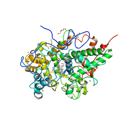
 | | Crystal Structure of GyraseB N-terminal at 1.93A Resolution | | 分子名称: | ADENOSINE-5'-DIPHOSPHATE, DI(HYDROXYETHYL)ETHER, DNA gyrase subunit B, ... | | 著者 | Tiwari, P, Gupta, D, Sachdeva, E, Sharma, S, Singh, T.P, Ethayathulla, A.S, Kaur, P. | | 登録日 | 2018-05-21 | | 公開日 | 2019-05-22 | | 最終更新日 | 2023-11-22 | | 実験手法 | X-RAY DIFFRACTION (1.938 Å) | | 主引用文献 | Structural insights into the transient closed conformation and pH dependent ATPase activity of S.Typhi GyraseB N- terminal domain.
Arch.Biochem.Biophys., 701, 2021
|
|
2DXR
 | | Crystal structure of the complex formed between C-terminal half of bovine lactoferrin and sorbitol at 2.85 A resolution | | 分子名称: | 2-acetamido-2-deoxy-beta-D-glucopyranose-(1-4)-2-acetamido-2-deoxy-beta-D-glucopyranose, CARBONATE ION, FE (III) ION, ... | | 著者 | Mir, R, Prem kumar, R, Sinha, M, Singh, N, Sharma, S, Bhushan, A, Kaur, P, Singh, T.P. | | 登録日 | 2006-08-30 | | 公開日 | 2006-09-12 | | 最終更新日 | 2023-10-25 | | 実験手法 | X-RAY DIFFRACTION (2.85 Å) | | 主引用文献 | Crystal structure of the complex formed between C-terminal half of bovine lactoferrin and sorbitol at 2.85 A resolution
To be Published
|
|
2DYX
 | | Structure of the complex of lactoferrin C-lobe with melibiose at 2.0 A resolution | | 分子名称: | 2-acetamido-2-deoxy-beta-D-glucopyranose-(1-4)-2-acetamido-2-deoxy-beta-D-glucopyranose, CARBONATE ION, FE (III) ION, ... | | 著者 | Mir, R, Prem kumar, R, Sinha, M, Singh, N, Sharma, S, Kaur, P, Bhushan, A, Singh, T.P. | | 登録日 | 2006-09-19 | | 公開日 | 2006-10-03 | | 最終更新日 | 2023-10-25 | | 実験手法 | X-RAY DIFFRACTION (2 Å) | | 主引用文献 | Structure of the complex of lactoferrin C-lobe with melibiose at 2.0 A resolution
To be Published
|
|
2E0S
 | | Carbohydrate recognition of C-terminal half of lactoferrin: Crystal structure of the complex of C-lobe with rhamnose at 2.15 A resolution | | 分子名称: | 2-acetamido-2-deoxy-beta-D-glucopyranose-(1-4)-2-acetamido-2-deoxy-beta-D-glucopyranose, CARBONATE ION, FE (III) ION, ... | | 著者 | Mir, R, Prem Kumar, R, Singh, N, Sinha, M, Sharma, S, Bhushan, A, Kaur, P, Singh, T.P. | | 登録日 | 2006-10-11 | | 公開日 | 2006-10-24 | | 最終更新日 | 2023-10-25 | | 実験手法 | X-RAY DIFFRACTION (2.15 Å) | | 主引用文献 | Carbohydrate recognition of C-terminal half of lactoferrin: Crystal structure of the complex of C-lobe with rhamnose at 2.15 A resolution
To be Published
|
|
5YRR
 | | The crystal structure of Phosphopantetheine adenylyltransferase from Acinetobacter baumannii with Coenzyme A at 2.88 A resolution | | 分子名称: | COENZYME A, Phosphopantetheine adenylyltransferase, SULFATE ION | | 著者 | Bairagya, H.R, Gupta, A, Iqbal, N, Kaur, P, Sharma, S, Singh, T.P. | | 登録日 | 2017-11-09 | | 公開日 | 2017-11-22 | | 最終更新日 | 2023-11-22 | | 実験手法 | X-RAY DIFFRACTION (2.88 Å) | | 主引用文献 | The crystal structure of Phosphopantetheine adenylyltransferase from Acinetobacter baumannii with Coenzyme A at 2.88 A resolution
To Be Published
|
|
5Z9A
 | |
7WYJ
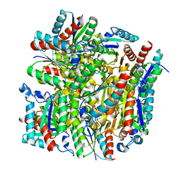
 | | Structure of the complex of lactoperoxidase with nitric oxide catalytic product nitrite at 1.89 A resolution | | 分子名称: | 1,2-ETHANEDIOL, 2-acetamido-2-deoxy-beta-D-glucopyranose, 2-acetamido-2-deoxy-beta-D-glucopyranose-(1-4)-2-acetamido-2-deoxy-beta-D-glucopyranose, ... | | 著者 | Viswanathan, V, Pandey, N, Singh, A.K, Sinha, M, Singh, R.P, Sharma, P, Kaur, P, Sharma, S, Singh, T.P. | | 登録日 | 2022-02-16 | | 公開日 | 2023-01-11 | | 最終更新日 | 2023-11-29 | | 実験手法 | X-RAY DIFFRACTION (1.89 Å) | | 主引用文献 | Structural evidence of the conversion of nitric oxide (NO) to nitrite ion (NO2-) by lactoperoxidase (LPO): Structure of the complex of LPO with NO2- at 1.89 angstrom resolution
J.Inorg.Biochem., 247, 2023
|
|
5YPQ
 | |
3UBA
 | | Crystal structure of the complex of bovine lactoperoxidase with p-hydroxycinnamic acid at 2.6 A resolution | | 分子名称: | 1,2-ETHANEDIOL, 2-acetamido-2-deoxy-beta-D-glucopyranose, 2-acetamido-2-deoxy-beta-D-glucopyranose-(1-4)-2-acetamido-2-deoxy-beta-D-glucopyranose, ... | | 著者 | Pandey, N, Singh, A.K, Singh, R.P, Sinha, M, Kaur, P, Sharma, S, Singh, T.P. | | 登録日 | 2011-10-24 | | 公開日 | 2011-12-28 | | 最終更新日 | 2023-11-01 | | 実験手法 | X-RAY DIFFRACTION (2.65 Å) | | 主引用文献 | Crystal structure of the complex of bovine lactoperoxidase with p-hydroxycinnamic acid at 2.6 A resolution
to be published
|
|
3TGY
 | | Crystal structure of the complex of Bovine Lactoperoxidase with Ascorbic acid at 2.35 A resolution | | 分子名称: | (4S)-2-METHYL-2,4-PENTANEDIOL, 1,2-ETHANEDIOL, 2-acetamido-2-deoxy-beta-D-glucopyranose, ... | | 著者 | Yamini, S, Singh, R.P, Singh, A.K, Pandey, N, Sinha, M, Kaur, P, Sharma, S, Singh, T.P. | | 登録日 | 2011-08-18 | | 公開日 | 2011-09-21 | | 最終更新日 | 2023-11-01 | | 実験手法 | X-RAY DIFFRACTION (2.35 Å) | | 主引用文献 | Structure of bovine lactoperoxidase with a partially linked heme moiety at 1.98 angstrom resolution.
Biochim.Biophys.Acta, 1865, 2017
|
|
2O92
 | | Crystal structure of a signalling protein (SPG-40) complex with tetrasaccharide at 3.0A resolution | | 分子名称: | 2-acetamido-2-deoxy-beta-D-glucopyranose-(1-4)-2-acetamido-2-deoxy-beta-D-glucopyranose-(1-4)-2-acetamido-2-deoxy-beta-D-glucopyranose-(1-4)-2-acetamido-2-deoxy-beta-D-glucopyranose, Chitinase-3-like protein 1, alpha-D-mannopyranose-(1-4)-2-acetamido-2-deoxy-beta-D-glucopyranose-(1-4)-2-acetamido-2-deoxy-beta-D-glucopyranose | | 著者 | Sharma, P, Singh, N, Sharma, S, Kaur, P, Singh, T.P. | | 登録日 | 2006-12-13 | | 公開日 | 2006-12-26 | | 最終更新日 | 2023-10-25 | | 実験手法 | X-RAY DIFFRACTION (3 Å) | | 主引用文献 | Crystal structure of a signalling protein (SPG-40) complex with tetrasaccharide at 3.0A resolution
To be Published
|
|
2OLH
 | | Crystal structure of a signalling protein (SPG-40) complex with cellobiose at 2.78 A resolution | | 分子名称: | Chitinase-3-like protein 1, alpha-D-mannopyranose-(1-4)-2-acetamido-2-deoxy-beta-D-glucopyranose-(1-4)-2-acetamido-2-deoxy-beta-D-glucopyranose, beta-D-glucopyranose-(1-4)-beta-D-glucopyranose | | 著者 | Sharma, P, Singh, N, Sharma, S, Bhushan, A, Kaur, P, Singh, T.P. | | 登録日 | 2007-01-19 | | 公開日 | 2007-02-13 | | 最終更新日 | 2023-10-25 | | 実験手法 | X-RAY DIFFRACTION (2.78 Å) | | 主引用文献 | Crystal structure of a signalling protein (SPG-40) complex with cellobiose at 2.78 A resolution
To be Published
|
|
3QV4
 | | Crystal structure of the complex of peptidoglycan recognition protein (PGRP-S) with dipeptide L-ALA D-GLU at 2.7 A resolution | | 分子名称: | 1,2-ETHANEDIOL, 2-acetamido-2-deoxy-beta-D-glucopyranose-(1-4)-2-acetamido-2-deoxy-beta-D-glucopyranose-(1-4)-2-acetamido-2-deoxy-beta-D-glucopyranose, ALANINE, ... | | 著者 | Shukla, P.K, Sharma, P, Sinha, M, Kaur, P, Sharma, S, Singh, T.P. | | 登録日 | 2011-02-25 | | 公開日 | 2011-03-30 | | 最終更新日 | 2023-11-01 | | 実験手法 | X-RAY DIFFRACTION (2.7 Å) | | 主引用文献 | Crystal structure of the complex of peptidoglycan recognition protein (PGRP-S) with dipeptide L-ALA D-GLU at 2.7 A resolution
To be Published
|
|
2PI6
 | | Crystal structure of the sheep signalling glycoprotein (SPS-40) complex with 2-methyl-2-4-pentanediol at 1.65A resolution reveals specific binding characteristics of SPS-40 | | 分子名称: | (4S)-2-METHYL-2,4-PENTANEDIOL, Chitinase-3-like protein 1, ETHANOL, ... | | 著者 | Sharma, P, Singh, N, Sharma, S, Kaur, P, Betzel, C, Singh, T.P. | | 登録日 | 2007-04-13 | | 公開日 | 2007-05-01 | | 最終更新日 | 2023-10-25 | | 実験手法 | X-RAY DIFFRACTION (1.65 Å) | | 主引用文献 | Tryptophan as a three-way switch in regulating the function of the secretory signalling glycoprotein (SPS-40) from mammary glands: structure of SPS-40 complexed with 2-methylpentane-2,4-diol at 1.6 A resolution.
Acta Crystallogr.,Sect.D, 65, 2009
|
|
2PT3
 | | Crystal structure of bovine lactoperoxidase at 2.34 A resolution reveals multiple anion binding sites | | 分子名称: | 2-acetamido-2-deoxy-beta-D-glucopyranose-(1-4)-2-acetamido-2-deoxy-beta-D-glucopyranose, CALCIUM ION, Lactoperoxidase, ... | | 著者 | Singh, A.K, Singh, N, Sharma, S, Kaur, P, Betzel, C, Singh, T.P. | | 登録日 | 2007-05-08 | | 公開日 | 2007-05-22 | | 最終更新日 | 2023-08-30 | | 実験手法 | X-RAY DIFFRACTION (2.34 Å) | | 主引用文献 | Crystal structure of bovine lactoperoxidase at 2.34 A resolution reveals multiple anion binding sites
To be Published
|
|
3TOD
 | | Crystal Structure of C-lobe of Bovine lactoferrin Complexed with 1-Butyl-1H-Pyrazole-5-carboxylic acid at 1.38 A Resolution | | 分子名称: | 1-butyl-1H-pyrazole-5-carboxylic acid, 2-acetamido-2-deoxy-beta-D-glucopyranose-(1-4)-2-acetamido-2-deoxy-beta-D-glucopyranose, CARBONATE ION, ... | | 著者 | Shukla, P.K, Gautam, L, Sinha, M, Kaur, P, Sharma, S, Singh, T.P. | | 登録日 | 2011-09-05 | | 公開日 | 2011-09-21 | | 最終更新日 | 2023-11-01 | | 実験手法 | X-RAY DIFFRACTION (1.38 Å) | | 主引用文献 | Crystal Structure of C-lobe of Bovine lactoferrin Complexed with 1-Butyl-1H-Pyrazole-5-carboxylic acid at 1.38 A Resolution
TO BE PUBLISHED
|
|
3O97
 | | Crystal Structure of the complex of C-lobe of lactoferrin with indole acetic acid at 2.68 A Resolution | | 分子名称: | 1H-INDOL-3-YLACETIC ACID, 2-acetamido-2-deoxy-beta-D-glucopyranose, 2-acetamido-2-deoxy-beta-D-glucopyranose-(1-4)-2-acetamido-2-deoxy-beta-D-glucopyranose, ... | | 著者 | Shukla, P.K, Sinha, M, Bhushan, A, Vikram, G, Kaur, P, Sharma, S, Singh, T.P. | | 登録日 | 2010-08-04 | | 公開日 | 2010-09-08 | | 最終更新日 | 2023-11-01 | | 実験手法 | X-RAY DIFFRACTION (2.68 Å) | | 主引用文献 | Crystal Structure of the complex of C-lobe of lactoferrin with indole acetic acid at 2.68 A Resolution
To be Published
|
|
3OSH
 | | Crystal Structure of The Complex of Group 1 Phospholipase A2 With Atropin At 1.5 A Resolution | | 分子名称: | (1R,5S)-8-METHYL-8-AZABICYCLO[3.2.1]OCT-3-YL (2R)-3-HYDROXY-2-PHENYLPROPANOATE, CALCIUM ION, Phospholipase A2 isoform 3 | | 著者 | Shukla, P.K, Kaushik, S, Sinha, M, Bhushan, A, Kaur, P, Sharma, S, Singh, T.P. | | 登録日 | 2010-09-09 | | 公開日 | 2010-11-17 | | 最終更新日 | 2023-11-01 | | 実験手法 | X-RAY DIFFRACTION (1.5 Å) | | 主引用文献 | Crystal Structure of The Complex of Group 1 Phospholipase A2 With Atropin At 1.5 A Resolution
To be Published
|
|
3PTL
 | | Crystal structure of proteinase K inhibited by a lactoferrin nonapeptide, Lys-Gly-Glu-Ala-Asp-Ala-Leu-Ser-Leu-Asp at 1.3 A resolution. | | 分子名称: | 10-mer peptide from Lactoferrin, Proteinase K | | 著者 | Shukla, P.K, Sinha, M, Kaur, P, Sharma, S, Singh, T.P. | | 登録日 | 2010-12-03 | | 公開日 | 2010-12-22 | | 最終更新日 | 2023-11-01 | | 実験手法 | X-RAY DIFFRACTION (1.3 Å) | | 主引用文献 | Crystal structure of proteinase K inhibited by a lactoferrin nonapeptide, Lys-Gly-Glu-Ala-Asp-Ala-Leu-Ser-Leu-Asp at 1.3 A resolution.
TO BE PUBLISHED
|
|
3Q4Y
 | | Crystal structure of group I phospholipase A2 at 2.3 A resolution in 40% ethanol revealed the critical elements of hydrophobicity of the substrate-binding site | | 分子名称: | CALCIUM ION, ETHANOL, Phospholipase A2 isoform 3 | | 著者 | Shukla, P.K, Kaushik, S, Sinha, M, Kaur, P, Sharma, S, Singh, T.P. | | 登録日 | 2010-12-26 | | 公開日 | 2011-01-26 | | 最終更新日 | 2023-11-01 | | 実験手法 | X-RAY DIFFRACTION (2.3 Å) | | 主引用文献 | Crystal structure of group I phospholipase A2 at 2.3 A resolution in 40% ethanol revealed the critical elements of hydrophobicity of the substrate-binding site
To be Published
|
|
3Q4P
 | | Crystal structure of the complex of type I ribosome inactivating protein with 7n-methyl -8-hydroguanosine-5-p-diphosphate at 1.8 A resolution | | 分子名称: | 2-acetamido-2-deoxy-beta-D-glucopyranose, 7N-METHYL-8-HYDROGUANOSINE-5'-DIPHOSPHATE, DI(HYDROXYETHYL)ETHER, ... | | 著者 | Kushwaha, G.S, Yamini, S, Sinha, M, Kaur, P, Sharma, S, Singh, T.P. | | 登録日 | 2010-12-24 | | 公開日 | 2011-01-12 | | 最終更新日 | 2023-11-01 | | 実験手法 | X-RAY DIFFRACTION (1.8 Å) | | 主引用文献 | Crystal structure of the complex of type I ribosome inactivating protein with 7n-methyl-8-hydroguanosine-5-p-diphosphate at 1.8 A resolution
To be Published
|
|
3QF1
 | | Crystal structure of the complex of caprine lactoperoxidase with diethylenediamine at 2.6A resolution | | 分子名称: | 1,2-ETHANEDIOL, 2-acetamido-2-deoxy-beta-D-glucopyranose, 2-acetamido-2-deoxy-beta-D-glucopyranose-(1-4)-2-acetamido-2-deoxy-beta-D-glucopyranose, ... | | 著者 | Pandey, N, Singh, R.P, Singh, A.K, Sinha, M, Kaur, P, Sharma, S, Singh, T.P. | | 登録日 | 2011-01-21 | | 公開日 | 2011-04-27 | | 最終更新日 | 2023-11-01 | | 実験手法 | X-RAY DIFFRACTION (2.6 Å) | | 主引用文献 | Crystal structure of the complex of caprine lactoperoxidase with diethylenediamine at 2.6A resolution
To be Published
|
|
2NQX
 | | Crystal Structure of bovine lactoperoxidase with iodide ions at 2.9A resolution | | 分子名称: | 1-(OXIDOSULFANYL)METHANAMINE, 2-acetamido-2-deoxy-beta-D-glucopyranose-(1-4)-2-acetamido-2-deoxy-beta-D-glucopyranose, CALCIUM ION, ... | | 著者 | Singh, A.K, Kaur, P, Singh, N, Bhushan, A, Sharma, S, Singh, T.P. | | 登録日 | 2006-11-01 | | 公開日 | 2006-11-21 | | 最終更新日 | 2023-10-25 | | 実験手法 | X-RAY DIFFRACTION (2.95 Å) | | 主引用文献 | Crystal Structure of bovine lactoperoxidase with iodide ions at 2.9A resolution
To be Published
|
|
3PY4
 | | Crystal structure of bovine lactoperoxidase in complex with paracetamol at 2.4A resolution | | 分子名称: | (4S)-2-METHYL-2,4-PENTANEDIOL, 1,2-ETHANEDIOL, 2-acetamido-2-deoxy-beta-D-glucopyranose, ... | | 著者 | Pandey, N, Sing, R.P, Singh, A.K, Sinha, M, Kaur, P, Sharma, S, Singh, T.P. | | 登録日 | 2010-12-11 | | 公開日 | 2011-01-05 | | 最終更新日 | 2023-11-01 | | 実験手法 | X-RAY DIFFRACTION (2.42 Å) | | 主引用文献 | Crystal structure of bovine lactoperoxidase in complex with paracetamol at 2.4A resolution
To be published
|
|


