Loading
PDBj
MenuPDBj@FacebookPDBj@TwitterPDBj@YouTubewwPDB FoundationwwPDB
RCSB PDBPDBeBMRBAdv. SearchSearch help
PDB: 1714 results

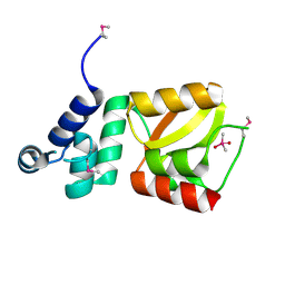
3M70
DownloadVisualize
BU of 3m70 by Molmil
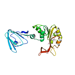
Crystal Structure of TehB from Haemophilus influenzae
Descriptor: Tellurite resistance protein tehB homolog
Authors:Chen, Y.-H, Hendrickson, W.A, New York Consortium on Membrane Protein Structure (NYCOMPS)
Deposit date:2010-03-16
Release date:2010-05-12
Last modified:2017-11-08
Method:X-RAY DIFFRACTION (1.95 Å)
Cite:Crystal Structure of TehB from Haemophilus influenzae
To be Published
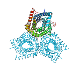
3M75
DownloadVisualize
BU of 3m75 by Molmil
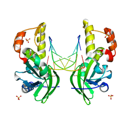
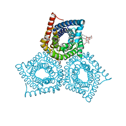
Crystal Structure of Plant SLAC1 homolog TehA
Descriptor: Tellurite resistance protein tehA homolog, octyl beta-D-glucopyranoside
Authors:Chen, Y.-H, Hendrickson, W.A, New York Consortium on Membrane Protein Structure (NYCOMPS)
Deposit date:2010-03-16
Release date:2010-05-12
Last modified:2024-02-21
Method:X-RAY DIFFRACTION (1.6 Å)
Cite:Homologue structure of the SLAC1 anion channel for closing stomata in leaves.
Nature, 467, 2010
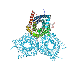
3M73
DownloadVisualize
BU of 3m73 by Molmil
Crystal Structure of Plant SLAC1 homolog TehA
Descriptor: Tellurite resistance protein tehA homolog, octyl beta-D-glucopyranoside
Authors:Chen, Y.-H, Hendrickson, W.A, New York Consortium on Membrane Protein Structure (NYCOMPS)
Deposit date:2010-03-16
Release date:2010-05-12
Last modified:2024-02-21
Method:X-RAY DIFFRACTION (1.15 Å)
Cite:Homologue structure of the SLAC1 anion channel for closing stomata in leaves.
Nature, 467, 2010
3M7L
DownloadVisualize
BU of 3m7l by Molmil
Crystal Structure of Plant SLAC1 homolog TehA
Descriptor: Tellurite resistance protein tehA homolog, octyl beta-D-glucopyranoside
Authors:Chen, Y.-H, Hendrickson, W.A, New York Consortium on Membrane Protein Structure (NYCOMPS)
Deposit date:2010-03-16
Release date:2010-05-12
Last modified:2020-07-29
Method:X-RAY DIFFRACTION (1.5 Å)
Cite:Homologue structure of the SLAC1 anion channel for closing stomata in leaves.
Nature, 467, 2010
2D28
DownloadVisualize
BU of 2d28 by Molmil
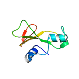
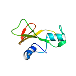
Structure of the N-terminal domain of XpsE (crystal form P43212)
Descriptor: CACODYLATE ION, type II secretion ATPase XpsE
Authors:Chen, Y, Shiue, S.-J, Huang, C.-W, Chang, J.-L, Chien, Y.-L, Hu, N.-T, Chan, N.-L.
Deposit date:2005-09-03
Release date:2005-09-20
Last modified:2024-10-16
Method:X-RAY DIFFRACTION (2 Å)
Cite:Structure and Function of the XpsE N-Terminal Domain, an Essential Component of the Xanthomonas campestris Type II Secretion System
J.Biol.Chem., 280, 2005
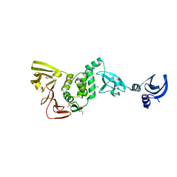
4YPT
DownloadVisualize
BU of 4ypt by Molmil
X-ray structural of three tandemly linked domains of nsp3 from murine hepatitis virus at 2.60 Angstroms resolution
Descriptor: GLYCEROL, Replicase polyprotein 1ab, ZINC ION
Authors:Chen, Y, Savinov, S.N, Mielech, A.M, Cao, T, Baker, S.C, Mesecar, A.D.
Deposit date:2015-03-13
Release date:2015-08-26
Last modified:2023-12-27
Method:X-RAY DIFFRACTION (2.6009 Å)
Cite:X-ray Structural and Functional Studies of the Three Tandemly Linked Domains of Non-structural Protein 3 (nsp3) from Murine Hepatitis Virus Reveal Conserved Functions.
J.Biol.Chem., 290, 2015
3FWN
DownloadVisualize
BU of 3fwn by Molmil
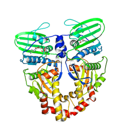
Dimeric 6-phosphogluconate dehydrogenase complexed with 6-phosphogluconate and 2'-monophosphoadenosine-5'-diphosphate
Descriptor: 2'-MONOPHOSPHOADENOSINE-5'-DIPHOSPHATE, 6-PHOSPHOGLUCONIC ACID, 6-phosphogluconate dehydrogenase, ...
Authors:Chen, Y.-Y, Ko, T.-P, Lo, L.-P, Lin, C.-H, Wang, A.H.-J.
Deposit date:2009-01-19
Release date:2009-09-01
Last modified:2023-11-01
Method:X-RAY DIFFRACTION (1.5 Å)
Cite:Conformational changes associated with cofactor/substrate binding of 6-phosphogluconate dehydrogenase from Escherichia coli and Klebsiella pneumoniae: Implications for enzyme mechanism
J.Struct.Biol., 169, 2010
4HJE
DownloadVisualize
BU of 4hje by Molmil
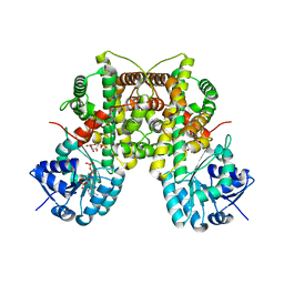
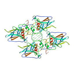
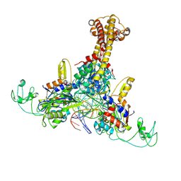
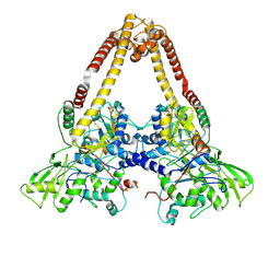
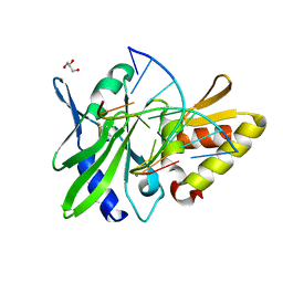
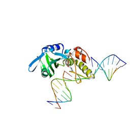
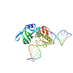
Crystal structure of p53 core domain in complex with DNA
Descriptor: Cellular tumor antigen p53, DNA (5'-D(*AP*GP*GP*CP*TP*TP*GP*TP*CP*TP*CP*TP*AP*AP*CP*TP*TP*GP*TP*GP*A)-3'), DNA (5'-D(*TP*CP*AP*CP*AP*AP*GP*TP*TP*AP*GP*AP*GP*AP*CP*AP*AP*GP*CP*CP*T)-3'), ...
Authors:Chen, Y, Chen, L.
Deposit date:2012-10-12
Release date:2013-07-17
Last modified:2024-02-28
Method:X-RAY DIFFRACTION (1.907 Å)
Cite:Structure of p53 binding to the BAX response element reveals DNA unwinding and compression to accommodate base-pair insertion.
Nucleic Acids Res., 41, 2013
8YO4
DownloadVisualize
BU of 8yo4 by Molmil
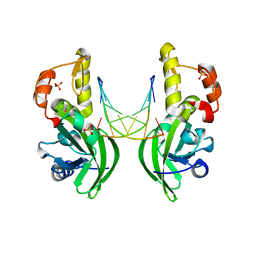
structure of phage T4 topoisomerase II central domain bound with DNA
Descriptor: DNA (5'-D(P*AP*TP*AP*TP*AP*TP*GP*TP*GP*TP*AP*TP*AP*TP*AP*TP*AP*CP*AP*CP*AP*CP*AP*T)-3'), DNA (5'-D(P*TP*GP*TP*GP*TP*GP*TP*AP*TP*AP*TP*AP*TP*AP*CP*AP*CP*AP*TP*AP*TP*AP*TP*A)-3'), DNA topoisomerase medium subunit, ...
Authors:Chen, Y.T, Xin, Y.H, Xian, R.Q.
Deposit date:2024-03-12
Release date:2024-09-25
Method:ELECTRON MICROSCOPY (3.2 Å)
Cite:structure of phage T4 topoisomerase II central domain bound with DNA
To be published
8YO5
DownloadVisualize
BU of 8yo5 by Molmil
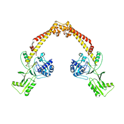
structure of phage T6 topoisomerase II central domain
Descriptor: DNA topoisomerase (ATP-hydrolyzing), DNA topoisomerase medium subunit
Authors:Chen, Y.T, Xin, Y.H, Xian, R.Q.
Deposit date:2024-03-12
Release date:2024-09-25
Method:ELECTRON MICROSCOPY (3.93 Å)
Cite:structure of phage T6 topoisomerase II central domain
To be published
8YON
DownloadVisualize
BU of 8yon by Molmil
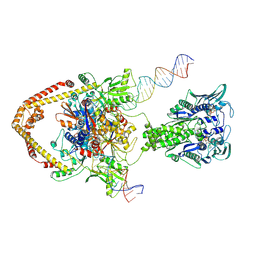
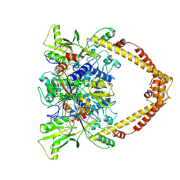
structure of phage T6 full-length topoisomerase II bound with DNA
Descriptor: DNA (50-MER), DNA topoisomerase (ATP-hydrolyzing), DNA topoisomerase medium subunit, ...
Authors:Chen, Y.T, Xin, Y.H, Xian, R.Q.
Deposit date:2024-03-13
Release date:2024-09-25
Method:ELECTRON MICROSCOPY (6.73 Å)
Cite:structure of phage T6 full-length topoisomerase II bound with DNA
To be published
8YO3
DownloadVisualize
BU of 8yo3 by Molmil
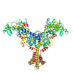
structure of phage T4 topoisomerase II central domain
Descriptor: DNA topoisomerase medium subunit, phage T4 topoisomerase II gp39-gp60 subunit
Authors:Chen, Y.T, Xin, Y.H, Xian, R.Q.
Deposit date:2024-03-12
Release date:2024-09-25
Method:ELECTRON MICROSCOPY (3.62 Å)
Cite:structure of phage T4 topoisomerase II central domain
To be published
8YOD
DownloadVisualize
BU of 8yod by Molmil
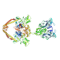
structure of phage T6 apo full-length topoisomerase II
Descriptor: DNA topoisomerase (ATP-hydrolyzing), DNA topoisomerase medium subunit, PHOSPHOAMINOPHOSPHONIC ACID-ADENYLATE ESTER
Authors:Chen, Y.T, Xin, Y.H, Xian, R.Q.
Deposit date:2024-03-13
Release date:2024-09-25
Method:ELECTRON MICROSCOPY (6.8 Å)
Cite:structure of phage T6 apo full-length topoisomerase II
To be published
8YO1
DownloadVisualize
BU of 8yo1 by Molmil
structure of phage T6 topoisomerase II ATPase domain bound with AMPPNP
Descriptor: DNA topoisomerase (ATP-hydrolyzing), MAGNESIUM ION, PHOSPHOAMINOPHOSPHONIC ACID-ADENYLATE ESTER
Authors:Chen, Y.T, Xin, Y.H.
Deposit date:2024-03-12
Release date:2024-09-25
Method:ELECTRON MICROSCOPY (3.55 Å)
Cite:structure of phage T6 topoisomerase II ATPase domain bound with AMPPNP
To be published
8YLU
DownloadVisualize
BU of 8ylu by Molmil
structure of phage T6 topoisomerase II central domain bound with DNA
Descriptor: DNA (5'-D(P*TP*AP*TP*AP*TP*GP*TP*GP*TP*AP*TP*AP*TP*AP*TP*AP*CP*AP*CP*AP*CP*A)-3'), DNA topoisomerase (ATP-hydrolyzing), DNA topoisomerase medium subunit, ...
Authors:Chen, Y.T, Xin, Y.H.
Deposit date:2024-03-06
Release date:2024-09-25
Method:ELECTRON MICROSCOPY (2.8 Å)
Cite:structure of phage T6 topoisomerase II central domain bound with DNA
To be published
8YO9
DownloadVisualize
BU of 8yo9 by Molmil
structure of phage T4 topoisomerase II gp52 subunit WHD-open state
Descriptor: DNA topoisomerase medium subunit
Authors:Chen, Y.T, Xin, Y.H, Xian, R.Q.
Deposit date:2024-03-13
Release date:2024-09-25
Method:ELECTRON MICROSCOPY (6.12 Å)
Cite:structure of phage T4 topoisomerase II gp52 subunit WHD-open state
To be published
1BAN
DownloadVisualize
BU of 1ban by Molmil
THE CONTRIBUTION OF BURIED HYDROGEN BONDS TO PROTEIN STABILITY: THE CRYSTAL STRUCTURES OF TWO BARNASE MUTANTS
Descriptor: BARNASE
Authors:Chen, Y.W, Fersht, A.R, Henrick, K.
Deposit date:1993-05-19
Release date:1993-10-31
Last modified:2024-02-07
Method:X-RAY DIFFRACTION (2.2 Å)
Cite:Contribution of buried hydrogen bonds to protein stability. The crystal structures of two barnase mutants.
J.Mol.Biol., 234, 1993
5HRB
DownloadVisualize
BU of 5hrb by Molmil
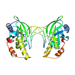
The crystal structure of AsfvPolX:DNA1 binary complex
Descriptor: BETA-MERCAPTOETHANOL, DNA (5'-D(*CP*GP*GP*AP*TP*AP*TP*CP*C)-3'), DNA polymerase beta-like protein, ...
Authors:Chen, Y.Q, Zhang, J, Gan, J.H.
Deposit date:2016-01-23
Release date:2017-01-18
Last modified:2023-11-08
Method:X-RAY DIFFRACTION (1.7 Å)
Cite:The crystal structure of Se-AsfvPolX(L52/163M mutant) in complex with 1nt-gap DNA1
To Be Published
5HRH
DownloadVisualize
BU of 5hrh by Molmil
The crystal structure of AsfvPolX(H115F/R127A mutant): 1nt-gap(P) DNA2:dGTP ternary complex
Descriptor: 2'-DEOXYGUANOSINE-5'-TRIPHOSPHATE, DNA (5'-D(*CP*GP*TP*TP*CP*TP*AP*TP*GP*TP*GP*TP*AP*CP*TP*CP*AP*C)-3'), DNA (5'-D(*GP*TP*GP*AP*GP*TP*AP*CP*A*)-3'), ...
Authors:Chen, Y.Q, Zhang, J, Gan, J.H.
Deposit date:2016-01-23
Release date:2017-01-18
Last modified:2024-03-20
Method:X-RAY DIFFRACTION (3 Å)
Cite:The crystal structure of Se-AsfvPolX(L52/163M mutant) in complex with 1nt-gap DNA1
To Be Published
5HRD
DownloadVisualize
BU of 5hrd by Molmil
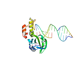
The crystal structure of AsfvPolX:DNA2 binary complex
Descriptor: DNA (5'-D(*GP*CP*GP*AP*TP*CP*GP*G)-3'), DNA polymerase beta-like protein, SULFATE ION
Authors:Chen, Y.Q, Zhang, J, Gan, J.H.
Deposit date:2016-01-23
Release date:2017-01-18
Last modified:2023-11-08
Method:X-RAY DIFFRACTION (1.8 Å)
Cite:The crystal structure of Se-AsfvPolX(L52/163M mutant) in complex with 1nt-gap DNA1
To Be Published
5HRF
DownloadVisualize
BU of 5hrf by Molmil
The crystal structure of AsfvPolX: DNA5: dGTP ternary complex
Descriptor: 2'-DEOXYGUANOSINE-5'-TRIPHOSPHATE, DNA (5'-D(*CP*AP*GP*GP*AP*TP*CP*CP*T*)-3'), DNA polymerase beta-like protein, ...
Authors:Chen, Y.Q, Zhang, J, Gan, J.H.
Deposit date:2016-01-23
Release date:2017-01-18
Last modified:2024-03-20
Method:X-RAY DIFFRACTION (2.25 Å)
Cite:The crystal structure of Se-AsfvPolX(L52/163M mutant) in complex with 1nt-gap DNA1
To Be Published
5HR9
DownloadVisualize
BU of 5hr9 by Molmil
The crystal structure of Se-AsfvPolX(L52/163M mutant) in complex with 1nt-gap DNA1
Descriptor: DNA (5'-D(*GP*GP*AP*CP*AP*AP*CP*GP*GP*GP*AP*CP*AP*AP*C)-3'), DNA (5'-D(*GP*TP*TP*GP*TP*CP*C)-3'), DNA polymerase beta-like protein
Authors:Chen, Y.Q, Zhang, J, Gan, J.H.
Deposit date:2016-01-23
Release date:2017-01-18
Method:X-RAY DIFFRACTION (2.2 Å)
Cite:The crystal structure of Se-AsfvPolX(L52/163M mutant) in complex with 1nt-gap DNA1
To Be Published
5HRL
DownloadVisualize
BU of 5hrl by Molmil
The crystal structure of AsfvPolX: 1nt-gap(P) DNA2: dGTP ternary complex.
Descriptor: 2'-DEOXYGUANOSINE-5'-TRIPHOSPHATE, DNA (5'-D(*CP*GP*TP*TP*CP*TP*AP*TP*GP*TP*GP*TP*AP*CP*TP*CP*AP*C)-3'), DNA (5'-D(*GP*TP*GP*AP*GP*TP*AP*CP*A)-3'), ...
Authors:Chen, Y.Q, Zhang, J, Gan, J.H.
Deposit date:2016-01-23
Release date:2017-01-18
Last modified:2023-11-08
Method:X-RAY DIFFRACTION (2.4 Å)
Cite:The crystal structure of Se-AsfvPolX(L52/163M mutant) in complex with 1nt-gap DNA1
To Be Published
5HRG
DownloadVisualize
BU of 5hrg by Molmil
The crystal structure of AsfvPolX(D51N mutant):DNA4 binary complex
Descriptor: DNA (5'-D(*GP*CP*GP*AP*TP*CP*GP*C)-3'), DNA polymerase beta-like protein, MANGANESE (II) ION, ...
Authors:Chen, Y.Q, Zhang, J, Gan, J.H.
Deposit date:2016-01-23
Release date:2017-01-18
Last modified:2023-11-08
Method:X-RAY DIFFRACTION (2 Å)
Cite:The crystal structure of Se-AsfvPolX(L52/163M mutant) in complex with 1nt-gap DNA1
To Be Published
1BNS
DownloadVisualize
BU of 1bns by Molmil
STRUCTURAL STUDIES OF BARNASE MUTANTS
Descriptor: BARNASE
Authors:Chen, Y.W.
Deposit date:1994-04-11
Release date:1994-06-22
Last modified:2024-02-07
Method:X-RAY DIFFRACTION (2.05 Å)
Cite:Contribution of buried hydrogen bonds to protein stability. The crystal structures of two barnase mutants.
J.Mol.Biol., 234, 1993

226707

PDB entries from 2024-10-30

PDB statisticsPDBj update infoContact PDBjnumon