Loading
PDBj
MenuPDBj@FacebookPDBj@TwitterPDBj@YouTubewwPDB FoundationwwPDB
RCSB PDBPDBeBMRBAdv. SearchSearch help
PDB: 87 results

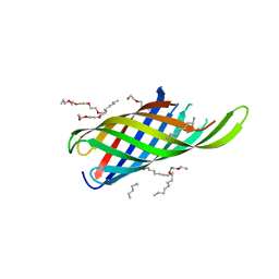
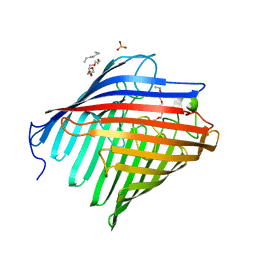
5DL8
DownloadVisualize
BU of 5dl8 by Molmil
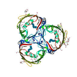
Crystal structure of Acinetobacter baumannii OccAB4
Descriptor: (HYDROXYETHYLOXY)TRI(ETHYLOXY)OCTANE, Benzoate transport porin BenP, CALCIUM ION
Authors:Zahn, M, Basle, A, van den Berg, B.
Deposit date:2015-09-04
Release date:2016-02-03
Last modified:2024-01-10
Method:X-RAY DIFFRACTION (2.2 Å)
Cite:Structural Insights into Outer Membrane Permeability of Acinetobacter baumannii.
Structure, 24, 2016
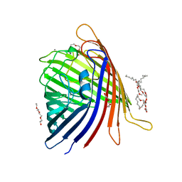
5DL5
DownloadVisualize
BU of 5dl5 by Molmil
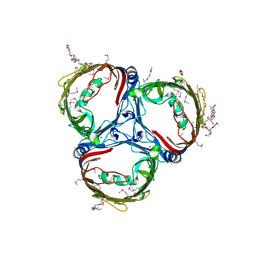
Crystal structure of Acinetobacter baumannii OccAB1
Descriptor: (HYDROXYETHYLOXY)TRI(ETHYLOXY)OCTANE, Membrane protein, PHOSPHATE ION
Authors:Zahn, M, Basle, A, van den Berg, B.
Deposit date:2015-09-04
Release date:2016-02-03
Last modified:2024-01-10
Method:X-RAY DIFFRACTION (2.05 Å)
Cite:Structural Insights into Outer Membrane Permeability of Acinetobacter baumannii.
Structure, 24, 2016
4EZN
DownloadVisualize
BU of 4ezn by Molmil
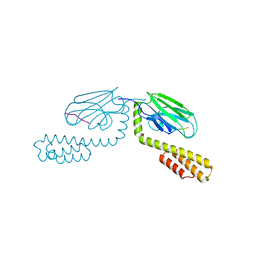
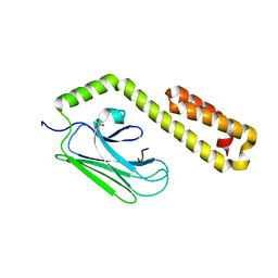
Crystal structure of the substrate binding domain of E.coli DnaK in complex with pyrrhocoricin
Descriptor: Chaperone protein DnaK, Pyrrhocoricin
Authors:Zahn, M, Straeter, N.
Deposit date:2012-05-03
Release date:2013-04-17
Last modified:2024-02-28
Method:X-RAY DIFFRACTION (1.8 Å)
Cite:Structural Studies on the Forward and Reverse Binding Modes of Peptides to the Chaperone DnaK.
J.Mol.Biol., 425, 2013
4EZX
DownloadVisualize
BU of 4ezx by Molmil
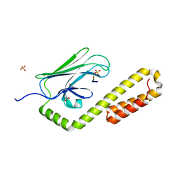
Crystal structure of the substrate binding domain of E.coli DnaK in complex with the designer peptide NRLMLTG
Descriptor: Chaperone protein DnaK, SULFATE ION, synthetic peptide NRLMLTG
Authors:Zahn, M, Straeter, N.
Deposit date:2012-05-03
Release date:2013-04-17
Last modified:2024-02-28
Method:X-RAY DIFFRACTION (1.7 Å)
Cite:Structural Studies on the Forward and Reverse Binding Modes of Peptides to the Chaperone DnaK.
J.Mol.Biol., 425, 2013
4EZV
DownloadVisualize
BU of 4ezv by Molmil
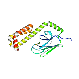
Crystal structure of the substrate binding domain of E.coli DnaK in complex with PR-bombesin in space group P21212
Descriptor: Chaperone protein DnaK, Proline rich bombesin-related protein
Authors:Zahn, M, Straeter, N.
Deposit date:2012-05-03
Release date:2013-04-24
Last modified:2024-02-28
Method:X-RAY DIFFRACTION (2.1 Å)
Cite:Structural studies of DnaK in complex with proline rich antimicrobial peptides reveal two different peptide binding modes
To be Published
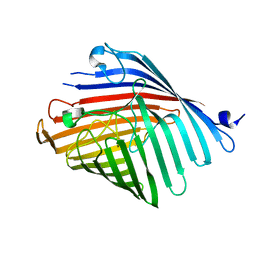
8C65
DownloadVisualize
BU of 8c65 by Molmil
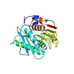
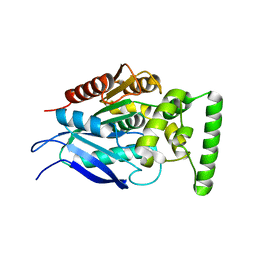
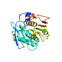
Crystal structure of cutinase AdCut from Acidovorax delafieldii (PBS depolymerase)
Descriptor: PBS(A) depolymerase
Authors:Zahn, M, Clark, M.
Deposit date:2023-01-11
Release date:2024-01-24
Method:X-RAY DIFFRACTION (1.491 Å)
Cite:Predictions of the substrate specificities of the plastic-degrading enzymes IsPETase and AdCut
To Be Published
4JWC
DownloadVisualize
BU of 4jwc by Molmil
Crystal structure of the substrate binding domain of E.coli DnaK in complex with bovine Bac7(1-16)
Descriptor: Cathelicidin-3, Chaperone protein DnaK, SULFATE ION
Authors:Zahn, M, Straeter, N.
Deposit date:2013-03-27
Release date:2013-11-13
Last modified:2023-09-20
Method:X-RAY DIFFRACTION (1.8 Å)
Cite:Structural Identification of DnaK Binding Sites within Bovine and Sheep Bactenecin Bac7.
Protein Pept.Lett., 21, 2014
8AYV
DownloadVisualize
BU of 8ayv by Molmil
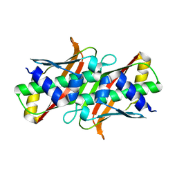
Crystal structure of the Malonyl-ACP Decarboxylase MadB from Pseudomonas putida
Descriptor: YiiD_C domain-containing protein
Authors:Zahn, M, Kuatsjah, E, Beckham, G.T, McGeehan, J.E.
Deposit date:2022-09-03
Release date:2023-03-01
Last modified:2024-05-01
Method:X-RAY DIFFRACTION (1.044 Å)
Cite:Initiation of fatty acid biosynthesis in Pseudomonas putida KT2440.
Metab Eng, 76, 2023
7QJR
DownloadVisualize
BU of 7qjr by Molmil
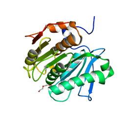
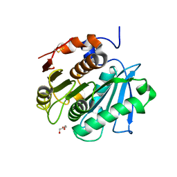
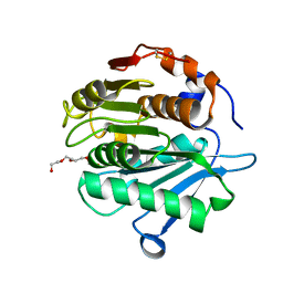
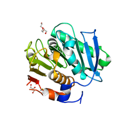
Crystal structure of cutinase 1 from Thermobifida fusca DSM44342 (703)
Descriptor: Cutinase 1, TETRAETHYLENE GLYCOL
Authors:Zahn, M, Avilan, L, Beckham, G.T, McGeehan, J.E.
Deposit date:2021-12-17
Release date:2022-12-28
Last modified:2024-01-31
Method:X-RAY DIFFRACTION (1.51 Å)
Cite:Sourcing thermotolerant poly(ethylene terephthalate) hydrolase scaffolds from natural diversity
Nat Commun, 13, 2022
7QJT
DownloadVisualize
BU of 7qjt by Molmil
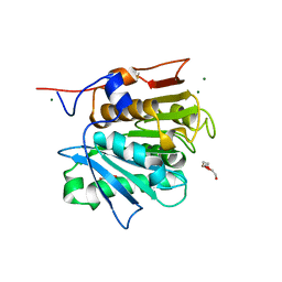
Crystal structure of a cutinase enzyme from Thermobifida cellulosilytica TB100 (711)
Descriptor: GLYCEROL, MAGNESIUM ION, TETRAETHYLENE GLYCOL, ...
Authors:Zahn, M, Shakespeare, T.J, Beckham, G.T, McGeehan, J.E.
Deposit date:2021-12-17
Release date:2022-12-28
Last modified:2024-01-31
Method:X-RAY DIFFRACTION (1.78 Å)
Cite:Sourcing thermotolerant poly(ethylene terephthalate) hydrolase scaffolds from natural diversity
Nat Commun, 13, 2022
7QJQ
DownloadVisualize
BU of 7qjq by Molmil
Crystal structure of a cutinase enzyme from Thermobifida fusca NTU22 (702)
Descriptor: Acetylxylan esterase, DI(HYDROXYETHYL)ETHER
Authors:Zahn, M, Gill, R.S, Avilan, L, Beckham, G.T, McGeehan, J.E.
Deposit date:2021-12-17
Release date:2022-12-28
Last modified:2024-01-31
Method:X-RAY DIFFRACTION (1.64 Å)
Cite:Sourcing thermotolerant poly(ethylene terephthalate) hydrolase scaffolds from natural diversity
Nat Commun, 13, 2022
7QJM
DownloadVisualize
BU of 7qjm by Molmil
Crystal structure of an alpha/beta-hydrolase enzyme from Chloroflexus sp. MS-G (202)
Descriptor: alpha/beta-hydrolase (202)
Authors:Zahn, M, Graham, R, Beckham, G.T, McGeehan, J.E.
Deposit date:2021-12-17
Release date:2022-12-28
Last modified:2024-01-31
Method:X-RAY DIFFRACTION (2.19 Å)
Cite:Sourcing thermotolerant poly(ethylene terephthalate) hydrolase scaffolds from natural diversity
Nat Commun, 13, 2022
7QJP
DownloadVisualize
BU of 7qjp by Molmil
Crystal structure of a cutinase enzyme from Saccharopolyspora flava (611)
Descriptor: Cutinase, TETRAETHYLENE GLYCOL
Authors:Zahn, M, Avilan, L, Beckham, G.T, McGeehan, J.E.
Deposit date:2021-12-17
Release date:2022-12-28
Last modified:2024-04-24
Method:X-RAY DIFFRACTION (1.561 Å)
Cite:Sourcing thermotolerant poly(ethylene terephthalate) hydrolase scaffolds from natural diversity
Nat Commun, 13, 2022
7QJN
DownloadVisualize
BU of 7qjn by Molmil
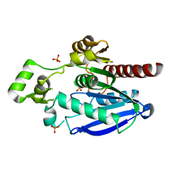
Crystal structure of an alpha/beta-hydrolase enzyme from Candidatus Kryptobacter tengchongensis (306)
Descriptor: Dienelactone hydrolase, PHOSPHATE ION
Authors:Zahn, M, Gill, R.S, Erickson, E, Beckham, G.T, McGeehan, J.E.
Deposit date:2021-12-17
Release date:2022-12-28
Last modified:2024-05-01
Method:X-RAY DIFFRACTION (1.885 Å)
Cite:Sourcing thermotolerant poly(ethylene terephthalate) hydrolase scaffolds from natural diversity
Nat Commun, 13, 2022
7QJO
DownloadVisualize
BU of 7qjo by Molmil
Crystal structure of a cutinase enzyme from Marinactinospora thermotolerans DSM45154 (606)
Descriptor: Cutinase
Authors:Zahn, M, Shakespeare, T.J, Beckham, G.T, McGeehan, J.E.
Deposit date:2021-12-17
Release date:2022-12-28
Last modified:2024-01-31
Method:X-RAY DIFFRACTION (1.933 Å)
Cite:Sourcing thermotolerant poly(ethylene terephthalate) hydrolase scaffolds from natural diversity
Nat Commun, 13, 2022
7QJS
DownloadVisualize
BU of 7qjs by Molmil
Crystal structure of a cutinase enzyme from Thermobifida fusca YX (705)
Descriptor: Cutinase 2, DI(HYDROXYETHYL)ETHER, GLYCEROL, ...
Authors:Zahn, M, Shakespeare, T.J, Beckham, G.T, McGeehan, J.E.
Deposit date:2021-12-17
Release date:2022-12-28
Last modified:2024-01-31
Method:X-RAY DIFFRACTION (1.429 Å)
Cite:Sourcing thermotolerant poly(ethylene terephthalate) hydrolase scaffolds from natural diversity
Nat Commun, 13, 2022
4RLB
DownloadVisualize
BU of 4rlb by Molmil
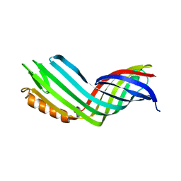
Crystal structure of Acinetobacter baumannii CarO2
Descriptor: Carbapenem-associated resistance protein
Authors:Zahn, M, Basle, A, van den Berg, B.
Deposit date:2014-10-16
Release date:2015-04-22
Last modified:2024-02-28
Method:X-RAY DIFFRACTION (2.7 Å)
Cite:Small-Molecule Transport by CarO, an Abundant Eight-Stranded beta-Barrel Outer Membrane Protein from Acinetobacter baumannii.
J.Mol.Biol., 427, 2015
4RL9
DownloadVisualize
BU of 4rl9 by Molmil
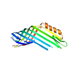
Crystal structure of Acinetobacter baumannii CarO1
Descriptor: Carbapenem-associated resistance protein
Authors:Zahn, M, Basle, A, van den Berg, B.
Deposit date:2014-10-16
Release date:2015-04-22
Last modified:2024-02-28
Method:X-RAY DIFFRACTION (2.7 Å)
Cite:Small-Molecule Transport by CarO, an Abundant Eight-Stranded beta-Barrel Outer Membrane Protein from Acinetobacter baumannii.
J.Mol.Biol., 427, 2015
4RLC
DownloadVisualize
BU of 4rlc by Molmil
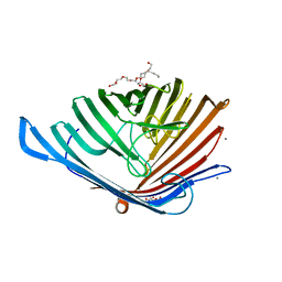
Crystal structure of the N-terminal beta-barrel domain of Pseudomonas aeruginosa OprF
Descriptor: (HYDROXYETHYLOXY)TRI(ETHYLOXY)OCTANE, Outer membrane porin F
Authors:Zahn, M, Basle, A, van den Berg, B.
Deposit date:2014-10-16
Release date:2015-04-22
Last modified:2024-02-28
Method:X-RAY DIFFRACTION (1.6 Å)
Cite:Small-Molecule Transport by CarO, an Abundant Eight-Stranded beta-Barrel Outer Membrane Protein from Acinetobacter baumannii.
J.Mol.Biol., 427, 2015
5DL7
DownloadVisualize
BU of 5dl7 by Molmil
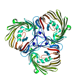
Crystal structure of Acinetobacter baumannii OccAB3
Descriptor: (HYDROXYETHYLOXY)TRI(ETHYLOXY)OCTANE, CALCIUM ION, Porin
Authors:Zahn, M, Basle, A, van den Berg, B.
Deposit date:2015-09-04
Release date:2016-02-03
Last modified:2024-01-10
Method:X-RAY DIFFRACTION (1.75 Å)
Cite:Structural Insights into Outer Membrane Permeability of Acinetobacter baumannii.
Structure, 24, 2016
5DL6
DownloadVisualize
BU of 5dl6 by Molmil
Crystal structure of Acinetobacter baumannii OccAB2
Descriptor: Putative porin for vanillate trafficking (VanP)
Authors:Zahn, M, Basle, A, van den Berg, B.
Deposit date:2015-09-04
Release date:2016-02-03
Last modified:2024-01-10
Method:X-RAY DIFFRACTION (2.9 Å)
Cite:Structural Insights into Outer Membrane Permeability of Acinetobacter baumannii.
Structure, 24, 2016
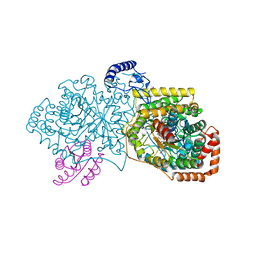
4R3U
DownloadVisualize
BU of 4r3u by Molmil
Crystal structure of 2-Hydroxyisobutyryl-CoA Mutase
Descriptor: 2-hydroxyisobutyryl-CoA mutase large subunit, 2-hydroxyisobutyryl-CoA mutase small subunit, 3-HYDROXYBUTANOYL-COENZYME A, ...
Authors:Zahn, M, Kurteva-Yaneva, N, Rohwerder, T, Straeter, N.
Deposit date:2014-08-18
Release date:2015-03-11
Last modified:2024-02-28
Method:X-RAY DIFFRACTION (2.5 Å)
Cite:Structural basis of the stereospecificity of bacterial B12-dependent 2-hydroxyisobutyryl-CoA mutase.
J.Biol.Chem., 290, 2015
5MDR
DownloadVisualize
BU of 5mdr by Molmil
Crystal structure of in vitro folded Chitoporin VhChip from Vibrio harveyi in complex with chitohexaose
Descriptor: (HYDROXYETHYLOXY)TRI(ETHYLOXY)OCTANE, 2-acetamido-2-deoxy-beta-D-glucopyranose-(1-4)-2-acetamido-2-deoxy-beta-D-glucopyranose-(1-4)-2-acetamido-2-deoxy-beta-D-glucopyranose-(1-4)-2-acetamido-2-deoxy-beta-D-glucopyranose-(1-4)-2-acetamido-2-deoxy-beta-D-glucopyranose-(1-4)-2-acetamido-2-deoxy-beta-D-glucopyranose, Chitoporin, ...
Authors:Zahn, M, van den Berg, B.
Deposit date:2016-11-13
Release date:2017-12-20
Last modified:2024-01-17
Method:X-RAY DIFFRACTION (1.9 Å)
Cite:Structural basis for chitin acquisition by marine Vibrio species.
Nat Commun, 9, 2018
5MDO
DownloadVisualize
BU of 5mdo by Molmil
Crystal structure of in vitro folded Chitoporin VhChip from Vibrio harveyi (crystal form I)
Descriptor: (HYDROXYETHYLOXY)TRI(ETHYLOXY)OCTANE, Chitoporin, SODIUM ION
Authors:Zahn, M, van den Berg, B.
Deposit date:2016-11-13
Release date:2017-12-20
Last modified:2018-01-24
Method:X-RAY DIFFRACTION (1.95 Å)
Cite:Structural basis for chitin acquisition by marine Vibrio species.
Nat Commun, 9, 2018
5MDS
DownloadVisualize
BU of 5mds by Molmil
Crystal structure of outer membrane expressed Chitoporin VhChip from Vibrio harveyi in complex with chitotetraose
Descriptor: 2-acetamido-2-deoxy-beta-D-glucopyranose-(1-4)-2-acetamido-2-deoxy-beta-D-glucopyranose-(1-4)-2-acetamido-2-deoxy-beta-D-glucopyranose-(1-4)-2-acetamido-2-deoxy-beta-D-glucopyranose, Chitoporin
Authors:Zahn, M, van den Berg, B.
Deposit date:2016-11-13
Release date:2017-12-20
Last modified:2024-01-17
Method:X-RAY DIFFRACTION (2.6 Å)
Cite:Structural basis for chitin acquisition by marine Vibrio species.
Nat Commun, 9, 2018


1234>

222415

건을2024-07-10부터공개중

PDB statisticsPDBj update infoContact PDBjnumon