Loading
PDBj
MenuPDBj@FacebookPDBj@TwitterPDBj@YouTubewwPDB FoundationwwPDB
RCSB PDBPDBeBMRBAdv. SearchSearch help
PDB: 7 results

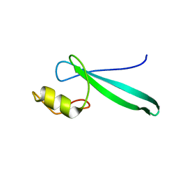
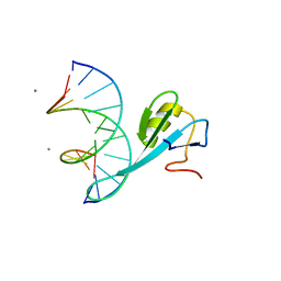
2KY8
DownloadVisualize
BU of 2ky8 by Molmil
Solution structure and dynamic analysis of chicken MBD2 methyl binding domain bound to a target methylated DNA sequence
Descriptor: DNA (5'-D(*GP*AP*GP*CP*(5CM)P*GP*AP*TP*(TED)P*CP*C)-3'), DNA (5'-D(*GP*GP*AP*AP*TP*(5CM)P*GP*GP*CP*(TED)P*C)-3'), MANGANESE (II) ION, ...
Authors:Williams Jr, D.C, Scarsdale Jr, J.N.
Deposit date:2010-05-18
Release date:2011-05-18
Last modified:2024-05-01
Method:SOLUTION NMR
Cite:Solution structure and dynamic analysis of chicken MBD2 methyl binding domain bound to a target-methylated DNA sequence.
Nucleic Acids Res., 39, 2011
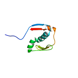
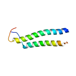
2L2L
DownloadVisualize
BU of 2l2l by Molmil
Solution structure of the coiled-coil complex between MBD2 and p66alpha
Descriptor: Methyl-CpG-binding domain protein 2, Transcriptional repressor p66-alpha
Authors:Williams Jr, D.C, Scarsdale Jr, N.
Deposit date:2010-08-20
Release date:2011-05-04
Last modified:2024-05-01
Method:SOLUTION NMR
Cite:p66Alpha-MBD2 coiled-coil interaction and recruitment of Mi-2 are critical for globin gene silencing by the MBD2-NuRD complex.
Proc.Natl.Acad.Sci.USA, 108, 2011
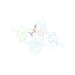
2MB7
DownloadVisualize
BU of 2mb7 by Molmil
Solution structure of MBD3 methylcytosine binding domain
Descriptor: Methyl-CpG-binding domain protein 3
Authors:Williams Jr, D.C, Scarsdale, J.N.
Deposit date:2013-07-26
Release date:2013-12-11
Last modified:2024-05-15
Method:SOLUTION NMR
Cite:Probing the Dynamic Distribution of Bound States for Methylcytosine-binding Domains on DNA.
J.Biol.Chem., 289, 2014
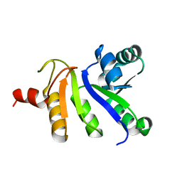
2N2Y
DownloadVisualize
BU of 2n2y by Molmil
Solution structure of the meiosis-expressed gene 1 (Meig1)
Descriptor: Meiosis-expressed gene 1 protein
Authors:Williams Jr, D.C, Walavalkar, N.M, Buchwald, W.A.
Deposit date:2015-05-16
Release date:2016-01-20
Last modified:2024-05-15
Method:SOLUTION NMR
Cite:Dissecting the structural basis of MEIG1 interaction with PACRG.
Sci Rep, 6, 2016
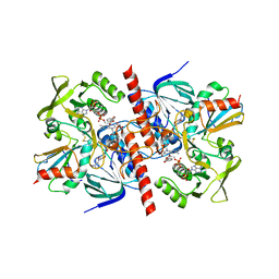
1CL0
DownloadVisualize
BU of 1cl0 by Molmil
CRYSTAL STRUCTURE OF REDUCED THIOREDOXIN REDUCTASE FROM ESCHERICHIA COLI.
Descriptor: FLAVIN-ADENINE DINUCLEOTIDE, THIOREDOXIN REDUCTASE
Authors:Lennon, B.W, Williams Jr, C.H, Ludwig, M.L.
Deposit date:1999-05-04
Release date:1999-12-03
Last modified:2023-08-09
Method:X-RAY DIFFRACTION (2.5 Å)
Cite:Crystal structure of reduced thioredoxin reductase from Escherichia coli: structural flexibility in the isoalloxazine ring of the flavin adenine dinucleotide cofactor.
Protein Sci., 8, 1999
2JZH
DownloadVisualize
BU of 2jzh by Molmil
structure of IIB domain of the mannose transporter of E. coli
Descriptor: PTS system mannose-specific EIIAB component
Authors:Komlosh, M, Williams Jr, D.C.
Deposit date:2008-01-08
Release date:2008-02-19
Last modified:2024-05-29
Method:SOLUTION NMR
Cite:Solution NMR Structures of Productive and Non-productive Complexes between the A and B Domains of the Cytoplasmic Subunit of the Mannose Transporter of the Escherichia coli Phosphotransferase System.
J.Biol.Chem., 283, 2008
1F6M
DownloadVisualize
BU of 1f6m by Molmil
CRYSTAL STRUCTURE OF A COMPLEX BETWEEN THIOREDOXIN REDUCTASE, THIOREDOXIN, AND THE NADP+ ANALOG, AADP+
Descriptor: 3-AMINOPYRIDINE-ADENINE DINUCLEOTIDE PHOSPHATE, FLAVIN-ADENINE DINUCLEOTIDE, THIOREDOXIN 1, ...
Authors:Lennon, B.W, Williams Jr, C.H, Ludwig, M.L.
Deposit date:2000-06-22
Release date:2000-08-30
Last modified:2024-03-13
Method:X-RAY DIFFRACTION (2.95 Å)
Cite:Twists in catalysis: alternating conformations of Escherichia coli thioredoxin reductase.
Science, 289, 2000

226707

数据于2024-10-30公开中

PDB statisticsPDBj update infoContact PDBjnumon