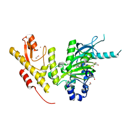
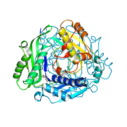
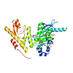
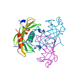
3P52
 
 | | NH3-dependent NAD synthetase from Campylobacter jejuni subsp. jejuni NCTC 11168 in complex with the nitrate ion | | 分子名称: | NH(3)-dependent NAD(+) synthetase, NITRATE ION | | 著者 | Filippova, E.V, Wawrzak, Z, Onopriyenko, O, Skarina, T, Edwards, A, Savchenko, A, Anderson, W.F, Center for Structural Genomics of Infectious Diseases (CSGID) | | 登録日 | 2010-10-07 | | 公開日 | 2010-10-27 | | 最終更新日 | 2023-12-06 | | 実験手法 | X-RAY DIFFRACTION (2.74 Å) | | 主引用文献 | NH3-dependent NAD synthetase from Campylobacter jejuni subsp. jejuni NCTC 11168 in complex with the nitrate ion
To be Published
|
|
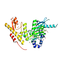
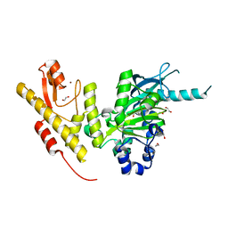
5ENJ
 | | Crystal structure of the second bromodomain of Pleckstrin homology domain interacting protein (PHIP) in complex with compound-14 N11530 (SGC - Diamond I04-1 fragment screening) | | 分子名称: | MAGNESIUM ION, PH-interacting protein, ~{N}-[[2,6-bis(chloranyl)phenyl]methyl]-~{N}-methyl-ethanamide | | 著者 | Krojer, T, Talon, R, Collins, P, Bradley, A, Cox, O, Szykowska, A, Burgess-Brown, N, Brennan, P, Bountra, C, Arrowsmith, C.H, Edwards, A, von Delft, F, Structural Genomics Consortium (SGC) | | 登録日 | 2015-11-09 | | 公開日 | 2016-04-27 | | 最終更新日 | 2024-01-10 | | 実験手法 | X-RAY DIFFRACTION (1.63 Å) | | 主引用文献 | A poised fragment library enables rapid synthetic expansion yielding the first reported inhibitors of PHIP(2), an atypical bromodomain.
Chem Sci, 7, 2016
|
|
3OOY
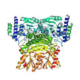 | | Crystal structure of human Transketolase (TKT) | | 分子名称: | 1,2-ETHANEDIOL, ACETATE ION, CALCIUM ION, ... | | 著者 | Krojer, T, Krysztofinska, E, Guo, K, Pilka, E, Kochan, G, Chaikuad, A, Vollmar, M, von Delft, F, Bountra, C, Arrowsmith, C.H, Weigelt, J, Edwards, A, Kavanagh, K, Oppermann, U, Structural Genomics Consortium (SGC) | | 登録日 | 2010-08-31 | | 公開日 | 2010-11-17 | | 最終更新日 | 2024-02-21 | | 実験手法 | X-RAY DIFFRACTION (2.05 Å) | | 主引用文献 | Crystal structure of human Transketolase (TKT)
To be Published
|
|
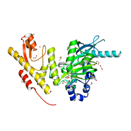
5ENE
 | | Crystal structure of the second bromodomain of Pleckstrin homology domain interacting protein (PHIP) in complex with 5-Amino-2-benzyl-1,3-oxazole-4-carbonitrile (SGC - Diamond I04-1 fragment screening) | | 分子名称: | 5-azanyl-2-(phenylmethyl)-1,3-oxazole-4-carbonitrile, PH-interacting protein | | 著者 | Krojer, T, Talon, R, Collins, P, Bradley, A, Cox, O, Amin, J, Szykowska, A, Burgess-Brown, N, Spencer, J, Brennan, P, Bountra, C, Arrowsmith, C.H, Edwards, A, von Delft, F, Structural Genomics Consortium (SGC) | | 登録日 | 2015-11-09 | | 公開日 | 2016-04-27 | | 最終更新日 | 2024-01-10 | | 実験手法 | X-RAY DIFFRACTION (1.49 Å) | | 主引用文献 | A poised fragment library enables rapid synthetic expansion yielding the first reported inhibitors of PHIP(2), an atypical bromodomain.
Chem Sci, 7, 2016
|
|
3OOW
 | | Octameric structure of the phosphoribosylaminoimidazole carboxylase catalytic subunit from Francisella tularensis subsp. tularensis SCHU S4. | | 分子名称: | (4S)-2-METHYL-2,4-PENTANEDIOL, CHLORIDE ION, FORMIC ACID, ... | | 著者 | Filippova, E.V, Wawrzak, Z, Kudritska, M, Edwards, A, Savchenko, A, Anderson, F.W, Center for Structural Genomics of Infectious Diseases (CSGID) | | 登録日 | 2010-08-31 | | 公開日 | 2010-09-15 | | 最終更新日 | 2023-12-06 | | 実験手法 | X-RAY DIFFRACTION (1.75 Å) | | 主引用文献 | Octameric structure of the phosphoribosylaminoimidazole carboxylase catalytic subunit from Francisella tularensis subsp. tularensis SCHU S4.
To be Published
|
|
3OXP
 | | Structure of phosphotransferase enzyme II, A component from Yersinia pestis CO92 at 1.2 A resolution | | 分子名称: | GLYCEROL, Phosphotransferase enzyme II, A component, ... | | 著者 | Filippova, E.V, Wawrzak, Z, Kudritska, M, Edwards, A, Savchenko, A, Anderson, W.F, Center for Structural Genomics of Infectious Diseases (CSGID) | | 登録日 | 2010-09-21 | | 公開日 | 2010-10-13 | | 最終更新日 | 2017-11-08 | | 実験手法 | X-RAY DIFFRACTION (1.2 Å) | | 主引用文献 | Structure of phosphotransferase enzyme II, A component from Yersinia pestis CO92 at 1.2 A resolution
To be Published
|
|
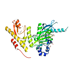
5ENC
 | | Crystal structure of the second bromodomain of Pleckstrin homology domain interacting protein (PHIP) in complex with N-(2,6-Dichlorobenzyl)acetamide (SGC - Diamond I04-1 fragment screening) | | 分子名称: | 1,2-ETHANEDIOL, PH-interacting protein, ~{N}-[[2,6-bis(chloranyl)phenyl]methyl]ethanamide | | 著者 | Krojer, T, Talon, R, Collins, P, Bradley, A, Cox, O, Szykowska, A, Burgess-Brown, N, Brennan, P, Bountra, C, Arrowsmith, C.H, Edwards, A, von Delft, F, Structural Genomics Consortium (SGC) | | 登録日 | 2015-11-09 | | 公開日 | 2016-04-27 | | 最終更新日 | 2024-01-10 | | 実験手法 | X-RAY DIFFRACTION (1.59 Å) | | 主引用文献 | A poised fragment library enables rapid synthetic expansion yielding the first reported inhibitors of PHIP(2), an atypical bromodomain.
Chem Sci, 7, 2016
|
|
3PJ9
 | | Crystal structure of a Nucleoside Diphosphate Kinase from Campylobacter jejuni | | 分子名称: | Nucleoside diphosphate kinase, SULFATE ION | | 著者 | Filippova, E.V, Wawrzak, Z, Onopriyenko, O, Edwards, A, Savchenko, A, Anderson, W.F, Center for Structural Genomics of Infectious Diseases (CSGID) | | 登録日 | 2010-11-09 | | 公開日 | 2010-11-24 | | 最終更新日 | 2023-09-06 | | 実験手法 | X-RAY DIFFRACTION (2.1 Å) | | 主引用文献 | Crystal structure of a Nucleoside Diphosphate Kinase from Campylobacter jejuni
To be Published
|
|
2O6T
 | | Crystal structure of the PA5185 protein from Pseudomonas Aeruginosa strain PAO1- orthorhombic form (P2221). | | 分子名称: | CHLORIDE ION, THIOESTERASE | | 著者 | Chruszcz, M, Koclega, K.D, Evdokimova, E, Cymborowski, M, Kudritska, M, Savchenko, A, Edwards, A, Minor, W. | | 登録日 | 2006-12-08 | | 公開日 | 2007-12-11 | | 最終更新日 | 2023-08-30 | | 実験手法 | X-RAY DIFFRACTION (2.55 Å) | | 主引用文献 | Function-biased choice of additives for optimization of protein crystallization - the case of the putative thioesterase PA5185 from Pseudomonas aeruginosa PAO1.
Cryst.Growth Des., 8, 2008
|
|
2O9X
 | | Crystal Structure Of A Putative Redox Enzyme Maturation Protein From Archaeoglobus Fulgidus | | 分子名称: | Reductase, assembly protein | | 著者 | Kirillova, O, Chruszcz, M, Skarina, T, Gorodichtchenskaia, E, Cymborowski, M, Shumilin, I, Savchenko, A, Edwards, A, Minor, W, Midwest Center for Structural Genomics (MCSG) | | 登録日 | 2006-12-14 | | 公開日 | 2007-01-16 | | 最終更新日 | 2023-12-27 | | 実験手法 | X-RAY DIFFRACTION (3.4 Å) | | 主引用文献 | An extremely SAD case: structure of a putative redox-enzyme maturation protein from Archaeoglobus fulgidus at 3.4 A resolution.
Acta Crystallogr.,Sect.D, 63, 2007
|
|
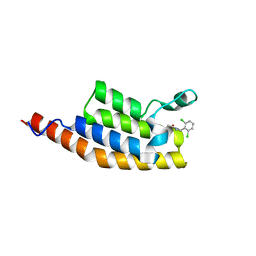
3PS4
 | | PDZ domain from Human microtubule-associated serine/threonine-protein kinase 1 | | 分子名称: | 1,2-ETHANEDIOL, IMIDAZOLE, Microtubule-associated serine/threonine-protein kinase 1 | | 著者 | Ugochukwu, E, Wang, J, Krojer, T, Muniz, J.R.C, Sethi, R, Pike, A.C.W, Roos, A, Salah, E, Cocking, R, Savitsky, P, Doyle, D.A, von Delft, F, Bountra, C, Arrowsmith, C.H, Weigelt, J, Edwards, A, Knapp, S, Elkins, J.M, Structural Genomics Consortium (SGC) | | 登録日 | 2010-11-30 | | 公開日 | 2010-12-29 | | 最終更新日 | 2023-09-06 | | 実験手法 | X-RAY DIFFRACTION (1.85 Å) | | 主引用文献 | PDZ domain from Human microtubule-associated serine/threonine-protein kinase 1
TO BE PUBLISHED
|
|
2QIS
 | | Crystal structure of human farnesyl pyrophosphate synthase T210S mutant bound to risedronate | | 分子名称: | 1-HYDROXY-2-(3-PYRIDINYL)ETHYLIDENE BIS-PHOSPHONIC ACID, Farnesyl pyrophosphate synthetase, MAGNESIUM ION | | 著者 | Kavanagh, K.L, Dunford, J.E, Hozjan, V, Evdokimov, A, Gileadi, O, von Delft, F, Weigelt, J, Arrowsmith, C.H, Sundstrom, M, Edwards, A, Oppermann, U, Structural Genomics Consortium (SGC) | | 登録日 | 2007-07-05 | | 公開日 | 2007-07-24 | | 最終更新日 | 2023-08-30 | | 実験手法 | X-RAY DIFFRACTION (1.8 Å) | | 主引用文献 | Crystal structure of human farnesyl pyrophosphate synthase T210S mutant bound to risedronate.
To be Published
|
|
5FY7
 | | Crystal structure of JmjC domain of human histone demethylase UTY in complex with succinate | | 分子名称: | 1,2-ETHANEDIOL, HISTONE DEMETHYLASE UTY, MANGANESE (II) ION, ... | | 著者 | Nowak, R, Krojer, T, Johansson, C, Gileadi, C, Kupinska, K, von Delft, F, Arrowsmith, C.H, Bountra, C, Edwards, A, Oppermann, U. | | 登録日 | 2016-03-04 | | 公開日 | 2016-03-16 | | 最終更新日 | 2024-01-10 | | 実験手法 | X-RAY DIFFRACTION (1.86 Å) | | 主引用文献 | Crystal Structure of Jmjc Domain of Human Histone Demethylase Uty in Complex with Succinate
To be Published
|
|
2P02
 | | Crystal structure of the alpha subunit of human S-adenosylmethionine synthetase 2 | | 分子名称: | CHLORIDE ION, S-ADENOSYLMETHIONINE, S-adenosylmethionine synthetase isoform type-2 | | 著者 | Papagrigoriou, E, Shafqat, N, Rojkova, A, Niessen, F.H, Kavanagh, K.L, von Delft, F, Gorrec, F, Ugochukwu, E, Arrowsmith, C.H, Edwards, A, Weigelt, J, Sundstrom, M, Oppermann, U, Structural Genomics Consortium (SGC) | | 登録日 | 2007-02-28 | | 公開日 | 2007-03-13 | | 最終更新日 | 2023-08-30 | | 実験手法 | X-RAY DIFFRACTION (1.21 Å) | | 主引用文献 | Crystal structure of the alpha subunit of human S-adenosylmethionine synthetase 2
To be Published
|
|
5FYM
 | | Crystal structure of JmjC domain of human histone demethylase UTY in complex with D-2-hydroxyglutarate | | 分子名称: | (2R)-2-hydroxypentanedioic acid, 1,2-ETHANEDIOL, HISTONE DEMETHYLASE UTY, ... | | 著者 | Nowak, R, Krojer, T, Johansson, C, Gileadi, C, Kupinska, K, von Delft, F, Arrowsmith, C.H, Bountra, C, Edwards, A, Oppermann, U. | | 登録日 | 2016-03-08 | | 公開日 | 2016-03-16 | | 最終更新日 | 2024-01-10 | | 実験手法 | X-RAY DIFFRACTION (2 Å) | | 主引用文献 | Crystal Structure of Jmjc Domain of Human Histone Demethylase Uty in Complex with D-2- Hydroxyglutarate
To be Published
|
|
5FY1
 | | Crystal structure of JmjC domain of human histone demethylase UTY in complex with N08619b | | 分子名称: | 1,2-ETHANEDIOL, 2-hydroxybenzonitrile, DIMETHYL SULFOXIDE, ... | | 著者 | Nowak, R, Krojer, T, Johansson, C, Gileadi, C, Kupinska, K, Pearce, N.M, von Delft, F, Arrowsmith, C.H, Bountra, C, Edwards, A, Oppermann, U. | | 登録日 | 2016-03-03 | | 公開日 | 2016-03-16 | | 最終更新日 | 2024-01-10 | | 実験手法 | X-RAY DIFFRACTION (1.78 Å) | | 主引用文献 | Crystal Structure of Jmjc Domain of Human Histone Demethylase Uty in Complex with N08619B
To be Published
|
|
5FXX
 | | Crystal structure of JmjC domain of human histone demethylase UTY in complex with oxaloacetate | | 分子名称: | 1,2-ETHANEDIOL, HISTONE DEMETHYLASE UTY, MANGANESE (II) ION, ... | | 著者 | Nowak, R, Krojer, T, Johansson, C, Gileadi, C, Kupinska, K, von Delft, F, Arrowsmith, C.H, Bountra, C, Edwards, A, Oppermann, U. | | 登録日 | 2016-03-03 | | 公開日 | 2016-03-16 | | 最終更新日 | 2024-01-10 | | 実験手法 | X-RAY DIFFRACTION (1.99 Å) | | 主引用文献 | Crystal Structure of Jmjc Domain of Human Histone Demethylase Uty in Complex with Oxaloacetate
To be Published
|
|
5FXZ
 | | Crystal structure of JmjC domain of human histone demethylase UTY in complex with citrate | | 分子名称: | 1,2-ETHANEDIOL, CITRATE ANION, HISTONE DEMETHYLASE UTY, ... | | 著者 | Nowak, R, Krojer, T, Johansson, C, Gileadi, C, Kupinska, K, von Delft, F, Arrowsmith, C.H, Bountra, C, Edwards, A, Oppermann, U. | | 登録日 | 2016-03-03 | | 公開日 | 2016-03-16 | | 最終更新日 | 2024-01-10 | | 実験手法 | X-RAY DIFFRACTION (1.98 Å) | | 主引用文献 | Crystal Structure of Jmjc Domain of Human Histone Demethylase Uty in Complex with Citrate
To be Published
|
|
5FXV
 | | Crystal structure of JmjC domain of human histone demethylase UTY in complex with N05859b | | 分子名称: | 1,2-ETHANEDIOL, 4-CARBOXYPIPERIDINE, HISTONE DEMETHYLASE UTY, ... | | 著者 | Nowak, R, Krojer, T, Johansson, C, Gileadi, C, Kupinska, K, Srikannathasan, V, Oerum, S, Pearce, N.M, von Delft, F, Arrowsmith, C.H, Bountra, C, Edwards, A, Oppermann, U. | | 登録日 | 2016-03-03 | | 公開日 | 2016-03-16 | | 最終更新日 | 2024-01-10 | | 実験手法 | X-RAY DIFFRACTION (1.91 Å) | | 主引用文献 | Crystal Structure of Jmjc Domain of Human Histone Demethylase Uty in Complex with N05859B
To be Published
|
|
5FXW
 | | Crystal structure of JmjC domain of human histone demethylase UTY in complex with fumarate | | 分子名称: | 1,2-ETHANEDIOL, FUMARIC ACID, HISTONE DEMETHYLASE UTY, ... | | 著者 | Nowak, R, Krojer, T, Johansson, C, Gileadi, C, Kupinska, K, von Delft, F, Arrowsmith, C.H, Bountra, C, Edwards, A, Oppermann, U. | | 登録日 | 2016-03-03 | | 公開日 | 2016-03-16 | | 最終更新日 | 2024-01-10 | | 実験手法 | X-RAY DIFFRACTION (2.09 Å) | | 主引用文献 | Crystal Structure of Jmjc Domain of Human Histone Demethylase Uty in Complex with Fumarate
To be Published
|
|
2O6U
 | | Crystal structure of the PA5185 protein from Pseudomonas Aeruginosa strain PAO1- new crystal form. | | 分子名称: | THIOESTERASE | | 著者 | Chruszcz, M, Koclega, K.D, Evdokimova, E, Cymborowski, M, Kudritska, M, Savchenko, A, Edwards, A, Minor, W. | | 登録日 | 2006-12-08 | | 公開日 | 2007-12-11 | | 最終更新日 | 2023-08-30 | | 実験手法 | X-RAY DIFFRACTION (3.01 Å) | | 主引用文献 | Function-biased choice of additives for optimization of protein crystallization - the case of the putative thioesterase PA5185 from Pseudomonas aeruginosa PAO1.
Cryst.Growth Des., 8, 2008
|
|
2OBV
 | | Crystal structure of the human S-adenosylmethionine synthetase 1 in complex with the product | | 分子名称: | S-ADENOSYLMETHIONINE, S-adenosylmethionine synthetase isoform type-1, SODIUM ION, ... | | 著者 | Pilka, E.S, Shafqat, N, Kavanagh, K.L, Cooper, C, Hozjan, V, Turnbull, A, von Delft, F, Arrowsmith, C.H, Edwards, A, Weigelt, J, Sundstrom, M, Oppermann, U, Structural Genomics Consortium (SGC) | | 登録日 | 2006-12-20 | | 公開日 | 2007-01-02 | | 最終更新日 | 2023-08-30 | | 実験手法 | X-RAY DIFFRACTION (2.05 Å) | | 主引用文献 | Crystal structure of the human S-adenosylmethionine synthetase 1 in complex with the product.
To be Published
|
|
2O6B
 | | Crystal structure of the PA5185 protein from Pseudomonas Aeruginosa strain PAO1- new crystal form. | | 分子名称: | THIOESTERASE | | 著者 | Chruszcz, M, Koclega, K.D, Evdokimova, E, Cymborowski, M, Kudritska, M, Savchenko, A, Edwards, A, Minor, W. | | 登録日 | 2006-12-07 | | 公開日 | 2007-12-11 | | 最終更新日 | 2023-08-30 | | 実験手法 | X-RAY DIFFRACTION (3.21 Å) | | 主引用文献 | Function-biased choice of additives for optimization of protein crystallization - the case of the putative thioesterase PA5185 from Pseudomonas aeruginosa PAO1.
Cryst.Growth Des., 8, 2008
|
|
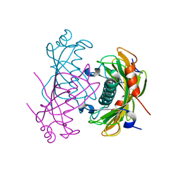
2ODT
 | | Structure of human Inositol 1,3,4-trisphosphate 5/6-kinase | | 分子名称: | Inositol-tetrakisphosphate 1-kinase | | 著者 | Busam, R.D, Arrowsmith, C, Berglund, H, Collins, R, Edwards, A, Ericsson, U.B, Flodin, S, Flores, A, Hammarstrom, M, Holmberg, S.L, Johansson, I, Karlberg, T, Kotenyova, T, Moche, M, Nilsson, M.E, Nordlund, P, Nyman, T, Ogg, D, Sagemark, J, Sundstrom, M, Uppenberg, J, Van Den Berg, S, Weigelt, J, Persson, C, Thorsell, A.G, Hallberg, B.M, Structural Genomics Consortium (SGC) | | 登録日 | 2006-12-26 | | 公開日 | 2007-02-13 | | 最終更新日 | 2023-12-27 | | 実験手法 | X-RAY DIFFRACTION (2.01 Å) | | 主引用文献 | Structure of human Inositol 1,3,4-trisphosphate 5/6-kinase
To be Published
|
|
3PP9
 | | 1.6 Angstrom resolution crystal structure of putative streptothricin acetyltransferase from Bacillus anthracis str. Ames in complex with acetyl coenzyme A | | 分子名称: | ACETYL COENZYME *A, PHOSPHATE ION, POTASSIUM ION, ... | | 著者 | Halavaty, A.S, Wawrzak, Z, Onopriyenko, O, Edwards, A, Savchenko, A, Peterson, S, Anderson, W.F, Center for Structural Genomics of Infectious Diseases (CSGID) | | 登録日 | 2010-11-24 | | 公開日 | 2011-01-12 | | 最終更新日 | 2024-04-03 | | 実験手法 | X-RAY DIFFRACTION (1.6 Å) | | 主引用文献 | 1.6 Angstrom resolution crystal structure of putative streptothricin acetyltransferase from Bacillus anthracis str. Ames in complex with acetyl coenzyme A
To be Published
|
|


