Loading
PDBj
メニューPDBj@FacebookPDBj@TwitterPDBj@YouTubewwPDB FoundationwwPDB
RCSB PDBPDBeBMRBAdv. SearchSearch help
PDB: 538 件

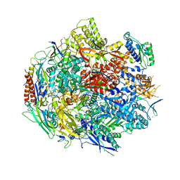
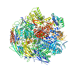
6BLP
DownloadVisualize
BU of 6blp by Molmil
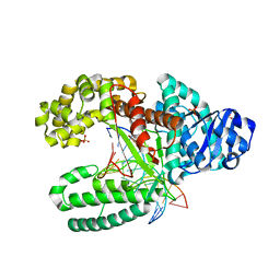
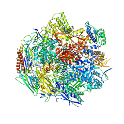
Pol II elongation complex with an abasic lesion at i+1 position, soaking AMPCPP
分子名称: DIPHOSPHOMETHYLPHOSPHONIC ACID ADENOSYL ESTER, DNA (5'-D(P*AP*(3DR)P*CP*TP*CP*TP*CP*GP*AP*TP*G)-3'), DNA-directed RNA polymerase II subunit RPB1, ...
著者Wang, W, Wang, D.
登録日2017-11-11
公開日2018-02-28
最終更新日2024-03-13
実験手法X-RAY DIFFRACTION (3.203 Å)
主引用文献Structural basis of transcriptional stalling and bypass of abasic DNA lesion by RNA polymerase II.
Proc. Natl. Acad. Sci. U.S.A., 115, 2018
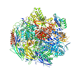
6BM2
DownloadVisualize
BU of 6bm2 by Molmil
Pol II elongation complex with an abasic lesion at i-1 position
分子名称: DNA (5'-D(P*CP*AP*(3DR)P*CP*TP*CP*TP*TP*GP*AP*TP*G)-3'), DNA-directed RNA polymerase II subunit RPB1, DNA-directed RNA polymerase II subunit RPB11, ...
著者Wang, W, Wang, D.
登録日2017-11-13
公開日2018-02-28
最終更新日2024-03-13
実験手法X-RAY DIFFRACTION (3.403 Å)
主引用文献Structural basis of transcriptional stalling and bypass of abasic DNA lesion by RNA polymerase II.
Proc. Natl. Acad. Sci. U.S.A., 115, 2018
6BM4
DownloadVisualize
BU of 6bm4 by Molmil
Pol II elongation complex with an abasic lesion at i-1 position,soaking UMPNPP
分子名称: 5'-O-[(S)-hydroxy{[(S)-hydroxy(phosphonooxy)phosphoryl]amino}phosphoryl]uridine, DNA (5'-D(P*CP*AP*(3DR)P*CP*TP*CP*TP*TP*GP*AP*TP*G)-3'), DNA-directed RNA polymerase II subunit RPB1, ...
著者Wang, W, Wang, D.
登録日2017-11-13
公開日2018-02-28
最終更新日2024-03-13
実験手法X-RAY DIFFRACTION (2.951 Å)
主引用文献Structural basis of transcriptional stalling and bypass of abasic DNA lesion by RNA polymerase II.
Proc. Natl. Acad. Sci. U.S.A., 115, 2018
6BLO
DownloadVisualize
BU of 6blo by Molmil
Pol II elongation complex with an abasic lesion at i+1 position
分子名称: DNA (5'-D(P*AP*(3DR)P*CP*TP*CP*TP*CP*GP*AP*TP*G)-3'), DNA-directed RNA polymerase II subunit RPB1, DNA-directed RNA polymerase II subunit RPB11, ...
著者Wang, W, Wang, D.
登録日2017-11-10
公開日2018-02-28
最終更新日2024-03-13
実験手法X-RAY DIFFRACTION (3.401 Å)
主引用文献Structural basis of transcriptional stalling and bypass of abasic DNA lesion by RNA polymerase II.
Proc. Natl. Acad. Sci. U.S.A., 115, 2018
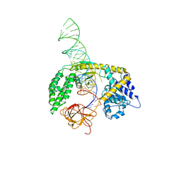
8SCZ
DownloadVisualize
BU of 8scz by Molmil
Cryo-EM structure of 14aa-GS RIG-I in complex with p3SLR30
分子名称: Antiviral innate immune response receptor RIG-I, ZINC ION, p3SLR30
著者Wang, W, Pyle, A.M.
登録日2023-04-06
公開日2024-03-13
実験手法ELECTRON MICROSCOPY (3.4 Å)
主引用文献Cryo-EM structure of 14aa-GS RIG-I in complex with p3SLR30
To Be Published
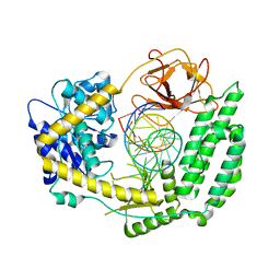
8SD0
DownloadVisualize
BU of 8sd0 by Molmil
Cryo-EM structure of RIG-I in complex with p3SLR14
分子名称: Antiviral innate immune response receptor RIG-I, ZINC ION, p3SLR14
著者Wang, W, Pyle, A.M.
登録日2023-04-06
公開日2024-03-13
実験手法ELECTRON MICROSCOPY (3.8 Å)
主引用文献Cryo-EM structure of RIG-I in complex with p3SLR14
To Be Published
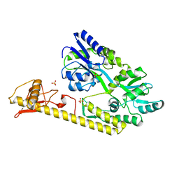
5WVM
DownloadVisualize
BU of 5wvm by Molmil
Crystal structure of baeS cocrystallized with 2 mM indole
分子名称: Maltose-binding periplasmic protein,Two-component system sensor kinase, SULFATE ION
著者Wang, W, Zhang, Y, Rang, T, Xu, D.
登録日2016-12-26
公開日2018-01-03
最終更新日2024-03-20
実験手法X-RAY DIFFRACTION (2.9 Å)
主引用文献Crystal structure of the sensor domain of BaeS from Serratia marcescens FS14
Proteins, 85, 2017
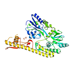
5WVN
DownloadVisualize
BU of 5wvn by Molmil
Crystal structure of MBS-BaeS fusion protein
分子名称: Maltose-binding periplasmic protein,Two-component system sensor kinase, SULFATE ION
著者Wang, W, Zhang, Y, Ran, T, Xu, D.
登録日2016-12-26
公開日2018-01-03
最終更新日2024-03-20
実験手法X-RAY DIFFRACTION (2.8 Å)
主引用文献Crystal structure of the sensor domain of BaeS from Serratia marcescens FS14
Proteins, 85, 2017
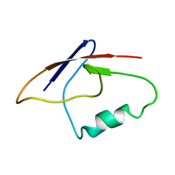
2MBE
DownloadVisualize
BU of 2mbe by Molmil
Backbone 1H and 15N Chemical Shift Assignments for the first domain of FAT10
分子名称: Ubiquitin D
著者Wang, W, Lim, L, Qin, H.
登録日2013-07-30
公開日2014-08-27
最終更新日2024-05-15
実験手法SOLUTION NMR
主引用文献Disruption of FAT10-MAD2 binding inhibits tumor progression.
Proc.Natl.Acad.Sci.USA, 111, 2014
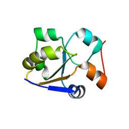
2MRM
DownloadVisualize
BU of 2mrm by Molmil
Solution structure of the rhodanese domain of YgaP from E. coli
分子名称: Membrane protein
著者Wang, W, Zhou, P, Tian, C, Wu, F.
登録日2014-07-12
公開日2014-10-08
最終更新日2024-05-01
実験手法SOLUTION NMR
主引用文献Fast conformational exchange between the sulfur-free and persulfide-bound rhodanese domain of E. coli YgaP
Biochem.Biophys.Res.Commun., 452, 2014
4L67
DownloadVisualize
BU of 4l67 by Molmil
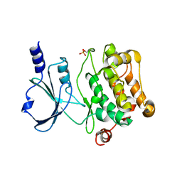
Crystal Structure of Catalytic Domain of PAK4
分子名称: Serine/threonine-protein kinase PAK 4
著者Wang, W, Song, J.
登録日2013-06-12
公開日2013-08-14
最終更新日2023-11-08
実験手法X-RAY DIFFRACTION (2.8 Å)
主引用文献NMR binding and crystal structure reveal that intrinsically-unstructured regulatory domain auto-inhibits PAK4 by a mechanism different for that of PAK1
Biochem.Biophys.Res.Commun., 438, 2013
3SSP
DownloadVisualize
BU of 3ssp by Molmil
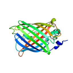
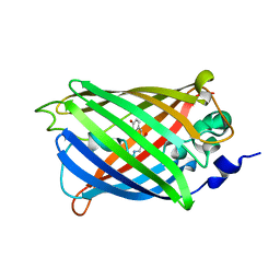
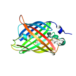
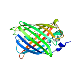
Engineered low-affinity halide-binding protein derived from YFP: halide-free
分子名称: Green fluorescent protein
著者Wang, W, Grimley, J.S, Beese, L.S, Hellinga, H.W.
登録日2011-07-08
公開日2012-07-11
最終更新日2023-11-15
実験手法X-RAY DIFFRACTION (1.628 Å)
主引用文献Determination of engineered chloride-binding site structures in fluorescent proteins reveals principles of halide recognition
To be Published
3SOV
DownloadVisualize
BU of 3sov by Molmil
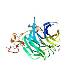
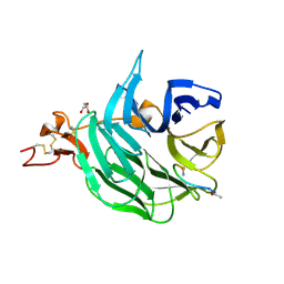
The structure of a beta propeller domain in complex with peptide S
分子名称: 2-acetamido-2-deoxy-beta-D-glucopyranose, GLYCEROL, Low-density lipoprotein receptor-related protein 6, ...
著者Wang, W, Bourhis, E, Zhang, Y, Rouge, L, Wu, Y, Franke, Y, Cochran, A.G.
登録日2011-06-30
公開日2011-09-21
最終更新日2020-07-29
実験手法X-RAY DIFFRACTION (1.27 Å)
主引用文献Wnt antagonists bind through a short peptide to the first beta-propeller domain of LRP5/6.
Structure, 19, 2011
3TAR
DownloadVisualize
BU of 3tar by Molmil
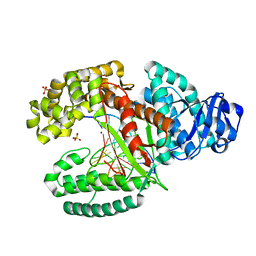
Crystal Structure of Bacillus DNA Polymerase I Large Fragment Bound to Duplex DNA with Cytosine-Adenine Mismatch at (n-6) Position
分子名称: 5'-D(*GP*AP*CP*GP*TP*AP*CP*GP*TP*GP*AP*TP*CP*GP*CP*A)-3', 5'-D(*GP*CP*GP*AP*TP*CP*AP*CP*GP*CP*AP*CP*GP*TP*C)-3', DNA polymerase I, ...
著者Wang, W, Beese, L.S.
登録日2011-08-04
公開日2011-10-19
最終更新日2023-09-13
実験手法X-RAY DIFFRACTION (1.6 Å)
主引用文献Structural evidence for the rare tautomer hypothesis of spontaneous mutagenesis.
Proc.Natl.Acad.Sci.USA, 108, 2011
3SSV
DownloadVisualize
BU of 3ssv by Molmil
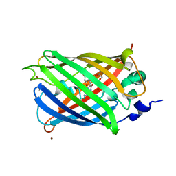
Engineered low-affinity halide-binding protein derived from YFP: fluoride complex
分子名称: FLUORIDE ION, Green fluorescent protein
著者Wang, W, Grimley, J.S, Beese, L.S, Hellinga, H.W.
登録日2011-07-08
公開日2012-07-11
最終更新日2023-11-15
実験手法X-RAY DIFFRACTION (1.861 Å)
主引用文献Determination of engineered chloride-binding site structures in fluorescent proteins reveals principles of halide recognition
To be Published
3SVD
DownloadVisualize
BU of 3svd by Molmil
Engineered medium-affinity halide-binding protein derived from YFP: bromide complex
分子名称: 1,2-ETHANEDIOL, BROMIDE ION, Green fluorescent protein
著者Wang, W, Grimley, J.S, Beese, L.S, Hellinga, H.W.
登録日2011-07-12
公開日2012-07-18
最終更新日2023-11-15
実験手法X-RAY DIFFRACTION (1.78 Å)
主引用文献Determination of engineered chloride-binding site structures in fluorescent proteins reveals principles of halide recognition
To be Published
3TAP
DownloadVisualize
BU of 3tap by Molmil
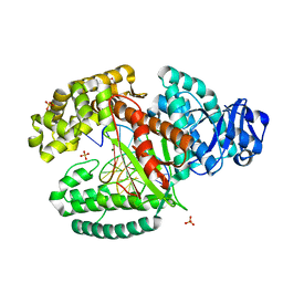
Crystal Structure of Bacillus DNA Polymerase I Large Fragment Bound to Duplex DNA with Cytosine-Adenine Mismatch at (n-3) Position
分子名称: 5'-D(*G*CP*GP*AP*TP*CP*AP*CP*GP*CP*AP*C)-3', 5'-D(*GP*A*CP*GP*TP*AP*CP*GP*TP*GP*AP*TP*CP*GP*CP*A)-3', DNA polymerase I, ...
著者Wang, W, Beese, L.S.
登録日2011-08-04
公開日2011-10-19
最終更新日2023-09-13
実験手法X-RAY DIFFRACTION (1.655 Å)
主引用文献Structural evidence for the rare tautomer hypothesis of spontaneous mutagenesis.
Proc.Natl.Acad.Sci.USA, 108, 2011
3TAQ
DownloadVisualize
BU of 3taq by Molmil
Crystal Structure of Bacillus DNA Polymerase I Large Fragment Bound to Duplex DNA with Cytosine-Adenine Mismatch at (n-4) Position
分子名称: 5'-D(*GP*AP*CP*GP*TP*AP*CP*GP*TP*GP*AP*TP*CP*GP*CP*A)-3', 5'-D(*GP*C*GP*AP*TP*CP*AP*CP*GP*CP*AP*CP*G)-3', DNA polymerase I, ...
著者Wang, W, Beese, L.S.
登録日2011-08-04
公開日2011-10-19
最終更新日2023-09-13
実験手法X-RAY DIFFRACTION (1.65 Å)
主引用文献Structural evidence for the rare tautomer hypothesis of spontaneous mutagenesis.
Proc.Natl.Acad.Sci.USA, 108, 2011
3SOB
DownloadVisualize
BU of 3sob by Molmil
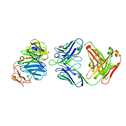
The structure of the first YWTD beta propeller domain of LRP6 in complex with a FAB
分子名称: CALCIUM ION, Low-density lipoprotein receptor-related protein 6, antibody heavy chain, ...
著者Wang, W, Bourhis, E, Tam, C, Zhang, Y, Rouge, L, Wu, Y, Franke, Y, Cochran, A.G.
登録日2011-06-30
公開日2011-09-21
最終更新日2014-05-14
実験手法X-RAY DIFFRACTION (1.9 Å)
主引用文献Wnt antagonists bind through a short peptide to the first beta-propeller domain of LRP5/6.
Structure, 19, 2011
3SRY
DownloadVisualize
BU of 3sry by Molmil
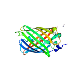
Engineered high-affinity halide-binding protein derived from YFP: halide-free
分子名称: Green fluorescent protein
著者Wang, W, Grimley, J.S, Beese, L.S, Hellinga, H.W.
登録日2011-07-07
公開日2012-07-11
最終更新日2023-11-15
実験手法X-RAY DIFFRACTION (1.159 Å)
主引用文献Determination of engineered chloride-binding site structures in fluorescent proteins reveals principles of halide recognition
To be Published
3SVB
DownloadVisualize
BU of 3svb by Molmil
Engineered medium-affinity halide-binding protein derived from YFP: fluoride complex
分子名称: 1,2-ETHANEDIOL, Green fluorescent protein
著者Wang, W, Grimley, J.S, Beese, L.S, Hellinga, H.W.
登録日2011-07-12
公開日2012-07-18
最終更新日2023-11-15
実験手法X-RAY DIFFRACTION (1.3 Å)
主引用文献Determination of engineered chloride-binding site structures in fluorescent proteins reveals principles of halide recognition
To be Published
3SST
DownloadVisualize
BU of 3sst by Molmil
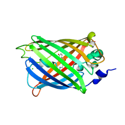
Engineered low-affinity halide-binding protein derived from YFP: chloride complex
分子名称: CHLORIDE ION, Green fluorescent protein
著者Wang, W, Grimley, J.S, Beese, L.S, Hellinga, H.W.
登録日2011-07-08
公開日2012-07-11
最終更新日2023-11-15
実験手法X-RAY DIFFRACTION (1.403 Å)
主引用文献Determination of engineered chloride-binding site structures in fluorescent proteins reveals principles of halide recognition
To be Published
3SVE
DownloadVisualize
BU of 3sve by Molmil
Engineered low-affinity halide-binding protein derived from YFP: bromide complex
分子名称: BROMIDE ION, Green fluorescent protein
著者Wang, W, Grimley, J.S, Beese, L.S, Hellinga, H.W.
登録日2011-07-12
公開日2012-07-18
最終更新日2023-11-15
実験手法X-RAY DIFFRACTION (1.492 Å)
主引用文献Determination of engineered chloride-binding site structures in fluorescent proteins reveals principles of halide recognition
To be Published
4FXW
DownloadVisualize
BU of 4fxw by Molmil
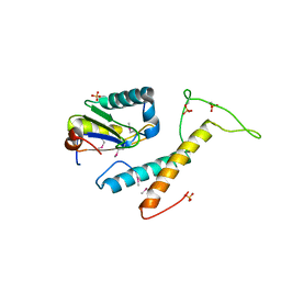
Structure of phosphorylated SF1 complex with U2AF65-UHM domain
分子名称: SULFATE ION, Splicing factor 1, Splicing factor U2AF 65 kDa subunit
著者Wang, W, Bauer, W.J, Wedekind, J.E, Kielkopf, C.L.
登録日2012-07-03
公開日2013-01-16
最終更新日2017-11-15
実験手法X-RAY DIFFRACTION (2.29 Å)
主引用文献Structure of Phosphorylated SF1 Bound to U2AF(65) in an Essential Splicing Factor Complex.
Structure, 21, 2013
3SOQ
DownloadVisualize
BU of 3soq by Molmil
The structure of the first YWTD beta propeller domain of LRP6 in complex with a DKK1 peptide
分子名称: 2-acetamido-2-deoxy-beta-D-glucopyranose, CALCIUM ION, Dickkopf-related protein 1, ...
著者Wang, W, Bourhis, E, Zhang, Y, Rouge, L, Wu, Y, Franke, Y, Cochran, A.G.
登録日2011-06-30
公開日2011-09-21
最終更新日2020-07-29
実験手法X-RAY DIFFRACTION (1.9 Å)
主引用文献Wnt antagonists bind through a short peptide to the first beta-propeller domain of LRP5/6.
Structure, 19, 2011

221716

件を2024-06-26に公開中

PDB statisticsPDBj update infoContact PDBjnumon