Loading
PDBj
MenuPDBj@FacebookPDBj@TwitterPDBj@YouTubewwPDB FoundationwwPDB
RCSB PDBPDBeBMRBAdv. SearchSearch help
PDB: 43 results

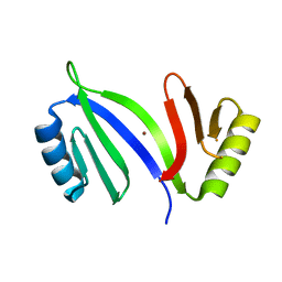
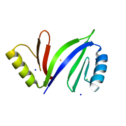
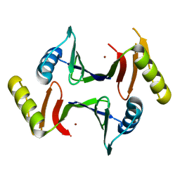
5HCW
DownloadVisualize
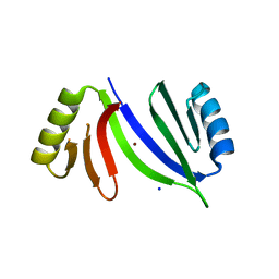
BU of 5hcw by Molmil
Crystal structure of C-As lyase with mutations Y100H and V102F (monoclinic form)
Descriptor: Glyoxalase/bleomycin resistance protein/dioxygenase
Authors:Venkadesh, S, Yoshinaga, M, Kandavelu, P, Sankaran, B, Rosen, B.P.
Deposit date:2016-01-04
Release date:2017-01-18
Last modified:2024-03-06
Method:X-RAY DIFFRACTION (2.785 Å)
Cite:Crystal structure of C-As lyase with mutations Y100H and V102F (monoclinic form)
To Be Published
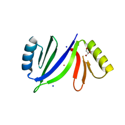
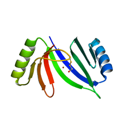
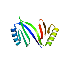
5V0F
DownloadVisualize
BU of 5v0f by Molmil
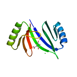
Crystal structure of C-As lyase with mutation K105A and substrate Roxarsone
Descriptor: 4-arsanyl-2-nitrophenol, CHLORIDE ION, FE (III) ION, ...
Authors:Venkadesh, S, Yoshinaga, M, Sankaran, B, Kandavelu, P, Rosen, B.P.
Deposit date:2017-02-28
Release date:2018-03-21
Last modified:2023-10-04
Method:X-RAY DIFFRACTION (2.23 Å)
Cite:Crystal structure of C-As lyase with mutation K105A and substrate Roxarsone.
To Be Published
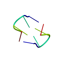
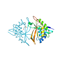
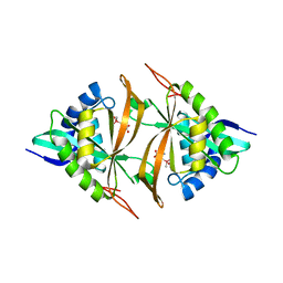
3Q5C
DownloadVisualize
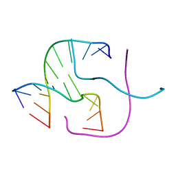
BU of 3q5c by Molmil
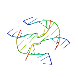
Crystal structure of four-way junction with sticky end
Descriptor: DNA (5'-D(*CP*GP*GP*CP*GP*GP*CP*CP*GP*C)-3')
Authors:Venkadesh, S, Mandal, P.K, Gautham, N.
Deposit date:2010-12-28
Release date:2011-05-18
Last modified:2023-11-01
Method:X-RAY DIFFRACTION (2.501 Å)
Cite:The sequence d(CGGCGGCCGC) self-assembles into a two dimensional rhombic DNA lattice
Biochem.Biophys.Res.Commun., 407, 2011
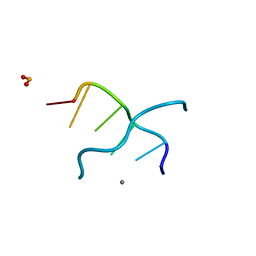
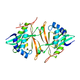
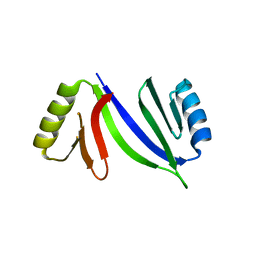
3FQ5
DownloadVisualize
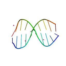
BU of 3fq5 by Molmil
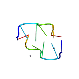
Crystal Structure of d(CACGCG).d(CGCGTG) with 10mM MnCl2
Descriptor: 5'-D(*CP*AP*CP*GP*CP*G)-3', 5'-D(*CP*GP*CP*GP*TP*G)-3', MANGANESE (II) ION
Authors:Venkadesh, S, Kannan, R, Mandal, P.K, Gautham, N.
Deposit date:2009-01-07
Release date:2009-02-10
Last modified:2024-03-20
Method:X-RAY DIFFRACTION (2.8 Å)
Cite:Crystal Structure of d(CACGCG).d(CGCGTG) with 10mM MnCl2
To be Published
3R86
DownloadVisualize
BU of 3r86 by Molmil
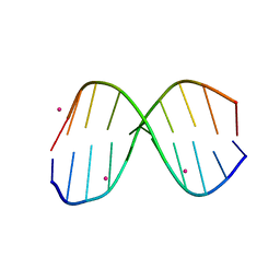
Crystal structure of d(CCGGTACCGG)2 as B-DNA duplex grown with 5 mM CoCl2
Descriptor: COBALT (II) ION, DNA (5'-D(*CP*CP*GP*GP*TP*AP*CP*CP*GP*G)-3')
Authors:Venkadesh, S, Mandal, P.K, Gautham, N.
Deposit date:2011-03-23
Release date:2011-04-06
Last modified:2024-04-03
Method:X-RAY DIFFRACTION (2.8 Å)
Cite:Crystal structure of d(CCGGTACCGG)2 as B-DNA duplex
To be Published
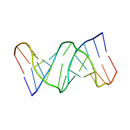
3E9W
DownloadVisualize
BU of 3e9w by Molmil
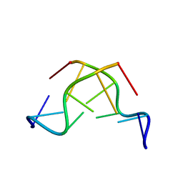
X-Ray Crystal Structure of the hexamer DCACACG:Crystal grown in the presence of cobalt(III)hexammine Chloride.
Descriptor: 5'-D(*CP*AP*CP*AP*CP*G)-3', 5'-D(*CP*GP*TP*GP*TP*G)-3'
Authors:Venkadesh, S, Mandal, P.K, Gautham, N.
Deposit date:2008-08-24
Release date:2008-09-30
Last modified:2024-04-03
Method:X-RAY DIFFRACTION (2.05 Å)
Cite:The structure of d(CACACG).d(CGTGTG).
Acta Crystallogr.,Sect.F, 65, 2009
3QK4
DownloadVisualize
BU of 3qk4 by Molmil
Crystal structure of d(CGCGGGTACCCGCG)2 as A-DNA duplex
Descriptor: DNA (5'-D(*CP*GP*CP*GP*GP*GP*TP*AP*CP*CP*CP*GP*CP*G)-3')
Authors:Venkadesh, S, Mandal, P.K, Gautham, N.
Deposit date:2011-01-31
Release date:2011-03-02
Last modified:2024-04-03
Method:X-RAY DIFFRACTION (2.6 Å)
Cite:The structure of a full turn of an A-DNA duplex d(CGCGGGTACCCGCG)(2)
Biochem.Biophys.Res.Commun., 407, 2011
3TCI
DownloadVisualize
BU of 3tci by Molmil
Crystal structure of the decameric sequence d(CGGGCGCCCG) as Z type duplex
Descriptor: DNA (5'-D(P*CP*GP*GP*GP*CP*GP*CP*CP*CP*G)-3')
Authors:Venkadesh, S, Mandal, P.K, Gautham, N.
Deposit date:2011-08-09
Release date:2011-10-05
Last modified:2024-03-20
Method:X-RAY DIFFRACTION (2.421 Å)
Cite:Crystal structure of the decameric sequence d(CGGGCGCCCG) as Z type duplex
To be Published
3G2R
DownloadVisualize
BU of 3g2r by Molmil
Crystal structure of d(CACGCG).d(CGCGTG) cocrystallized with MnCl2
Descriptor: 2'-DEOXYCYTIDINE-5'-MONOPHOSPHATE, 5'-D(*CP*GP*TP*GP*CP*G)-3', MANGANESE (II) ION
Authors:Venkadesh, S, Mandal, P.K, Kannan, R, Gautham, N.
Deposit date:2009-02-01
Release date:2009-03-03
Last modified:2024-03-20
Method:X-RAY DIFFRACTION (2.15 Å)
Cite:Crystal studies of d(CACGCG).d(CGCGTG) grown with various counter ions
To be Published
3G2A
DownloadVisualize
BU of 3g2a by Molmil
Crystal structure of d(CACGCG).d(CGCGTG) grown in presence of 1mM MnCl2
Descriptor: 5'-D(*CP*AP*CP*GP*CP*G)-3', 5'-D(*CP*GP*CP*GP*TP*G)-3'
Authors:Venkadesh, S, Mandal, P.K, Kannan, R, Gautham, N.
Deposit date:2009-01-31
Release date:2009-03-03
Last modified:2024-03-20
Method:X-RAY DIFFRACTION (2 Å)
Cite:Crystal Structures of d(CACGCG).d(CGCGTG) cocrystallized with MnCl2
To be Published
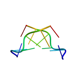
3HS1
DownloadVisualize
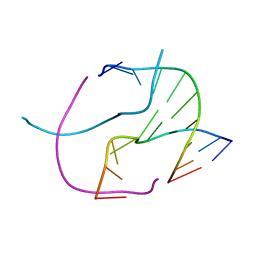
BU of 3hs1 by Molmil
Structure of Holliday junction formed by d(CCGGTACCGG); Crystal grown with CoCl2
Descriptor: 5'-D(*CP*CP*GP*GP*TP*AP*CP*CP*GP*G)-3'
Authors:Venkadesh, S, Mandal, P.K, Gautham, N.
Deposit date:2009-06-10
Release date:2010-06-16
Last modified:2023-11-01
Method:X-RAY DIFFRACTION (2.51 Å)
Cite:Crystal structure of Holliday junction
To be Published
3HQE
DownloadVisualize
BU of 3hqe by Molmil
Crystal Structure of the decamer CGGGCGCCCG forming a Holliday junction
Descriptor: 5'-D(*CP*GP*GP*GP*CP*GP*CP*CP*CP*G)-3'
Authors:Venkadesh, S, Mandal, P.K, Gautham, N.
Deposit date:2009-06-06
Release date:2010-06-09
Last modified:2023-11-01
Method:X-RAY DIFFRACTION (2.94 Å)
Cite:Sequence dependent structural variation of Holliday junction - crystal structure of d(CGGGCGCCCG)4
To be Published
3IXN
DownloadVisualize
BU of 3ixn by Molmil
Crystal structure of d(CCGGTACCGG) as B-DNA duplex
Descriptor: COBALT (II) ION, DNA (5'-D(*CP*CP*GP*GP*TP*AP*CP*CP*GP*G)-3')
Authors:Venkadesh, S, Mandal, P.K, Gautham, N.
Deposit date:2009-09-04
Release date:2010-09-15
Last modified:2024-04-03
Method:X-RAY DIFFRACTION (2.87 Å)
Cite:Crystal Structure of inverted repeat sequence d(CCGGTACCGG)as B-DNA duplex
To be Published
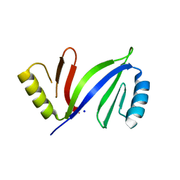
5C68
DownloadVisualize
BU of 5c68 by Molmil
Crystal structure of C-As lyase at 1.46 Angstroms resolution
Descriptor: Glyoxalase/bleomycin resistance protein/dioxygenase, NICKEL (II) ION, SODIUM ION
Authors:Venkadesh, S, Yoshinaga, M, Kandavelu, P, Rosen, B.P.
Deposit date:2015-06-22
Release date:2016-07-13
Last modified:2023-09-27
Method:X-RAY DIFFRACTION (1.4618 Å)
Cite:Crystal structure of C-As lyase at 1.46 Angstroms resolution
To Be Published
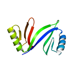
5C4P
DownloadVisualize
BU of 5c4p by Molmil
Crystal structure of ArsI C-As lyase solved by Ni-SAD phasing
Descriptor: Glyoxalase/bleomycin resistance protein/dioxygenase, NICKEL (II) ION
Authors:Venkadesh, S, Yoshinaga, M, Kandavelu, P, Rosen, B.P.
Deposit date:2015-06-18
Release date:2016-07-13
Last modified:2024-03-06
Method:X-RAY DIFFRACTION (1.97 Å)
Cite:Crystal structure of ArsI C-As lyase from Thermomonospora curvata
To Be Published
5C6X
DownloadVisualize
BU of 5c6x by Molmil
Crystal structure of C-As lyase with Co(II)
Descriptor: COBALT (II) ION, GLYCEROL, Glyoxalase/bleomycin resistance protein/dioxygenase, ...
Authors:Venkadesh, S, Yoshinaga, M, Sankaran, B, Kandavelu, P, Rosen, B.P.
Deposit date:2015-06-23
Release date:2016-07-13
Last modified:2023-09-27
Method:X-RAY DIFFRACTION (1.5 Å)
Cite:Crystal structure of C-As lyase with Co(II)
To Be Published
5DRH
DownloadVisualize
BU of 5drh by Molmil
Crystal structure of apo C-As lyase
Descriptor: Glyoxalase/bleomycin resistance protein/dioxygenase, SODIUM ION
Authors:Venkadesh, S, Yoshinaga, M, Sankaran, B, Kandavelu, P, Rosen, B.P.
Deposit date:2015-09-15
Release date:2016-09-21
Last modified:2023-09-27
Method:X-RAY DIFFRACTION (2.27 Å)
Cite:Crystal structure of apo C-As lyase
To Be Published
5DFG
DownloadVisualize
BU of 5dfg by Molmil
Crystal structure of C-As lyase with mutations Y100H and V102F
Descriptor: Glyoxalase/bleomycin resistance protein/dioxygenase
Authors:Venkadesh, S, Yoshinaga, M, Kandavelu, P, Sankaran, B, Rosen, B.P.
Deposit date:2015-08-26
Release date:2016-09-21
Last modified:2023-09-27
Method:X-RAY DIFFRACTION (1.9711 Å)
Cite:Crystal structure of C-As lyase with mutations Y100H and F102V
To Be Published
5D4F
DownloadVisualize
BU of 5d4f by Molmil
Crystal structure of C-As lyase with Fe(III)
Descriptor: CHLORIDE ION, FE (III) ION, Glyoxalase/bleomycin resistance protein/dioxygenase, ...
Authors:Venkadesh, S, Yoshinaga, M, Sankaran, B, Kandavelu, P, Rosen, B.P.
Deposit date:2015-08-07
Release date:2016-09-07
Last modified:2023-09-27
Method:X-RAY DIFFRACTION (1.72 Å)
Cite:Crystal structure of C-As lyase with mercaptoethonal
To Be Published
5CB9
DownloadVisualize
BU of 5cb9 by Molmil
Crystal structure of C-As lyase with mercaptoethonal
Descriptor: BETA-MERCAPTOETHANOL, CHLORIDE ION, Glyoxalase/bleomycin resistance protein/dioxygenase, ...
Authors:Venkadesh, S, Yoshinaga, M, Sankaran, B, Kandavelu, P, Rosen, B.P.
Deposit date:2015-06-30
Release date:2016-07-13
Last modified:2024-05-01
Method:X-RAY DIFFRACTION (1.95 Å)
Cite:Crystal structure of C-As lyase with mercaptoethonal
To Be Published
6M7G
DownloadVisualize
BU of 6m7g by Molmil
Crystal structure of ArsN, N-acetyltransferase with substrate phosphinothricin from Pseudomonas putida KT2440
Descriptor: PHOSPHINOTHRICIN, Phosphinothricin N-acetyltransferase
Authors:Venkadesh, S, Dheeman, D.S, Yoshinaga, M, Kandavelu, P, Rosen, B.P.
Deposit date:2018-08-20
Release date:2019-04-24
Last modified:2023-10-11
Method:X-RAY DIFFRACTION (2.657 Å)
Cite:Arsinothricin, an arsenic-containing non-proteinogenic amino acid analog of glutamate, is a broad-spectrum antibiotic.
Commun Biol, 2, 2019
5JTF
DownloadVisualize
BU of 5jtf by Molmil
Crystal structure of ArsN N-acetyltransferase from Pseudomonas putida KT2440
Descriptor: 2-AMINO-2-HYDROXYMETHYL-PROPANE-1,3-DIOL, Putative Phosphinothricin N-acetyltransferase
Authors:Venkadesh, S, Dheeman, D.S, Kandavelu, P, Rosen, B.P.
Deposit date:2016-05-09
Release date:2017-05-17
Last modified:2023-09-27
Method:X-RAY DIFFRACTION (2.156 Å)
Cite:Arsinothricin, an arsenic-containing non-proteinogenic amino acid analog of glutamate, is a broad-spectrum antibiotic.
Commun Biol, 2, 2019
6XA0
DownloadVisualize
BU of 6xa0 by Molmil
Crystal structure of C-As lyase with mutation K105R with Ni(II)
Descriptor: Glyoxalase/bleomycin resistance protein/dioxygenase, NICKEL (II) ION
Authors:Venkadesh, S, Yoshinaga, M, Kandavelu, P, Sankaran, B, Rosen, B.P.
Deposit date:2020-06-03
Release date:2021-06-09
Last modified:2023-10-18
Method:X-RAY DIFFRACTION (2.15 Å)
Cite:The ArsI C-As lyase: Elucidating the catalytic mechanism of degradation of organoarsenicals.
J.Inorg.Biochem., 232, 2022
6XCK
DownloadVisualize
BU of 6xck by Molmil
Crystal structure of C-As lyase with mutation K105E
Descriptor: Glyoxalase/bleomycin resistance protein/dioxygenase
Authors:Venkadesh, S, Yoshinaga, M, Kandavelu, P, Sankaran, B, Rosen, B.P.
Deposit date:2020-06-08
Release date:2021-06-16
Last modified:2023-10-18
Method:X-RAY DIFFRACTION (1.62 Å)
Cite:The ArsI C-As lyase: Elucidating the catalytic mechanism of degradation of organoarsenicals.
J.Inorg.Biochem., 232, 2022
5WPH
DownloadVisualize
BU of 5wph by Molmil
Crystal structure of ArsN, N-acetyltransferase with substrate AST from Pseudomonas putida KT2440
Descriptor: (2S)-2-amino-4-[hydroxy(methyl)arsoryl]butanoic acid, Phosphinothricin N-acetyltransferase, SODIUM ION
Authors:Venkadesh, S, Dheeman, D.S, Yoshinaga, M, Kandavelu, P, Rosen, B.P.
Deposit date:2017-08-04
Release date:2018-10-10
Last modified:2023-10-04
Method:X-RAY DIFFRACTION (2.19 Å)
Cite:Arsinothricin, an arsenic-containing non-proteinogenic amino acid analog of glutamate, is a broad-spectrum antibiotic.
Commun Biol, 2, 2019


12>

226707

건을2024-10-30부터공개중

PDB statisticsPDBj update infoContact PDBjnumon