Loading
PDBj
MenuPDBj@FacebookPDBj@TwitterPDBj@YouTubewwPDB FoundationwwPDB
RCSB PDBPDBeBMRBAdv. SearchSearch help
PDB: 11 results

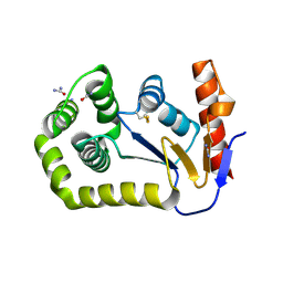
8D12
DownloadVisualize
BU of 8d12 by Molmil
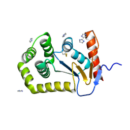
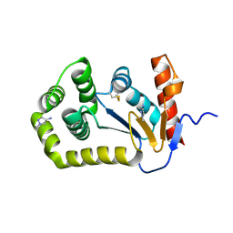
Crystal Structure of EcDsbA in a complex with 1-methyl-1H-pyrazol-4-amine
Descriptor: 1-methyl-1H-pyrazol-4-amine, COPPER (II) ION, Thiol:disulfide interchange protein DsbA
Authors:Whitehouse, R.L, Ilyichova, O.V, Taylor, A.J.
Deposit date:2022-05-26
Release date:2022-12-07
Last modified:2023-10-25
Method:X-RAY DIFFRACTION (1.6 Å)
Cite:Fragment screening libraries for the identification of protein hot spots and their minimal binding pharmacophores.
Rsc Med Chem, 14, 2023
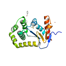
8CZM
DownloadVisualize
BU of 8czm by Molmil
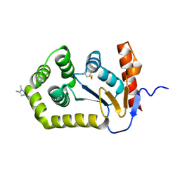
Crystal Structure of EcDsbA in a complex with 4-bromo-1H-pyrazole
Descriptor: 4-bromo-1H-pyrazole, TRIETHYLENE GLYCOL, Thiol:disulfide interchange protein DsbA
Authors:Whitehouse, R.L, Ilyichova, O.V, Taylor, A.J.
Deposit date:2022-05-25
Release date:2022-12-07
Last modified:2023-10-25
Method:X-RAY DIFFRACTION (1.8 Å)
Cite:Fragment screening libraries for the identification of protein hot spots and their minimal binding pharmacophores.
Rsc Med Chem, 14, 2023
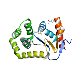
8DG0
DownloadVisualize
BU of 8dg0 by Molmil
Crystal Structure of EcDsbA in a complex with Urea
Descriptor: COPPER (II) ION, Thiol:disulfide interchange protein DsbA, UREA
Authors:Whitehouse, R.L, Ilyichova, O.V, Taylor, A.J.
Deposit date:2022-06-23
Release date:2022-12-07
Last modified:2023-10-25
Method:X-RAY DIFFRACTION (2.5 Å)
Cite:Fragment screening libraries for the identification of protein hot spots and their minimal binding pharmacophores.
Rsc Med Chem, 14, 2023
8CXD
DownloadVisualize
BU of 8cxd by Molmil
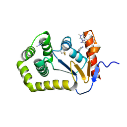
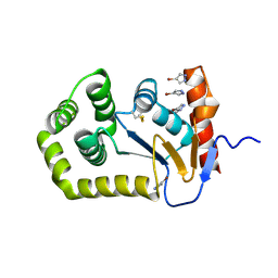
Crystal Structure of EcDsbA in a complex with phenylmethanol
Descriptor: COPPER (II) ION, Thiol:disulfide interchange protein DsbA, phenylmethanol
Authors:Whitehouse, R.L, Ilyichova, O.V, Taylor, A.J.
Deposit date:2022-05-20
Release date:2022-12-07
Last modified:2024-10-16
Method:X-RAY DIFFRACTION (1.8 Å)
Cite:Fragment screening libraries for the identification of protein hot spots and their minimal binding pharmacophores.
Rsc Med Chem, 14, 2023
8CZN
DownloadVisualize
BU of 8czn by Molmil
Crystal Structure of EcDsbA in a complex with 1H-pyrrole-3-carboxylic acid
Descriptor: 1H-pyrrole-3-carboxylic acid, COPPER (II) ION, Thiol:disulfide interchange protein DsbA
Authors:Whitehouse, R.L, Ilyichova, O.V, Taylor, A.J.
Deposit date:2022-05-25
Release date:2022-12-07
Last modified:2024-10-16
Method:X-RAY DIFFRACTION (1.7 Å)
Cite:Fragment screening libraries for the identification of protein hot spots and their minimal binding pharmacophores.
Rsc Med Chem, 14, 2023
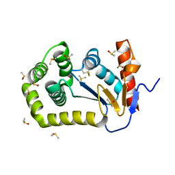
8DG1
DownloadVisualize
BU of 8dg1 by Molmil
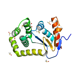
Crystal Structure of EcDsbA in a complex with DMSO
Descriptor: COPPER (II) ION, DIMETHYL SULFOXIDE, Thiol:disulfide interchange protein DsbA
Authors:Whitehouse, R.L, Ilyichova, O.V, Taylor, A.J.
Deposit date:2022-06-23
Release date:2022-12-07
Last modified:2024-11-06
Method:X-RAY DIFFRACTION (1.95 Å)
Cite:Fragment screening libraries for the identification of protein hot spots and their minimal binding pharmacophores.
Rsc Med Chem, 14, 2023
8CXE
DownloadVisualize
BU of 8cxe by Molmil
Crystal Structure of EcDsbA in a complex with 1H-imidazole
Descriptor: IMIDAZOLE, Thiol:disulfide interchange protein DsbA
Authors:Whitehouse, R.L, Ilyichova, O.V, Taylor, A.J.
Deposit date:2022-05-20
Release date:2022-12-07
Last modified:2024-10-30
Method:X-RAY DIFFRACTION (1.47 Å)
Cite:Fragment screening libraries for the identification of protein hot spots and their minimal binding pharmacophores.
Rsc Med Chem, 14, 2023
8D10
DownloadVisualize
BU of 8d10 by Molmil
Crystal Structure of EcDsbA in a complex with (1-methyl-1H-pyrazol-5-yl)methanamine
Descriptor: 1-(1-methyl-1H-pyrazol-5-yl)methanamine, COPPER (II) ION, Thiol:disulfide interchange protein DsbA
Authors:Whitehouse, R.L, Ilyichova, O.V, Taylor, A.J.
Deposit date:2022-05-26
Release date:2022-12-07
Last modified:2024-11-06
Method:X-RAY DIFFRACTION (1.6 Å)
Cite:Fragment screening libraries for the identification of protein hot spots and their minimal binding pharmacophores.
Rsc Med Chem, 14, 2023
8DG2
DownloadVisualize
BU of 8dg2 by Molmil
Crystal Structure of EcDsbA in a complex with DMSO
Descriptor: COPPER (II) ION, DIMETHYL SULFOXIDE, GLYCEROL, ...
Authors:Whitehouse, R.L, Ilyichova, O.V, Taylor, A.J.
Deposit date:2022-06-23
Release date:2022-12-07
Last modified:2023-10-25
Method:X-RAY DIFFRACTION (1.95 Å)
Cite:Fragment screening libraries for the identification of protein hot spots and their minimal binding pharmacophores.
Rsc Med Chem, 14, 2023
8D11
DownloadVisualize
BU of 8d11 by Molmil
Crystal Structure of EcDsbA in a complex with 1-methyl-1H-pyrazol-5-amine
Descriptor: 1-methyl-1H-pyrazol-5-amine, COPPER (II) ION, Thiol:disulfide interchange protein DsbA
Authors:Whitehouse, R.L, Ilyichova, O.V, Taylor, A.J.
Deposit date:2022-05-26
Release date:2022-12-07
Last modified:2023-10-25
Method:X-RAY DIFFRACTION (1.85 Å)
Cite:Fragment screening libraries for the identification of protein hot spots and their minimal binding pharmacophores.
Rsc Med Chem, 14, 2023
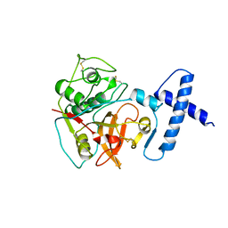
1PCI
DownloadVisualize
BU of 1pci by Molmil
PROCARICAIN
Descriptor: PROCARICAIN
Authors:Groves, M.R, Taylor, M.A.J, Scott, M, Cummings, N.J, Pickersgill, R.W, Jenkins, J.A.
Deposit date:1996-06-28
Release date:1997-04-01
Last modified:2023-08-09
Method:X-RAY DIFFRACTION (3.2 Å)
Cite:The prosequence of procaricain forms an alpha-helical domain that prevents access to the substrate-binding cleft.
Structure, 4, 1996

227111

數據於2024-11-06公開中

PDB statisticsPDBj update infoContact PDBjnumon