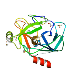
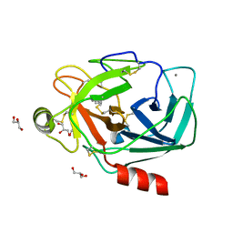
2R96
 
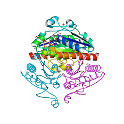 | | Crystal structure of E. coli WrbA in complex with FMN | | 分子名称: | 1,2-ETHANEDIOL, FLAVIN MONONUCLEOTIDE, Flavoprotein WrbA | | 著者 | Kuta Smatanova, I, Wolfova, J, Brynda, J, Mesters, J.R, Grandori, R, Carey, J. | | 登録日 | 2007-09-12 | | 公開日 | 2008-09-23 | | 最終更新日 | 2023-08-30 | | 実験手法 | X-RAY DIFFRACTION (2.6 Å) | | 主引用文献 | Structural organization of WrbA in apo- and holoprotein crystals.
Biochim.Biophys.Acta, 1794, 2009
|
|
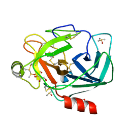
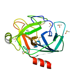
4FJ2
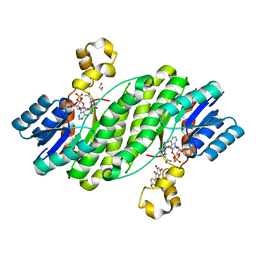 | | Crystal structure of the ternary complex between a fungal 17beta-hydroxysteroid dehydrogenase (Holo form) and biochanin A | | 分子名称: | 1,2-ETHANEDIOL, 17beta-hydroxysteroid dehydrogenase, 5,7-dihydroxy-3-(4-methoxyphenyl)-4H-chromen-4-one, ... | | 著者 | Cassetta, A, Lamba, D, Krastanova, I. | | 登録日 | 2012-06-11 | | 公開日 | 2013-07-03 | | 最終更新日 | 2023-09-13 | | 実験手法 | X-RAY DIFFRACTION (2.5 Å) | | 主引用文献 | Structural basis for inhibition of 17 beta-hydroxysteroid dehydrogenases by phytoestrogens: The case of fungal 17 beta-HSDcl.
J. Steroid Biochem. Mol. Biol., 171, 2017
|
|
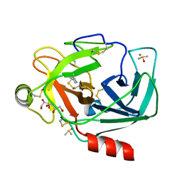
4FJ0
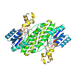 | | Crystal structure of the ternary complex between a fungal 17beta-hydroxysteroid dehydrogenase (Holo form) and 3,7-dihydroxy flavone | | 分子名称: | 1,2-ETHANEDIOL, 17beta-hydroxysteroid dehydrogenase, 3,7-dihydroxy-2-phenyl-4H-chromen-4-one, ... | | 著者 | Cassetta, A, Lamba, D, Krastanova, I. | | 登録日 | 2012-06-11 | | 公開日 | 2013-07-03 | | 最終更新日 | 2023-09-13 | | 実験手法 | X-RAY DIFFRACTION (2.2 Å) | | 主引用文献 | Structural basis for inhibition of 17 beta-hydroxysteroid dehydrogenases by phytoestrogens: The case of fungal 17 beta-HSDcl.
J. Steroid Biochem. Mol. Biol., 171, 2017
|
|
3AZV
 | | Crystal structure of the receptor binding domain | | 分子名称: | D/C mosaic neurotoxin, SULFATE ION | | 著者 | Nuemket, N, Tanaka, Y, Tsukamoto, K, Tsuji, T, Nakamura, K, Kozaki, S, Yao, M, Tanaka, I. | | 登録日 | 2011-06-02 | | 公開日 | 2011-12-28 | | 最終更新日 | 2017-10-11 | | 実験手法 | X-RAY DIFFRACTION (3.1 Å) | | 主引用文献 | Structural and mutational analyses of the receptor binding domain of botulinum D/C mosaic neurotoxin: insight into the ganglioside binding mechanism
Biochem.Biophys.Res.Commun., 411, 2011
|
|
3AZW
 | | Crystal structure of the receptor binding domain | | 分子名称: | D/C mosaic neurotoxin, SULFATE ION | | 著者 | Nuemket, N, Tanaka, Y, Tsukamoto, K, Tsuji, T, Nakamura, K, Kozaki, S, Yao, M, Tanaka, I. | | 登録日 | 2011-06-02 | | 公開日 | 2011-12-28 | | 最終更新日 | 2024-03-13 | | 実験手法 | X-RAY DIFFRACTION (2.99 Å) | | 主引用文献 | Structural and mutational analyses of the receptor binding domain of botulinum D/C mosaic neurotoxin: insight into the ganglioside binding mechanism
Biochem.Biophys.Res.Commun., 411, 2011
|
|
1V49
 | | Solution structure of microtubule-associated protein light chain-3 | | 分子名称: | Microtubule-associated proteins 1A/1B light chain 3B | | 著者 | Kouno, T, Mizuguchi, M, Tanida, I, Ueno, T, Kominami, E, Kawano, K. | | 登録日 | 2003-11-11 | | 公開日 | 2004-12-28 | | 最終更新日 | 2023-12-27 | | 実験手法 | SOLUTION NMR | | 主引用文献 | Solution structure of microtubule-associated protein light chain 3 and identification of its functional subdomains.
J.Biol.Chem., 280, 2005
|
|
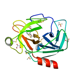
3RXP
 | | Crystal structure of Trypsin complexed with (1,5-dimethylpyrazol-3-yl)methanamine | | 分子名称: | 1-(1,5-dimethyl-1H-pyrazol-3-yl)methanamine, CALCIUM ION, Cationic trypsin, ... | | 著者 | Yamane, J, Yao, M, Zhou, Y, Tanaka, I. | | 登録日 | 2011-05-10 | | 公開日 | 2011-08-24 | | 最終更新日 | 2023-11-01 | | 実験手法 | X-RAY DIFFRACTION (1.6 Å) | | 主引用文献 | In-crystal affinity ranking of fragment hit compounds reveals a relationship with their inhibitory activities
J.Appl.Crystallogr., 44, 2011
|
|
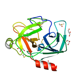
3RXM
 | | Crystal structure of Trypsin complexed with [2-(2-thienyl)thiazol-4-yl]methanamine | | 分子名称: | 1-[2-(thiophen-2-yl)-1,3-thiazol-4-yl]methanamine, CALCIUM ION, Cationic trypsin, ... | | 著者 | Yamane, J, Yao, M, Zhou, Y, Tanaka, I. | | 登録日 | 2011-05-10 | | 公開日 | 2011-08-24 | | 最終更新日 | 2023-11-01 | | 実験手法 | X-RAY DIFFRACTION (1.7 Å) | | 主引用文献 | In-crystal affinity ranking of fragment hit compounds reveals a relationship with their inhibitory activities
J.Appl.Crystallogr., 44, 2011
|
|
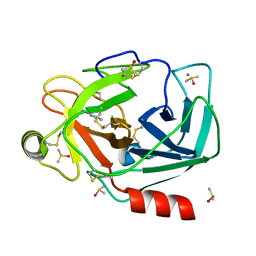
3RXH
 | | Crystal structure of Trypsin complexed with 2-(1H-imidazol-4-yl)ethanamine | | 分子名称: | CALCIUM ION, Cationic trypsin, DIMETHYL SULFOXIDE, ... | | 著者 | Yamane, J, Yao, M, Zhou, Y, Tanaka, I. | | 登録日 | 2011-05-10 | | 公開日 | 2011-08-24 | | 最終更新日 | 2023-11-01 | | 実験手法 | X-RAY DIFFRACTION (1.7 Å) | | 主引用文献 | In-crystal affinity ranking of fragment hit compounds reveals a relationship with their inhibitory activities
J.Appl.Crystallogr., 44, 2011
|
|
3RXF
 | | Crystal structure of Trypsin complexed with 4-aminopyridine | | 分子名称: | 4-AMINOPYRIDINE, CALCIUM ION, Cationic trypsin, ... | | 著者 | Yamane, J, Yao, M, Zhou, Y, Tanaka, I. | | 登録日 | 2011-05-10 | | 公開日 | 2011-08-24 | | 最終更新日 | 2023-11-01 | | 実験手法 | X-RAY DIFFRACTION (1.7 Å) | | 主引用文献 | In-crystal affinity ranking of fragment hit compounds reveals a relationship with their inhibitory activities
J.Appl.Crystallogr., 44, 2011
|
|
3RXG
 | | Crystal structure of Trypsin complexed with 4-aminocyclohexanol | | 分子名称: | CALCIUM ION, Cationic trypsin, DIMETHYL SULFOXIDE, ... | | 著者 | Yamane, J, Yao, M, Zhou, Y, Tanaka, I. | | 登録日 | 2011-05-10 | | 公開日 | 2011-08-24 | | 最終更新日 | 2023-11-01 | | 実験手法 | X-RAY DIFFRACTION (1.7 Å) | | 主引用文献 | In-crystal affinity ranking of fragment hit compounds reveals a relationship with their inhibitory activities
J.Appl.Crystallogr., 44, 2011
|
|
3RXC
 | | Crystal structure of Trypsin complexed with 2-aminopyridine | | 分子名称: | 2-AMINOPYRIDINE, CALCIUM ION, Cationic trypsin, ... | | 著者 | Yamane, J, Yao, M, Zhou, Y, Tanaka, I. | | 登録日 | 2011-05-10 | | 公開日 | 2011-08-24 | | 最終更新日 | 2023-11-01 | | 実験手法 | X-RAY DIFFRACTION (1.7 Å) | | 主引用文献 | In-crystal affinity ranking of fragment hit compounds reveals a relationship with their inhibitory activities
J.Appl.Crystallogr., 44, 2011
|
|
3RXQ
 | | Crystal structure of Trypsin complexed with benzamide (F01 and F05, cocktail experiment) | | 分子名称: | BENZAMIDINE, CALCIUM ION, Cationic trypsin, ... | | 著者 | Yamane, J, Yao, M, Zhou, Y, Tanaka, I. | | 登録日 | 2011-05-10 | | 公開日 | 2011-08-24 | | 最終更新日 | 2023-11-01 | | 実験手法 | X-RAY DIFFRACTION (1.68 Å) | | 主引用文献 | In-crystal affinity ranking of fragment hit compounds reveals a relationship with their inhibitory activities
J.Appl.Crystallogr., 44, 2011
|
|
3RXJ
 | | Crystal structure of Trypsin complexed with 4-guanidinobenzoic acid | | 分子名称: | 4-carbamimidamidobenzoic acid, CALCIUM ION, Cationic trypsin, ... | | 著者 | Yamane, J, Yao, M, Zhou, Y, Tanaka, I. | | 登録日 | 2011-05-10 | | 公開日 | 2011-08-24 | | 最終更新日 | 2023-11-01 | | 実験手法 | X-RAY DIFFRACTION (1.7 Å) | | 主引用文献 | In-crystal affinity ranking of fragment hit compounds reveals a relationship with their inhibitory activities
J.Appl.Crystallogr., 44, 2011
|
|
3RXA
 | | Crystal structure of Trypsin complexed with cycloheptanamine | | 分子名称: | CALCIUM ION, Cationic trypsin, GLYCEROL, ... | | 著者 | Yamane, J, Yao, M, Zhou, Y, Tanaka, I. | | 登録日 | 2011-05-10 | | 公開日 | 2011-08-24 | | 最終更新日 | 2023-11-01 | | 実験手法 | X-RAY DIFFRACTION (1.7 Å) | | 主引用文献 | In-crystal affinity ranking of fragment hit compounds reveals a relationship with their inhibitory activities
J.Appl.Crystallogr., 44, 2011
|
|
3RXK
 | | Crystal structure of Trypsin complexed with methyl 4-amino-1-methyl-pyrrolidine-2-carboxylate | | 分子名称: | CALCIUM ION, Cationic trypsin, DIMETHYL SULFOXIDE, ... | | 著者 | Yamane, J, Yao, M, Zhou, Y, Tanaka, I. | | 登録日 | 2011-05-10 | | 公開日 | 2011-08-24 | | 最終更新日 | 2023-11-01 | | 実験手法 | X-RAY DIFFRACTION (1.6 Å) | | 主引用文献 | In-crystal affinity ranking of fragment hit compounds reveals a relationship with their inhibitory activities
J.Appl.Crystallogr., 44, 2011
|
|
3RXR
 | | Crystal structure of Trypsin complexed with cycloheptanamine (F01 and F03, cocktail experiment) | | 分子名称: | CALCIUM ION, Cationic trypsin, DIMETHYL SULFOXIDE, ... | | 著者 | Yamane, J, Yao, M, Zhou, Y, Tanaka, I. | | 登録日 | 2011-05-10 | | 公開日 | 2011-08-24 | | 最終更新日 | 2023-11-01 | | 実験手法 | X-RAY DIFFRACTION (1.72 Å) | | 主引用文献 | In-crystal affinity ranking of fragment hit compounds reveals a relationship with their inhibitory activities
J.Appl.Crystallogr., 44, 2011
|
|
4BEB
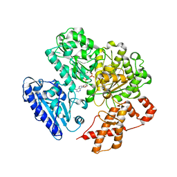 | | MUTANT (K220E) OF THE HSDR SUBUNIT OF THE ECOR124I RESTRICTION ENZYME IN COMPLEX WITH ATP | | 分子名称: | ADENOSINE-5'-TRIPHOSPHATE, MAGNESIUM ION, TYPE I RESTRICTION ENZYME HSDR | | 著者 | Csefalvay, E, Lapkouski, M, Guzanova, A, Csefalvay, L, Baikova, T, Shevelev, I, Janscak, P, Smatanova, I.K, Panjikar, S, Carey, J, Weiserova, M, Ettrich, R. | | 登録日 | 2013-03-07 | | 公開日 | 2014-03-26 | | 最終更新日 | 2023-12-20 | | 実験手法 | X-RAY DIFFRACTION (2.989 Å) | | 主引用文献 | Functional Coupling of Duplex Translocation to DNA Cleavage in a Type I Restriction Enzyme.
Plos One, 10, 2015
|
|
4FJ1
 | | Crystal Structure of the ternary complex between a fungal 17beta-hydroxysteroid dehydrogenase (Holo form) and genistein | | 分子名称: | 17beta-hydroxysteroid dehydrogenase, DIMETHYL SULFOXIDE, GENISTEIN, ... | | 著者 | Cassetta, A, Lamba, D, Krastanova, I. | | 登録日 | 2012-06-11 | | 公開日 | 2013-07-03 | | 最終更新日 | 2023-09-13 | | 実験手法 | X-RAY DIFFRACTION (2.3 Å) | | 主引用文献 | Structural basis for inhibition of 17 beta-hydroxysteroid dehydrogenases by phytoestrogens: The case of fungal 17 beta-HSDcl.
J. Steroid Biochem. Mol. Biol., 171, 2017
|
|
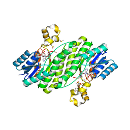
3AJ1
 | | The structure of AxCeSD octamer (N-terminal HIS-tag) from Acetobacter xylinum | | 分子名称: | Cellulose synthase operon protein D | | 著者 | Hu, S.Q, Tajima, K, Zhou, Y, Tanaka, I, Yao, M. | | 登録日 | 2010-05-20 | | 公開日 | 2010-10-06 | | 最終更新日 | 2011-07-13 | | 実験手法 | X-RAY DIFFRACTION (2.5 Å) | | 主引用文献 | Structure of bacterial cellulose synthase subunit D octamer with four inner passageways
Proc.Natl.Acad.Sci.USA, 107, 2010
|
|
4FIZ
 | |
3A8E
 | | The structure of AxCesD octamer complexed with cellopentaose | | 分子名称: | Cellulose synthase operon protein D, beta-D-glucopyranose-(1-4)-beta-D-glucopyranose-(1-4)-beta-D-glucopyranose-(1-4)-beta-D-glucopyranose-(1-4)-beta-D-glucopyranose | | 著者 | Hu, S.Q, Tajima, K, Zhou, Y, Yao, M, Tanaka, I. | | 登録日 | 2009-10-05 | | 公開日 | 2010-09-22 | | 最終更新日 | 2023-11-01 | | 実験手法 | X-RAY DIFFRACTION (3 Å) | | 主引用文献 | Structure of bacterial cellulose synthase subunit D octamer with four inner passageways
Proc.Natl.Acad.Sci.USA, 107, 2010
|
|
3B0P
 | | tRNA-dihydrouridine synthase from Thermus thermophilus | | 分子名称: | FLAVIN MONONUCLEOTIDE, tRNA-dihydrouridine synthase | | 著者 | Yu, F, Tanaka, Y, Yamashita, K, Nakamura, A, Yao, M, Tanaka, I. | | 登録日 | 2011-06-12 | | 公開日 | 2011-12-14 | | 最終更新日 | 2024-03-13 | | 実験手法 | X-RAY DIFFRACTION (1.7 Å) | | 主引用文献 | Molecular basis of dihydrouridine formation on tRNA
Proc.Natl.Acad.Sci.USA, 108, 2011
|
|
1RL2
 | |
3AJ2
 | | The structure of AxCeSD octamer (C-terminal HIS-tag) from Acetobacter xylinum | | 分子名称: | Cellulose synthase operon protein D | | 著者 | Hu, S.Q, Tajima, K, Zhou, Y, Tanaka, I, Yao, M. | | 登録日 | 2010-05-20 | | 公開日 | 2010-10-06 | | 最終更新日 | 2023-11-01 | | 実験手法 | X-RAY DIFFRACTION (2.7 Å) | | 主引用文献 | Structure of bacterial cellulose synthase subunit D octamer with four inner passageways
Proc.Natl.Acad.Sci.USA, 107, 2010
|
|


