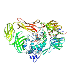
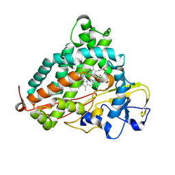
5D94
 
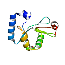 | | Crystal structure of LC3-LIR peptide complex | | 分子名称: | Microtubule-associated proteins 1A/1B light chain 3B, Peptide from FYVE and coiled-coil domain-containing protein 1 | | 著者 | Takagi, K, Mizushima, T, Johansen, T. | | 登録日 | 2015-08-18 | | 公開日 | 2015-10-21 | | 最終更新日 | 2024-03-20 | | 実験手法 | X-RAY DIFFRACTION (1.53 Å) | | 主引用文献 | FYCO1 Contains a C-terminally Extended, LC3A/B-preferring LC3-interacting Region (LIR) Motif Required for Efficient Maturation of Autophagosomes during Basal Autophagy
J.Biol.Chem., 290, 2015
|
|
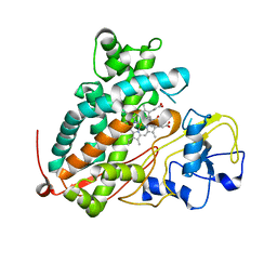
5B0N
 | | Structure of Shigella effector LRR domain | | 分子名称: | E3 ubiquitin-protein ligase ipaH9.8 | | 著者 | Takagi, K, Sasakawa, C, Kim, M, Mizushima, T. | | 登録日 | 2015-11-02 | | 公開日 | 2016-04-06 | | 最終更新日 | 2024-03-20 | | 実験手法 | X-RAY DIFFRACTION (1.8 Å) | | 主引用文献 | Crystal structure of the substrate-recognition domain of the Shigella E3 ligase IpaH9.8
Acta Crystallogr.,Sect.F, 72, 2016
|
|
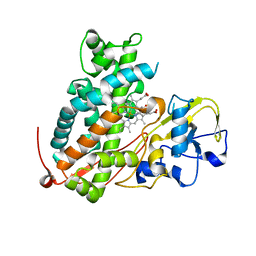
5B0T
 | | Structure of Shigella effector LRR domain | | 分子名称: | E3 ubiquitin-protein ligase ipaH9.8 | | 著者 | Takagi, K, Sasakawa, C, Kim, M, Mizushima, T. | | 登録日 | 2015-11-04 | | 公開日 | 2016-04-06 | | 最終更新日 | 2024-03-20 | | 実験手法 | X-RAY DIFFRACTION (2 Å) | | 主引用文献 | Crystal structure of the substrate-recognition domain of the Shigella E3 ligase IpaH9.8
Acta Crystallogr.,Sect.F, 72, 2016
|
|
3VLE
 | | Crystal structure of yeast proteasome interacting protein | | 分子名称: | DNA mismatch repair protein HSM3 | | 著者 | Takagi, K, Kim, S, Kato, K, Tanaka, K, Saeki, Y, Mizushima, T. | | 登録日 | 2011-12-01 | | 公開日 | 2012-02-22 | | 最終更新日 | 2023-11-08 | | 実験手法 | X-RAY DIFFRACTION (2.41 Å) | | 主引用文献 | Structural basis for specific recognition of Rpt1, an ATPase subunit of the 26S proteasome, by a proteasome-dedicated chaperone Hsm3
J.Biol.Chem., 287, 2012
|
|
3VLD
 | | Crystal structure of yeast proteasome interacting protein | | 分子名称: | DNA mismatch repair protein HSM3 | | 著者 | Takagi, K, Kim, S, Kato, K, Tanaka, K, Saeki, Y, Mizushima, T. | | 登録日 | 2011-12-01 | | 公開日 | 2012-02-22 | | 最終更新日 | 2012-04-18 | | 実験手法 | X-RAY DIFFRACTION (2.05 Å) | | 主引用文献 | Structural basis for specific recognition of Rpt1, an ATPase subunit of the 26S proteasome, by a proteasome-dedicated chaperone Hsm3
J.Biol.Chem., 287, 2012
|
|
3VLF
 | | Crystal structure of yeast proteasome interacting protein | | 分子名称: | 26S protease regulatory subunit 7 homolog, DNA mismatch repair protein HSM3 | | 著者 | Takagi, K, Kim, S, Kato, K, Tanaka, K, Saeki, Y, Mizushima, T. | | 登録日 | 2011-12-01 | | 公開日 | 2012-02-22 | | 最終更新日 | 2023-12-06 | | 実験手法 | X-RAY DIFFRACTION (3.8 Å) | | 主引用文献 | Structural basis for specific recognition of Rpt1, an ATPase subunit of the 26S proteasome, by a proteasome-dedicated chaperone Hsm3
J.Biol.Chem., 287, 2012
|
|
4XZX
 | | Shigella flexneri effector OspI C62S mutant | | 分子名称: | ACETATE ION, ORF169b | | 著者 | Nishide, A, Takagi, K, Minsoo, K, Sasakawa, C, Mizushima, T. | | 登録日 | 2015-02-05 | | 公開日 | 2016-02-10 | | 最終更新日 | 2023-11-08 | | 実験手法 | X-RAY DIFFRACTION (2.2 Å) | | 主引用文献 | New insights into the active site structure of Shigella effecter OspI
To Be Published
|
|
4ZY3
 | | Crystal Structure of Keap1 in Complex with a small chemical compound, K67 | | 分子名称: | FORMIC ACID, Kelch-like ECH-associated protein 1, N,N'-[2-(2-oxopropyl)naphthalene-1,4-diyl]bis(4-ethoxybenzenesulfonamide) | | 著者 | Fukutomi, T, Iso, T, Suzuki, T, Takagi, K, Mizushima, T, Komatsu, M, Yamamoto, M. | | 登録日 | 2015-05-21 | | 公開日 | 2016-05-25 | | 最終更新日 | 2023-11-08 | | 実験手法 | X-RAY DIFFRACTION (1.8 Å) | | 主引用文献 | p62/Sqstm1 promotes malignancy of HCV-positive hepatocellular carcinoma through Nrf2-dependent metabolic reprogramming
Nat Commun, 7, 2016
|
|
1JJU
 | | Structure of a Quinohemoprotein Amine Dehydrogenase with a Unique Redox Cofactor and Highly Unusual Crosslinking | | 分子名称: | PROTOPORPHYRIN IX CONTAINING FE, QUINOHEMOPROTEIN AMINE DEHYDROGENASE, SODIUM ION, ... | | 著者 | Datta, S, Mori, Y, Takagi, K, Kawaguchi, K, Chen, Z.-W, Kano, K, Ikeda, T, Okajima, T, Kuroda, S, Tanizawa, K, Mathews, F.S. | | 登録日 | 2001-07-09 | | 公開日 | 2001-12-12 | | 最終更新日 | 2011-07-13 | | 実験手法 | X-RAY DIFFRACTION (2.05 Å) | | 主引用文献 | Structure of a quinohemoprotein amine dehydrogenase with an uncommon redox cofactor and highly unusual crosslinking.
Proc.Natl.Acad.Sci.USA, 98, 2001
|
|
7YA8
 | | The crystal structure of IpaH2.5 LRR domain | | 分子名称: | RING-type E3 ubiquitin transferase | | 著者 | Hiragi, K, Nishide, A, Takagi, K, Iwai, K, Kim, M, Mizushima, T. | | 登録日 | 2022-06-27 | | 公開日 | 2023-02-08 | | 最終更新日 | 2023-11-29 | | 実験手法 | X-RAY DIFFRACTION (3.4 Å) | | 主引用文献 | Structural insight into the recognition of the linear ubiquitin assembly complex by Shigella E3 ligase IpaH1.4/2.5.
J.Biochem., 173, 2023
|
|
7YA7
 | | The crystal structure of IpaH1.4 LRR domain | | 分子名称: | RING-type E3 ubiquitin transferase | | 著者 | Hiragi, K, Nishide, A, Takagi, K, Iwai, K, Kim, M, Mizushima, T. | | 登録日 | 2022-06-27 | | 公開日 | 2023-02-08 | | 最終更新日 | 2023-11-29 | | 実験手法 | X-RAY DIFFRACTION (1.4 Å) | | 主引用文献 | Structural insight into the recognition of the linear ubiquitin assembly complex by Shigella E3 ligase IpaH1.4/2.5.
J.Biochem., 173, 2023
|
|
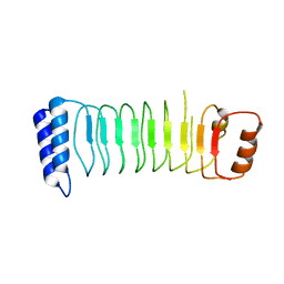
3ACP
 | | Crystal Structure of Yeast Rpn14, a Chaperone of the 19S Regulatory Particle of the Proteasome | | 分子名称: | WD repeat-containing protein YGL004C | | 著者 | Kim, S, Saeki, Y, Suzuki, A, Takagi, K, Fukunaga, K, Yamane, T, Kato, K, Tanaka, K, Mizushima, T. | | 登録日 | 2010-01-08 | | 公開日 | 2010-03-16 | | 最終更新日 | 2011-07-13 | | 実験手法 | X-RAY DIFFRACTION (2 Å) | | 主引用文献 | Crystal structure of yeast Rpn14, a chaperone of the 19S regulatory particle of the proteasome
J.Biol.Chem., 285, 2010
|
|
3WSO
 | | Crystal structure of the Skp1-FBG3 complex | | 分子名称: | F-box only protein 44, S-phase kinase-associated protein 1 | | 著者 | Kumanomidou, T, Nishio, K, Takagi, K, Nakagawa, T, Suzuki, A, Yamane, T, Tokunaga, F, Iwai, K, Murakami, A, Yoshida, Y, Tanaka, K, Mizushima, T. | | 登録日 | 2014-03-18 | | 公開日 | 2015-03-25 | | 最終更新日 | 2023-11-08 | | 実験手法 | X-RAY DIFFRACTION (2.6 Å) | | 主引用文献 | The Structural Differences between a Glycoprotein Specific F-Box Protein Fbs1 and Its Homologous Protein FBG3
Plos One, 10, 2015
|
|
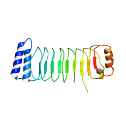
3VL1
 | | Crystal structure of yeast Rpn14 | | 分子名称: | 26S proteasome regulatory subunit RPN14 | | 著者 | Kim, S, Nishide, A, Saeki, Y, Takagi, K, Tanaka, K, Kato, K, Mizushima, T. | | 登録日 | 2011-11-28 | | 公開日 | 2012-05-02 | | 最終更新日 | 2023-11-08 | | 実験手法 | X-RAY DIFFRACTION (1.6 Å) | | 主引用文献 | New crystal structure of the proteasome-dedicated chaperone Rpn14 at 1.6 A resolution
Acta Crystallogr.,Sect.F, 68, 2012
|
|
3WRM
 | | Crystal structure of P450cam | | 分子名称: | CAMPHOR, Camphor 5-monooxygenase, POTASSIUM ION, ... | | 著者 | Kishimoto, A, Takagi, K, Amano, A, Sakurai, K, Mizushima, T, Shimada, H. | | 登録日 | 2014-02-25 | | 公開日 | 2015-03-18 | | 最終更新日 | 2023-11-08 | | 実験手法 | X-RAY DIFFRACTION (1.95 Å) | | 主引用文献 | Structure of P450cam intermedite
To be published
|
|
3WRJ
 | | Crystal structure of P450cam | | 分子名称: | CAMPHOR, Camphor 5-monooxygenase, POTASSIUM ION, ... | | 著者 | Kishimoto, A, Takagi, K, Amano, A, Sakurai, K, Mizushima, T, Shimada, H. | | 登録日 | 2014-02-25 | | 公開日 | 2015-03-18 | | 最終更新日 | 2023-11-08 | | 実験手法 | X-RAY DIFFRACTION (1.85 Å) | | 主引用文献 | Structure of P450cam intermedite
to be published
|
|
3WRL
 | | Crystal structure of P450cam | | 分子名称: | CAMPHOR, Camphor 5-monooxygenase, POTASSIUM ION, ... | | 著者 | Kishimoto, A, Takagi, K, Amano, A, Sakurai, K, Mizushima, T, Shimada, H. | | 登録日 | 2014-02-25 | | 公開日 | 2015-03-18 | | 最終更新日 | 2023-11-08 | | 実験手法 | X-RAY DIFFRACTION (1.65 Å) | | 主引用文献 | Structure of P450cam intermedite
To be published
|
|
3WRH
 | | Crystal structure of P450cam | | 分子名称: | CAMPHOR, Camphor 5-monooxygenase, POTASSIUM ION, ... | | 著者 | Kishimoto, A, Takagi, K, Amano, A, Sakurai, K, Mizushima, T, Shimada, H. | | 登録日 | 2014-02-25 | | 公開日 | 2015-03-18 | | 最終更新日 | 2023-11-08 | | 実験手法 | X-RAY DIFFRACTION (1.62 Å) | | 主引用文献 | Structure of P450cam intermedite
To be Published
|
|
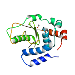
3WDZ
 | | Crystal Structure of Keap1 in Complex with phosphorylated p62 | | 分子名称: | Kelch-like ECH-associated protein 1, Peptide from Sequestosome-1 | | 著者 | Fukutomi, T, Takagi, K, Mizushima, T, Tanaka, K, Komatsu, M, Yamamoto, M. | | 登録日 | 2013-06-26 | | 公開日 | 2013-09-04 | | 最終更新日 | 2023-11-08 | | 実験手法 | X-RAY DIFFRACTION (2.6 Å) | | 主引用文献 | Phosphorylation of p62 activates the Keap1-Nrf2 pathway during selective autophagy.
Mol.Cell, 51, 2013
|
|
3WRI
 | | Crystal structure of P450cam | | 分子名称: | CAMPHOR, Camphor 5-monooxygenase, PROTOPORPHYRIN IX CONTAINING FE | | 著者 | Kishimoto, A, Takagi, K, Amano, A, Sakurai, K, Mizushima, T, Shimada, H. | | 登録日 | 2014-02-25 | | 公開日 | 2015-03-18 | | 最終更新日 | 2023-11-08 | | 実験手法 | X-RAY DIFFRACTION (2.9 Å) | | 主引用文献 | Structure of P450cam intermediate
To be Published
|
|
3WRK
 | | Crystal structure of P450cam | | 分子名称: | CAMPHOR, Camphor 5-monooxygenase, PROTOPORPHYRIN IX CONTAINING FE | | 著者 | Kishimoto, A, Takagi, K, Amano, A, Sakurai, K, Mizushima, T, Shimada, H. | | 登録日 | 2014-02-25 | | 公開日 | 2015-03-18 | | 最終更新日 | 2023-11-08 | | 実験手法 | X-RAY DIFFRACTION (2.609 Å) | | 主引用文献 | Structure of P450cam intermedite
To be Published
|
|
3WXR
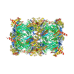 | | Yeast 20S proteasome with a mutation of alpha7 subunit | | 分子名称: | Probable proteasome subunit alpha type-7, Proteasome subunit alpha type-1, Proteasome subunit alpha type-2, ... | | 著者 | Yashiroda, H, Toda, Y, Otsu, S, Takagi, K, Mizushima, T, Murata, S. | | 登録日 | 2014-08-06 | | 公開日 | 2014-10-22 | | 最終更新日 | 2023-11-08 | | 実験手法 | X-RAY DIFFRACTION (3.15 Å) | | 主引用文献 | N-terminal alpha 7 deletion of the proteasome 20S core particle substitutes for yeast PI31 function
Mol.Cell.Biol., 35, 2015
|
|
3W31
 | | Structual basis for the recognition of Ubc13 by the Shigella flexneri effector OspI | | 分子名称: | IODIDE ION, ORF169b, Ubiquitin-conjugating enzyme E2 N | | 著者 | Nishide, A, Kim, M, Takagi, K, Sasakawa, C, Mizushima, T. | | 登録日 | 2012-12-07 | | 公開日 | 2013-03-27 | | 最終更新日 | 2023-11-08 | | 実験手法 | X-RAY DIFFRACTION (2.96 Å) | | 主引用文献 | Structural basis for the recognition of Ubc13 by the Shigella flexneri effector OspI.
J.Mol.Biol., 425, 2013
|
|
3W30
 | | Structual basis for the recognition of Ubc13 by the Shigella flexneri effector OspI | | 分子名称: | ORF169b | | 著者 | Nishide, A, Kim, M, Takagi, K, Sasakawa, C, Mizushima, T. | | 登録日 | 2012-12-07 | | 公開日 | 2013-03-27 | | 最終更新日 | 2023-11-08 | | 実験手法 | X-RAY DIFFRACTION (2.99 Å) | | 主引用文献 | Structural Basis for the Recognition of Ubc13 by the Shigella flexneri Effector OspI.
J.Mol.Biol., 425, 2013
|
|
3WN7
 | | Crystal Structure of Keap1 in Complex with the N-terminal region of the Nrf2 transcription factor | | 分子名称: | ACETATE ION, Kelch-like ECH-associated protein 1, Peptide from Nuclear factor erythroid 2-related factor 2 | | 著者 | Fukutomi, T, Takagi, K, Mizushima, T, Ohuchi, N, Yamamoto, M. | | 登録日 | 2013-12-05 | | 公開日 | 2013-12-25 | | 最終更新日 | 2023-11-08 | | 実験手法 | X-RAY DIFFRACTION (1.57 Å) | | 主引用文献 | Kinetic, thermodynamic, and structural characterizations of the association between Nrf2-DLGex degron and Keap1
Mol.Cell.Biol., 34, 2014
|
|


