6N8Z
 
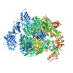 | | HSP104DWB extended conformation | | 分子名称: | ADENOSINE-5'-TRIPHOSPHATE, Heat shock protein 104 | | 著者 | Lee, S, Rho, S.H, Lee, J, Sung, N, Liu, J, Tsai, F.T.F. | | 登録日 | 2018-11-30 | | 公開日 | 2019-01-02 | | 最終更新日 | 2019-01-16 | | 実験手法 | ELECTRON MICROSCOPY (9.3 Å) | | 主引用文献 | Cryo-EM Structures of the Hsp104 Protein Disaggregase Captured in the ATP Conformation.
Cell Rep, 26, 2019
|
|
6N8T
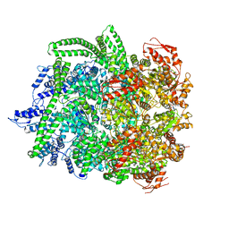 | | Hsp104DWB closed conformation | | 分子名称: | ADENOSINE-5'-TRIPHOSPHATE, Heat shock protein 104 | | 著者 | Lee, S, Rho, S.H, Lee, J, Sung, N, Liu, J, Tsai, F.T.F. | | 登録日 | 2018-11-30 | | 公開日 | 2019-01-02 | | 最終更新日 | 2024-03-20 | | 実験手法 | ELECTRON MICROSCOPY (7.7 Å) | | 主引用文献 | Cryo-EM Structures of the Hsp104 Protein Disaggregase Captured in the ATP Conformation.
Cell Rep, 26, 2019
|
|
8IYR
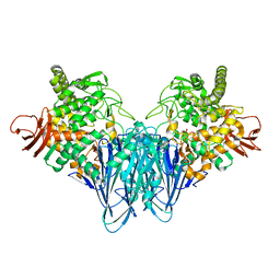 | |
5F5R
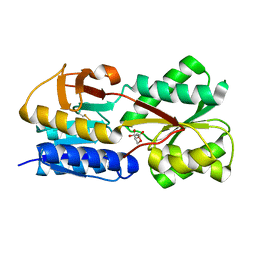
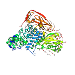
 | | TRAP1N-ADPNP | | 分子名称: | Heat shock protein 75 kDa, mitochondrial, MAGNESIUM ION, ... | | 著者 | Tsai, F.T.F, Lee, S, Sung, N, Lee, J, Chang, C, Joachimiak, A. | | 登録日 | 2015-12-04 | | 公開日 | 2016-03-02 | | 最終更新日 | 2023-09-27 | | 実験手法 | X-RAY DIFFRACTION (1.85 Å) | | 主引用文献 | Mitochondrial Hsp90 is a ligand-activated molecular chaperone coupling ATP binding to dimer closure through a coiled-coil intermediate.
Proc.Natl.Acad.Sci.USA, 113, 2016
|
|
5GTA
 | | 3D Crystal Structure of LsrB Bound to Furanosyl diester (R)-THMF, from Salmonella typhi | | 分子名称: | (2R,4S)-2-methyl-2,3,3,4-tetrahydroxytetrahydrofuran, Autoinducer 2-binding protein LsrB | | 著者 | Gopinath, S, Perumal, P, Rahul, R, Arockiasamy, A, SundaraBaalaji, N. | | 登録日 | 2016-08-19 | | 公開日 | 2017-08-23 | | 最終更新日 | 2023-11-08 | | 実験手法 | X-RAY DIFFRACTION (2.998 Å) | | 主引用文献 | 3D Crystal Structure of LsrB Bound to Furanosyl diester (R)-THMF, from Salmonella typhi
To Be Published
|
|
5E68
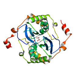 | | High resolution crystal structure of LuxS - Quorum sensor molecular complex from Salmonella typhi at 1.58 Angstroms | | 分子名称: | (2R,4S)-2-methyl-2,3,3,4-tetrahydroxytetrahydrofuran, METHIONINE, S-ribosylhomocysteine lyase, ... | | 著者 | Perumal, P, Raina, R, Arockiasamy, A, SundaraBaalaji, N. | | 登録日 | 2015-10-09 | | 公開日 | 2016-10-12 | | 最終更新日 | 2023-11-08 | | 実験手法 | X-RAY DIFFRACTION (1.58 Å) | | 主引用文献 | High resolution crystal structure of LuxS - Quorum sensor molecular complex from Salmonella typhi at 1.58 Angstroms
To Be Published
|
|
5V2W
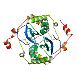 | | Crystal structure of a LuxS from salmonella typhi | | 分子名称: | S-ribosylhomocysteine lyase, ZINC ION | | 著者 | Perumal, P, Raina, R, Manoj Kumar, P, Arockisamy, A, SundaraBaalaji, N. | | 登録日 | 2017-03-06 | | 公開日 | 2017-08-23 | | 最終更新日 | 2023-11-08 | | 実験手法 | X-RAY DIFFRACTION (2.3 Å) | | 主引用文献 | Crystal structure of a LuxS from salmonella typhi
To Be Published
|
|
7VC6
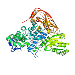 | | The structure of beta-xylosidase from Phanerochaete chrysosporium(PcBxl3) | | 分子名称: | 2-acetamido-2-deoxy-beta-D-glucopyranose, GLYCEROL, xylan 1,4-beta-xylosidase | | 著者 | Kojima, K, Sunagawa, N, Igarashi, K. | | 登録日 | 2021-09-01 | | 公開日 | 2022-02-09 | | 最終更新日 | 2023-11-29 | | 実験手法 | X-RAY DIFFRACTION (2.54 Å) | | 主引用文献 | Comparison of glycoside hydrolase family 3 beta-xylosidases from basidiomycetes and ascomycetes reveals evolutionarily distinct xylan degradation systems.
J.Biol.Chem., 298, 2022
|
|
7VC7
 | | The structure of beta-xylosidase from Phanerochaete chrysosporium(PcBxl3) | | 分子名称: | 1,2-ETHANEDIOL, 2-acetamido-2-deoxy-beta-D-glucopyranose, DI(HYDROXYETHYL)ETHER, ... | | 著者 | Kojima, K, Sunagawa, N, Igarashi, K. | | 登録日 | 2021-09-01 | | 公開日 | 2022-02-09 | | 最終更新日 | 2023-11-29 | | 実験手法 | X-RAY DIFFRACTION (3.08 Å) | | 主引用文献 | Comparison of glycoside hydrolase family 3 beta-xylosidases from basidiomycetes and ascomycetes reveals evolutionarily distinct xylan degradation systems.
J.Biol.Chem., 298, 2022
|
|
7EZZ
 | | Crystal structure of Salmonella typhi outer membrane phospholipase (OMPLA) dimer with bound calcium | | 分子名称: | CALCIUM ION, DODECANE, Phospholipase A1, ... | | 著者 | Perumal, P, Raina, R, Sreeshma, N.S, Arockiasamy, A, Sundarabaalaji, N. | | 登録日 | 2021-06-02 | | 公開日 | 2021-06-23 | | 最終更新日 | 2023-11-29 | | 実験手法 | X-RAY DIFFRACTION (2.76 Å) | | 主引用文献 | Crystal structure of Salmonella typhi outer membrane phospholipase (OMPLA) dimer with bound calcium
To Be Published
|
|


