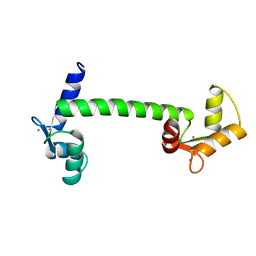
1KVW
 
 | |
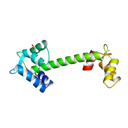
1CLM
 | |
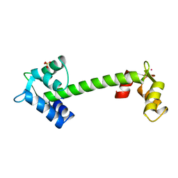
1NCY
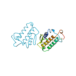
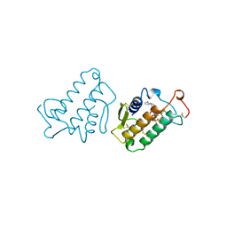
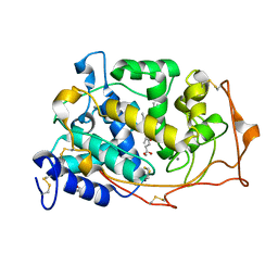
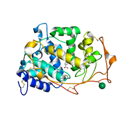
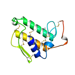
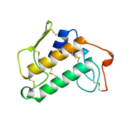
 | | TROPONIN-C, COMPLEX WITH MANGANESE | | 分子名称: | MANGANESE (II) ION, SULFATE ION, TROPONIN C | | 著者 | Sundaralingam, M, Rao, S.T. | | 登録日 | 1996-06-02 | | 公開日 | 1997-01-11 | | 最終更新日 | 2024-02-14 | | 実験手法 | X-RAY DIFFRACTION (2.1 Å) | | 主引用文献 | X-ray structures of Mn, Cd and Tb metal complexes of troponin C.
Acta Crystallogr.,Sect.D, 52, 1996
|
|
1NCZ
 | | TROPONIN C | | 分子名称: | SULFATE ION, TERBIUM(III) ION, TROPONIN C | | 著者 | Sundaralingam, M, Rao, S.T. | | 登録日 | 1996-06-02 | | 公開日 | 1996-12-07 | | 最終更新日 | 2024-02-14 | | 実験手法 | X-RAY DIFFRACTION (1.8 Å) | | 主引用文献 | X-ray structures of Mn, Cd and Tb metal complexes of troponin C.
Acta Crystallogr.,Sect.D, 52, 1996
|
|
1NCX
 | | TROPONIN C | | 分子名称: | CADMIUM ION, SULFATE ION, TROPONIN C | | 著者 | Sundaralingam, M, Rao, S.T. | | 登録日 | 1996-06-02 | | 公開日 | 1996-12-07 | | 最終更新日 | 2024-02-14 | | 実験手法 | X-RAY DIFFRACTION (1.8 Å) | | 主引用文献 | X-ray structures of Mn, Cd and Tb metal complexes of troponin C.
Acta Crystallogr.,Sect.D, 52, 1996
|
|
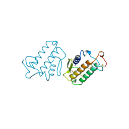
1IRB
 | | CARBOXYLIC ESTER HYDROLASE | | 分子名称: | CALCIUM ION, PHOSPHOLIPASE A2 | | 著者 | Sundaralingam, M. | | 登録日 | 1997-08-13 | | 公開日 | 1997-12-24 | | 最終更新日 | 2023-08-09 | | 実験手法 | X-RAY DIFFRACTION (1.9 Å) | | 主引用文献 | Phospholipase A2 engineering. Deletion of the C-terminus segment changes substrate specificity and uncouples calcium and substrate binding at the zwitterionic interface.
Biochemistry, 35, 1996
|
|
3AJL
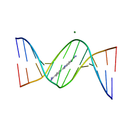 | | Crystal structure of d(CGCGGATf5UCGCG): 5-Formyluridine:guanosine Base-pair in B-DNA with DAPI | | 分子名称: | 5'-D(*CP*GP*CP*GP*GP*AP*TP*(UFR)P*CP*GP*CP*G)-3', 6-AMIDINE-2-(4-AMIDINO-PHENYL)INDOLE, MAGNESIUM ION | | 著者 | Tsunoda, M, Sakaue, T, Ueno, Y, Matsuda, A, Takenaka, A. | | 登録日 | 2010-06-07 | | 公開日 | 2011-04-27 | | 最終更新日 | 2023-11-01 | | 実験手法 | X-RAY DIFFRACTION (2.7 Å) | | 主引用文献 | Insights into the structures of DNA damaged by hydroxyl radical: crystal structures of DNA duplexes containing 5-formyluracil
J Nucleic Acids, 2010, 2010
|
|
1MKV
 | |
1G8V
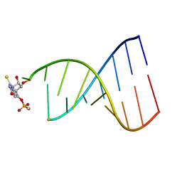
 | | MOLECULAR AND CRYSTAL STRUCTURE OF D(CGCGAATF5UCGCG):5-FORMYLURIDINE/ ADENOSINE BASE-PAIRS IN B-DNA | | 分子名称: | 5'-D(*CP*GP*CP*GP*AP*AP*TP*(UFR)P*CP*GP*CP*G)-3' | | 著者 | Tsunoda, M, Karino, N, Ueno, Y, Matsuda, A, Takenaka, A. | | 登録日 | 2000-11-21 | | 公開日 | 2001-02-05 | | 最終更新日 | 2023-08-09 | | 実験手法 | X-RAY DIFFRACTION (1.8 Å) | | 主引用文献 | Crystallization and preliminary X-ray analysis of a DNA dodecamer containing 2'-deoxy-5-formyluridine; what is the role of magnesium cation in crystallization of Dickerson-type DNA dodecamers?
Acta Crystallogr.,Sect.D, 57, 2001
|
|
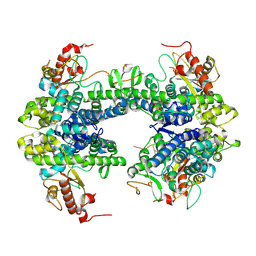
3M5Q
 | | 0.93 A Structure of Manganese-Bound Manganese Peroxidase | | 分子名称: | 2-acetamido-2-deoxy-beta-D-glucopyranose-(1-4)-2-acetamido-2-deoxy-beta-D-glucopyranose, CALCIUM ION, GLYCEROL, ... | | 著者 | Sundaramoorthy, M, Gold, M.H, Poulos, T.L. | | 登録日 | 2010-03-12 | | 公開日 | 2010-04-14 | | 最終更新日 | 2023-09-06 | | 実験手法 | X-RAY DIFFRACTION (0.93 Å) | | 主引用文献 | Ultrahigh (0.93A) resolution structure of manganese peroxidase from Phanerochaete chrysosporium: implications for the catalytic mechanism.
J.Inorg.Biochem., 104, 2010
|
|
1G8U
 | | MOLECULAR AND CRYSTAL STRUCTURE OF D(CGCGAATF5UCGCG):5-FORMYLURIDINE/ ADENOSINE BASE-PAIRS IN B-DNA | | 分子名称: | 5'-D(*CP*GP*CP*GP*AP*AP*TP*(UFR)P*CP*GP*CP*G)-3' | | 著者 | Tsunoda, M, Karino, N, Ueno, Y, Matsuda, A, Takenaka, A. | | 登録日 | 2000-11-21 | | 公開日 | 2001-02-05 | | 最終更新日 | 2023-08-09 | | 実験手法 | X-RAY DIFFRACTION (1.85 Å) | | 主引用文献 | Crystallization and preliminary X-ray analysis of a DNA dodecamer containing 2'-deoxy-5-formyluridine; what is the role of magnesium cation in crystallization of Dickerson-type DNA dodecamers?
Acta Crystallogr.,Sect.D, 57, 2001
|
|
1G75
 | | MOLECULAR AND CRYSTAL STRUCTURE OF D(CGCGAATF5UCGCG): 5-FORMYLURIDINE/ ADENOSINE BASE-PAIRS IN B-DNA | | 分子名称: | 5'-D(*CP*GP*CP*GP*AP*AP*TP*(UFR)P*CP*GP*CP*G)-3', MAGNESIUM ION, POTASSIUM ION | | 著者 | Tsunoda, M, Karino, N, Ueno, Y, Matsuda, A, Takenaka, A. | | 登録日 | 2000-11-09 | | 公開日 | 2001-02-05 | | 最終更新日 | 2023-08-09 | | 実験手法 | X-RAY DIFFRACTION (1.57 Å) | | 主引用文献 | Crystallization and preliminary X-ray analysis of a DNA dodecamer containing 2'-deoxy-5-formyluridine; what is the role of magnesium cation in crystallization of Dickerson-type DNA dodecamers?
Acta Crystallogr.,Sect.D, 57, 2001
|
|
3AJJ
 | | Crystal Structure of d(CGCGGATf5UCGCG): 5-Formyluridine/Guanosine Base-pair in B-DNA | | 分子名称: | 5'-D(*CP*GP*CP*GP*GP*AP*TP*(UFR)P*CP*GP*CP*G)-3' | | 著者 | Tsunoda, M, Sakaue, T, Ueno, Y, Matsuda, A, Takenaka, A. | | 登録日 | 2010-06-07 | | 公開日 | 2011-04-27 | | 最終更新日 | 2023-11-01 | | 実験手法 | X-RAY DIFFRACTION (3.02 Å) | | 主引用文献 | Insights into the structures of DNA damaged by hydroxyl radical: crystal structures of DNA duplexes containing 5-formyluracil
J Nucleic Acids, 2010, 2010
|
|
3M8M
 | | 1.05 A Structure of Manganese-free Manganese Peroxidase | | 分子名称: | 2-acetamido-2-deoxy-beta-D-glucopyranose-(1-4)-2-acetamido-2-deoxy-beta-D-glucopyranose, CALCIUM ION, GLYCEROL, ... | | 著者 | Sundaramoorthy, M, Gold, M.H, Poulos, T.L. | | 登録日 | 2010-03-18 | | 公開日 | 2010-04-14 | | 最終更新日 | 2020-07-29 | | 実験手法 | X-RAY DIFFRACTION (1.05 Å) | | 主引用文献 | Ultrahigh (0.93A) resolution structure of manganese peroxidase from Phanerochaete chrysosporium: implications for the catalytic mechanism.
J.Inorg.Biochem., 104, 2010
|
|
1FL8
 | |
1G8N
 | | MOLECULAR AND CRYSTAL STRUCTURE OF D(CGCGAATF5UCGCG):5-FORMYLURIDINE/ ADENOSINE BASE-PAIRS IN B-DNA | | 分子名称: | 5'-D(*CP*GP*CP*GP*AP*AP*TP*(UFR)P*CP*GP*CP*G)-3', MAGNESIUM ION | | 著者 | Tsunoda, M, Karino, N, Ueno, Y, Matsuda, A, Takenaka, A. | | 登録日 | 2000-11-20 | | 公開日 | 2001-02-05 | | 最終更新日 | 2023-08-09 | | 実験手法 | X-RAY DIFFRACTION (1.55 Å) | | 主引用文献 | Crystallization and preliminary X-ray analysis of a DNA dodecamer containing 2'-deoxy-5-formyluridine; what is the role of magnesium cation in crystallization of Dickerson-type DNA dodecamers?
Acta Crystallogr.,Sect.D, 57, 2001
|
|
3AJK
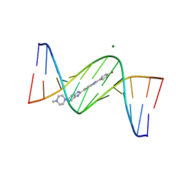 | | Crystal structure of d(CGCGGATf5UCGCG): 5-Formyluridine:Guanosine Base-pair in B-DNA with Hoechst33258 | | 分子名称: | 2'-(4-HYDROXYPHENYL)-5-(4-METHYL-1-PIPERAZINYL)-2,5'-BI-BENZIMIDAZOLE, 5'-D(*CP*GP*CP*GP*GP*AP*TP*(UFR)P*CP*GP*CP*G*)-3', MAGNESIUM ION | | 著者 | Tsunoda, M, Sakaue, T, Ueno, Y, Matsuda, A, Takenaka, A. | | 登録日 | 2010-06-07 | | 公開日 | 2011-04-27 | | 最終更新日 | 2023-11-01 | | 実験手法 | X-RAY DIFFRACTION (1.95 Å) | | 主引用文献 | Insights into the structures of DNA damaged by hydroxyl radical: crystal structures of DNA duplexes containing 5-formyluracil
J Nucleic Acids, 2010, 2010
|
|
3A07
 | | Crystal Structure of Actinohivin; Potent anti-HIV Protein | | 分子名称: | Actinohivin, SODIUM ION | | 著者 | Tsunoda, M, Suzuki, K, Sagara, T, Takenaka, A. | | 登録日 | 2009-03-04 | | 公開日 | 2009-08-25 | | 最終更新日 | 2017-10-11 | | 実験手法 | X-RAY DIFFRACTION (1.19 Å) | | 主引用文献 | Mechanism by which the lectin actinohivin blocks HIV infection of target cells
Proc.Natl.Acad.Sci.USA, 106, 2009
|
|
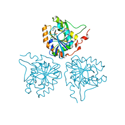
1M3D
 | | Structure of Type IV Collagen NC1 Domains | | 分子名称: | BROMIDE ION, GLYCEROL, LUTETIUM (III) ION, ... | | 著者 | Sundaramoorthy, M, Meiyappan, M, Todd, P, Hudson, B.G. | | 登録日 | 2002-06-27 | | 公開日 | 2003-01-07 | | 最終更新日 | 2011-07-13 | | 実験手法 | X-RAY DIFFRACTION (2 Å) | | 主引用文献 | Crystal Structure of NC1 Domains. Structural Basis for Type IV Collagen Assembly in Basement Membranes
J.Biol.Chem., 277, 2002
|
|
1MKT
 | |
1MKU
 | |
1WTA
 | |
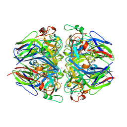
2TAA
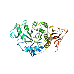 | | STRUCTURE AND POSSIBLE CATALYTIC RESIDUES OF TAKA-AMYLASE A | | 分子名称: | CALCIUM ION, TAKA-AMYLASE A | | 著者 | Kusunoki, M, Matsuura, Y, Tanaka, N, Kakudo, M. | | 登録日 | 1982-10-18 | | 公開日 | 1982-10-21 | | 最終更新日 | 2024-06-05 | | 実験手法 | X-RAY DIFFRACTION (3 Å) | | 主引用文献 | Structure and possible catalytic residues of Taka-amylase A
J.Biochem.(Tokyo), 95, 1984
|
|
1MKS
 | |
8TDW
 | | ssRNA bound SAMHD1 T open | | 分子名称: | Deoxynucleoside triphosphate triphosphohydrolase SAMHD1, FE (III) ION, RNA (5'-R(P*CP*CP*GP*AP*CP*C)-3'), ... | | 著者 | Sung, M, Huynh, K, Han, S. | | 登録日 | 2023-07-05 | | 公開日 | 2023-11-22 | | 最終更新日 | 2023-12-20 | | 実験手法 | ELECTRON MICROSCOPY (3.04 Å) | | 主引用文献 | Guanine-containing ssDNA and RNA induce dimeric and tetrameric structural forms of SAMHD1.
Nucleic Acids Res., 51, 2023
|
|


