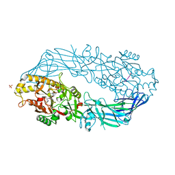
5XUU
 
 | | Crystal structure of Lachnospiraceae bacterium ND2006 Cpf1 in complex with crRNA and target DNA (TCCA PAM) | | 分子名称: | 1,2-ETHANEDIOL, DNA (29-MER), DNA (5'-D(*CP*GP*TP*CP*CP*TP*CP*CP*A)-3'), ... | | 著者 | Yamano, T, Nishimasu, H, Ishitani, R, Nureki, O. | | 登録日 | 2017-06-26 | | 公開日 | 2017-08-09 | | 最終更新日 | 2023-11-22 | | 実験手法 | X-RAY DIFFRACTION (2.5 Å) | | 主引用文献 | Structural Basis for the Canonical and Non-canonical PAM Recognition by CRISPR-Cpf1.
Mol. Cell, 67, 2017
|
|
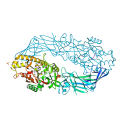
5XUS
 | | Crystal structure of Lachnospiraceae bacterium ND2006 Cpf1 in complex with crRNA and target DNA (TTTA PAM) | | 分子名称: | 1,2-ETHANEDIOL, DNA (29-MER), DNA (5'-D(*CP*GP*TP*CP*CP*TP*TP*TP*A)-3'), ... | | 著者 | Yamano, T, Nishimasu, H, Ishitani, R, Nureki, O. | | 登録日 | 2017-06-26 | | 公開日 | 2017-08-09 | | 最終更新日 | 2023-11-22 | | 実験手法 | X-RAY DIFFRACTION (2.5 Å) | | 主引用文献 | Structural Basis for the Canonical and Non-canonical PAM Recognition by CRISPR-Cpf1.
Mol. Cell, 67, 2017
|
|
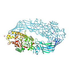
3WRI
 | | Crystal structure of P450cam | | 分子名称: | CAMPHOR, Camphor 5-monooxygenase, PROTOPORPHYRIN IX CONTAINING FE | | 著者 | Kishimoto, A, Takagi, K, Amano, A, Sakurai, K, Mizushima, T, Shimada, H. | | 登録日 | 2014-02-25 | | 公開日 | 2015-03-18 | | 最終更新日 | 2023-11-08 | | 実験手法 | X-RAY DIFFRACTION (2.9 Å) | | 主引用文献 | Structure of P450cam intermediate
To be Published
|
|
3WRM
 | | Crystal structure of P450cam | | 分子名称: | CAMPHOR, Camphor 5-monooxygenase, POTASSIUM ION, ... | | 著者 | Kishimoto, A, Takagi, K, Amano, A, Sakurai, K, Mizushima, T, Shimada, H. | | 登録日 | 2014-02-25 | | 公開日 | 2015-03-18 | | 最終更新日 | 2023-11-08 | | 実験手法 | X-RAY DIFFRACTION (1.95 Å) | | 主引用文献 | Structure of P450cam intermedite
To be published
|
|
3WRJ
 | | Crystal structure of P450cam | | 分子名称: | CAMPHOR, Camphor 5-monooxygenase, POTASSIUM ION, ... | | 著者 | Kishimoto, A, Takagi, K, Amano, A, Sakurai, K, Mizushima, T, Shimada, H. | | 登録日 | 2014-02-25 | | 公開日 | 2015-03-18 | | 最終更新日 | 2023-11-08 | | 実験手法 | X-RAY DIFFRACTION (1.85 Å) | | 主引用文献 | Structure of P450cam intermedite
to be published
|
|
3WRL
 | | Crystal structure of P450cam | | 分子名称: | CAMPHOR, Camphor 5-monooxygenase, POTASSIUM ION, ... | | 著者 | Kishimoto, A, Takagi, K, Amano, A, Sakurai, K, Mizushima, T, Shimada, H. | | 登録日 | 2014-02-25 | | 公開日 | 2015-03-18 | | 最終更新日 | 2023-11-08 | | 実験手法 | X-RAY DIFFRACTION (1.65 Å) | | 主引用文献 | Structure of P450cam intermedite
To be published
|
|
3WRK
 | | Crystal structure of P450cam | | 分子名称: | CAMPHOR, Camphor 5-monooxygenase, PROTOPORPHYRIN IX CONTAINING FE | | 著者 | Kishimoto, A, Takagi, K, Amano, A, Sakurai, K, Mizushima, T, Shimada, H. | | 登録日 | 2014-02-25 | | 公開日 | 2015-03-18 | | 最終更新日 | 2023-11-08 | | 実験手法 | X-RAY DIFFRACTION (2.609 Å) | | 主引用文献 | Structure of P450cam intermedite
To be Published
|
|
3WRH
 | | Crystal structure of P450cam | | 分子名称: | CAMPHOR, Camphor 5-monooxygenase, POTASSIUM ION, ... | | 著者 | Kishimoto, A, Takagi, K, Amano, A, Sakurai, K, Mizushima, T, Shimada, H. | | 登録日 | 2014-02-25 | | 公開日 | 2015-03-18 | | 最終更新日 | 2023-11-08 | | 実験手法 | X-RAY DIFFRACTION (1.62 Å) | | 主引用文献 | Structure of P450cam intermedite
To be Published
|
|
3AK8
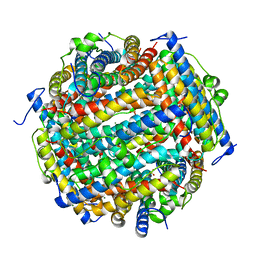 | | Crystal structure of the SEp22 dodecamer, a Dps-like protein from Salmonella enterica subsp. enterica serovar Enteritidis | | 分子名称: | DNA protection during starvation protein, MAGNESIUM ION, SULFATE ION | | 著者 | Miyamoto, T, Asahina, Y, Miyazaki, S, Shimizu, H, Ohto, U, Noguchi, S, Satow, Y. | | 登録日 | 2010-07-08 | | 公開日 | 2011-01-12 | | 最終更新日 | 2023-11-01 | | 実験手法 | X-RAY DIFFRACTION (1.25 Å) | | 主引用文献 | Structures of the SEp22 dodecamer, a Dps-like protein from Salmonella enterica subsp. enterica serovar Enteritidis
Acta Crystallogr.,Sect.F, 67, 2011
|
|
3AK9
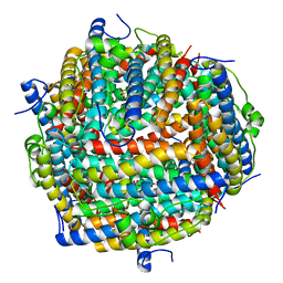 | | Crystal structure of the SEp22 dodecamer, a Dps-like protein from Salmonella enterica subsp. enterica serovar Enteritidis, FE-soaked form | | 分子名称: | DNA protection during starvation protein, FE (II) ION, MAGNESIUM ION, ... | | 著者 | Miyamoto, T, Asahina, Y, Miyazaki, S, Shimizu, H, Ohto, U, Noguchi, S, Satow, Y. | | 登録日 | 2010-07-08 | | 公開日 | 2011-01-12 | | 最終更新日 | 2023-11-01 | | 実験手法 | X-RAY DIFFRACTION (1.3 Å) | | 主引用文献 | Structures of the SEp22 dodecamer, a Dps-like protein from Salmonella enterica subsp. enterica serovar Enteritidis
Acta Crystallogr.,Sect.F, 67, 2011
|
|
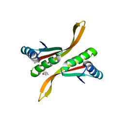
2CT9
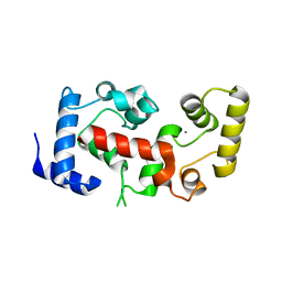 | | The crystal structure of calcineurin B homologous proein 1 (CHP1) | | 分子名称: | CALCIUM ION, Calcium-binding protein p22 | | 著者 | Naoe, Y, Arita, K, Hashimoto, H, Kanazawa, H, Sato, M, Shimizu, T. | | 登録日 | 2005-05-23 | | 公開日 | 2005-07-05 | | 最終更新日 | 2024-03-13 | | 実験手法 | X-RAY DIFFRACTION (2.2 Å) | | 主引用文献 | Structural characterization of calcineurin B homologous protein 1
J.Biol.Chem., 280, 2005
|
|
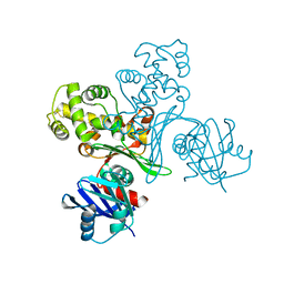
2CW8
 | | Crystal structure of intein homing endonuclease II | | 分子名称: | Endonuclease PI-PkoII, GLYCEROL, SULFATE ION | | 著者 | Matsumura, H, Takahashi, H, Inoue, T, Hashimoto, H, Nishioka, M, Fujiwara, S, Takagi, M, Imanaka, T, Kai, Y. | | 登録日 | 2005-06-17 | | 公開日 | 2006-04-18 | | 最終更新日 | 2011-07-13 | | 実験手法 | X-RAY DIFFRACTION (2.5 Å) | | 主引用文献 | Crystal structure of intein homing endonuclease II encoded in DNA polymerase gene from hyperthermophilic archaeon Thermococcus kodakaraensis strain KOD1
Proteins, 63, 2006
|
|
3VYV
 | | Crystal structure of subtilisin NAT at 1.36 | | 分子名称: | CALCIUM ION, GLYCEROL, Subtilisin NAT | | 著者 | Ushijima, H, Fuchita, N, Kajiwara, T, Motoshima, H, Ueno, G, Watanabe, K. | | 登録日 | 2012-10-03 | | 公開日 | 2013-10-09 | | 最終更新日 | 2023-11-08 | | 実験手法 | X-RAY DIFFRACTION (1.36 Å) | | 主引用文献 | Crystal structure of subtilisin NAT at 1.36
TO BE PUBLISHED
|
|
3VPZ
 | |
2CW7
 | | Crystal structure of intein homing endonuclease II | | 分子名称: | Endonuclease PI-PkoII, SULFATE ION | | 著者 | Matsumura, H, Takahashi, H, Inoue, T, Hashimoto, H, Nishioka, M, Fujiwara, S, Takagi, M, Imanaka, T, Kai, Y. | | 登録日 | 2005-06-17 | | 公開日 | 2006-04-18 | | 最終更新日 | 2024-04-03 | | 実験手法 | X-RAY DIFFRACTION (2.7 Å) | | 主引用文献 | Crystal structure of intein homing endonuclease II encoded in DNA polymerase gene from hyperthermophilic archaeon Thermococcus kodakaraensis strain KOD1
Proteins, 63, 2006
|
|
2DEW
 | | Crystal structure of human peptidylarginine deiminase 4 in complex with histone H3 N-terminal tail including Arg8 | | 分子名称: | 10-mer peptide from histone H3, CALCIUM ION, Protein-arginine deiminase type IV, ... | | 著者 | Arita, K, Shimizu, T, Hashimoto, H, Hidaka, Y, Yamada, M, Sato, M. | | 登録日 | 2006-02-18 | | 公開日 | 2006-04-11 | | 最終更新日 | 2023-10-25 | | 実験手法 | X-RAY DIFFRACTION (2.1 Å) | | 主引用文献 | Structural basis for histone N-terminal recognition by human peptidylarginine deiminase 4
Proc.Natl.Acad.Sci.Usa, 103, 2006
|
|
2DEX
 | | Crystal structure of human peptidylarginine deiminase 4 in complex with histone H3 N-terminal peptide including Arg17 | | 分子名称: | 10-mer peptide from histone H3, CALCIUM ION, Protein-arginine deiminase type IV, ... | | 著者 | Arita, K, Shimizu, T, Hashimoto, H, Hidaka, Y, Yamada, M, Sato, M. | | 登録日 | 2006-02-18 | | 公開日 | 2006-04-11 | | 最終更新日 | 2023-10-25 | | 実験手法 | X-RAY DIFFRACTION (2.1 Å) | | 主引用文献 | Structural basis for histone N-terminal recognition by human peptidylarginine deiminase 4
Proc.Natl.Acad.Sci.Usa, 103, 2006
|
|
2DEY
 | | Crystal structure of human peptidylarginine deiminase 4 in complex with histone H4 N-terminal tail including Arg3 | | 分子名称: | 10-mer peptide from histone H4, CALCIUM ION, Protein-arginine deiminase type IV, ... | | 著者 | Arita, K, Shimizu, T, Hashimoto, H, Hidaka, Y, Yamada, M, Sato, M. | | 登録日 | 2006-02-18 | | 公開日 | 2006-04-11 | | 最終更新日 | 2023-10-25 | | 実験手法 | X-RAY DIFFRACTION (2.25 Å) | | 主引用文献 | Structural basis for histone N-terminal recognition by human peptidylarginine deiminase 4
Proc.Natl.Acad.Sci.Usa, 103, 2006
|
|
2Z7C
 | | Crystal structure of chromatin protein alba from hyperthermophilic archaeon pyrococcus horikoshii | | 分子名称: | ARGININE, DNA/RNA-binding protein Alba | | 著者 | Hada, K, Nakashima, T, Osawa, T, Shimada, H, Kakuta, Y, Kimura, M. | | 登録日 | 2007-08-17 | | 公開日 | 2008-08-05 | | 最終更新日 | 2023-11-01 | | 実験手法 | X-RAY DIFFRACTION (2.8 Å) | | 主引用文献 | Crystal structure and functional analysis of an archaeal chromatin protein Alba from the hyperthermophilic archaeon Pyrococcus horikoshii OT3.
Biosci.Biotechnol.Biochem., 72, 2008
|
|
2ZUH
 | | Crystal Structure of Camphor-soaked Ferric Cytochrome P450cam Mutant (D297A) | | 分子名称: | 2-AMINO-2-HYDROXYMETHYL-PROPANE-1,3-DIOL, CAMPHOR, Camphor 5-monooxygenase, ... | | 著者 | Sakurai, K, Harada, K, Shimada, H, Shimokata, K, Hayashi, T, Tsukihara, T. | | 登録日 | 2008-10-18 | | 公開日 | 2009-10-20 | | 最終更新日 | 2023-11-01 | | 実験手法 | X-RAY DIFFRACTION (1.55 Å) | | 主引用文献 | Crystal Structure of Camphor-soaked Ferric Cytochrome P450cam Mutant
to be published
|
|
2ZUI
 | | Crystal Structure of Camphor-soaked Ferric Cytochrome P450cam Mutant (D297N) | | 分子名称: | CAMPHOR, Camphor 5-monooxygenase, POTASSIUM ION, ... | | 著者 | Sakurai, K, Harada, K, Shimada, H, Shimokata, K, Hayashi, T, Tsukihara, T. | | 登録日 | 2008-10-18 | | 公開日 | 2009-10-20 | | 最終更新日 | 2023-11-01 | | 実験手法 | X-RAY DIFFRACTION (1.5 Å) | | 主引用文献 | Crystal Structure of Camphor-soaked Ferric Cytochrome P450cam Mutant
TO BE PUBLISHED
|
|
2ZUJ
 | | Crystal Structure of Camphor-soaked Ferric Cytochrome P450cam Mutant(D297L) | | 分子名称: | CAMPHOR, Camphor 5-monooxygenase, POTASSIUM ION, ... | | 著者 | Sakurai, K, Harada, K, Shimada, H, Shimokata, K, Hayashi, T, Tsukihara, T. | | 登録日 | 2008-10-18 | | 公開日 | 2009-10-20 | | 最終更新日 | 2023-11-01 | | 実験手法 | X-RAY DIFFRACTION (1.6 Å) | | 主引用文献 | Crystal Structure of Camphor-soaked Ferric Cytochrome P450cam Mutant
to be published
|
|
2D0S
 | | Crystal structure of the Cytochrome C552 from moderate thermophilic bacterium, hydrogenophilus thermoluteolus | | 分子名称: | HEME C, cytochrome c | | 著者 | Nakamura, S, Ichiki, S.I, Takashima, H, Uchiyama, S, Hasegawa, J, Kobayashi, Y, Sambongi, Y, Ohkubo, T. | | 登録日 | 2005-08-08 | | 公開日 | 2006-05-23 | | 最終更新日 | 2011-07-13 | | 実験手法 | X-RAY DIFFRACTION (2.2 Å) | | 主引用文献 | Structure of Cytochrome c552 from a Moderate Thermophilic Bacterium, Hydrogenophilus thermoluteolus: Comparative Study on the Thermostability of Cytochrome c
Biochemistry, 45, 2006
|
|
2DJL
 | | Crystal structure of Trypanosoma cruzi dihydroorotate dehydrogenase in complex with succinate | | 分子名称: | COBALT HEXAMMINE(III), FLAVIN MONONUCLEOTIDE, GLYCEROL, ... | | 著者 | Inaoka, D.K, Shimizu, H, Sakamoto, K, Shiba, T, Kurisu, G, Nara, T, Aoki, T, Harada, S, Kita, K. | | 登録日 | 2006-04-04 | | 公開日 | 2007-06-26 | | 最終更新日 | 2023-10-25 | | 実験手法 | X-RAY DIFFRACTION (1.38 Å) | | 主引用文献 | Crystal structure of Trypanosoma cruzi dihydroorotate dehydrogenase in complex with succinate
To be Published
|
|
2DJX
 | | Crystal structure of native Trypanosoma cruzi dihydroorotate dehydrogenase | | 分子名称: | COBALT HEXAMMINE(III), Dihydroorotate Dehydrogenase, FLAVIN MONONUCLEOTIDE | | 著者 | Inaoka, D.K, Shimizu, H, Sakamoto, K, Shiba, T, Kurisu, G, Nara, T, Aoki, T, Harada, S, Kita, K. | | 登録日 | 2006-04-05 | | 公開日 | 2007-06-26 | | 最終更新日 | 2023-10-25 | | 実験手法 | X-RAY DIFFRACTION (1.58 Å) | | 主引用文献 | Crystal structure of native Trypanosoma cruzi dihydroorotate dehydrogenase
To be Published
|
|


