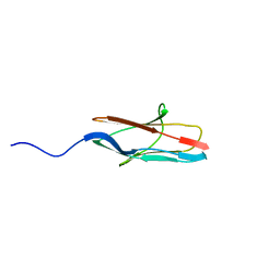
3W9A
 
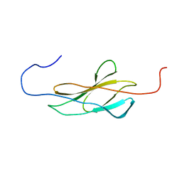
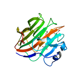 | | Crystal structure of the catalytic domain of the glycoside hydrolase family 131 protein from Coprinopsis cinerea | | 分子名称: | GLYCEROL, Putative uncharacterized protein | | 著者 | Miyazaki, T, Tanaka, Y, Tamura, M, Yoshida, M, Nishikawa, A, Tonozuka, T. | | 登録日 | 2013-04-01 | | 公開日 | 2013-05-22 | | 最終更新日 | 2013-07-10 | | 実験手法 | X-RAY DIFFRACTION (1.99 Å) | | 主引用文献 | Crystal structure of the N-terminal domain of a glycoside hydrolase family 131 protein from Coprinopsis cinerea
Febs Lett., 587, 2013
|
|
2CR7
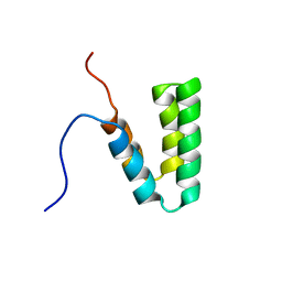 | |
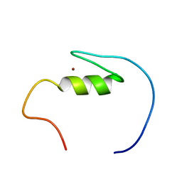
2CP9
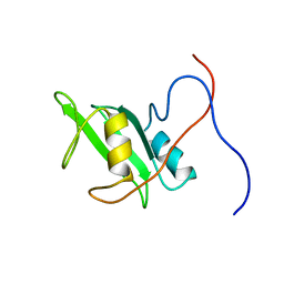
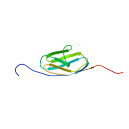
 | | Solution structure of RSGI RUH-042, a UBA domain from human mitochondrial elongation factor Ts | | 分子名称: | Elongation factor Ts, mitochondrial | | 著者 | Ohashi, W, Hirota, H, Izumi, K, Yoshida, M, Yokoyama, S, RIKEN Structural Genomics/Proteomics Initiative (RSGI) | | 登録日 | 2005-05-19 | | 公開日 | 2005-11-19 | | 最終更新日 | 2024-05-29 | | 実験手法 | SOLUTION NMR | | 主引用文献 | Solution structure of RSGI RUH-042, a UBA domain from human mitochondrial elongation factor Ts
To be Published
|
|
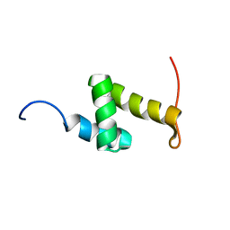
2CRA
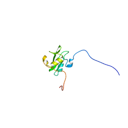
 | | Solution structure of the homeobox domain of human homeo box B13 | | 分子名称: | Homeobox protein Hox-B13 | | 著者 | Seimiya, K, Hayashi, F, Nagashima, T, Yoshida, M, Yokoyama, S, RIKEN Structural Genomics/Proteomics Initiative (RSGI) | | 登録日 | 2005-05-20 | | 公開日 | 2005-11-20 | | 最終更新日 | 2024-05-29 | | 実験手法 | SOLUTION NMR | | 主引用文献 | Solution structure of the homeobox domain of human homeo box B13
To be published
|
|
2CRM
 | |
2CS0
 | |
2CRH
 | |
3TK5
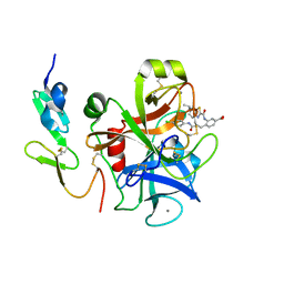 | | Factor Xa in complex with D102-4380 | | 分子名称: | 4-{3-[(4-chlorophenyl)amino]-3-oxopropyl}-3-({[5-(propan-2-yl)-4,5,6,7-tetrahydro[1,3]thiazolo[5,4-c]pyridin-2-yl]carbonyl}amino)benzoic acid, CALCIUM ION, Factor X heavy chain, ... | | 著者 | Suzuki, M, Mochizuki, A, Nagata, T, Takano, H, Kanno, H, Kishida, M, Ohta, T. | | 登録日 | 2011-08-25 | | 公開日 | 2012-08-29 | | 最終更新日 | 2017-11-08 | | 実験手法 | X-RAY DIFFRACTION (2.2 Å) | | 主引用文献 | Zwitter ionic potent durable orally active Factor Xa inhibitor.
To be Published
|
|
4EIW
 | | Whole cytosolic region of atp-dependent metalloprotease FtsH (G399L) | | 分子名称: | ADENOSINE-5'-DIPHOSPHATE, ATP-dependent zinc metalloprotease FtsH | | 著者 | Suno, R, Niwa, H, Tsuchiya, D, Yoshida, M, Morikawa, K. | | 登録日 | 2012-04-06 | | 公開日 | 2012-06-06 | | 最終更新日 | 2024-03-20 | | 実験手法 | X-RAY DIFFRACTION (3.9 Å) | | 主引用文献 | Structure of the whole cytosolic region of ATP-dependent protease FtsH
Mol.Cell, 22, 2006
|
|
2DHR
 | | Whole cytosolic region of ATP-dependent metalloprotease FtsH (G399L) | | 分子名称: | ADENOSINE-5'-DIPHOSPHATE, FtsH | | 著者 | Suno, R, Niwa, H, Tsuchiya, D, Zhang, X, Yoshida, M, Morikawa, K. | | 登録日 | 2006-03-24 | | 公開日 | 2006-06-27 | | 最終更新日 | 2024-05-29 | | 実験手法 | X-RAY DIFFRACTION (3.9 Å) | | 主引用文献 | Structure of the Whole Cytosolic Region of ATP-Dependent Protease FtsH
Mol.Cell, 22, 2006
|
|
3TK6
 | | factor Xa in complex with D46-5241 | | 分子名称: | CALCIUM ION, Factor X heavy chain, Factor X light chain, ... | | 著者 | Suzuki, M, Mochizuki, A, Nagata, T, Takano, H, Kanno, H, Kishida, M, Ohta, T. | | 登録日 | 2011-08-25 | | 公開日 | 2012-08-29 | | 最終更新日 | 2017-11-08 | | 実験手法 | X-RAY DIFFRACTION (1.8 Å) | | 主引用文献 | Zwitter ionic potent durable orally active Factor Xa inhibitor.
To be Published
|
|
2DI4
 | | Crystal structure of the FtsH protease domain | | 分子名称: | Cell division protein ftsH homolog, MERCURY (II) ION | | 著者 | Suno, R, Niwa, H, Tsuchiya, D, Zhang, X, Yoshida, M, Morikawa, K. | | 登録日 | 2006-03-28 | | 公開日 | 2006-06-27 | | 最終更新日 | 2011-07-13 | | 実験手法 | X-RAY DIFFRACTION (2.79 Å) | | 主引用文献 | Structure of the Whole Cytosolic Region of ATP-Dependent Protease FtsH
Mol.Cell, 22, 2006
|
|
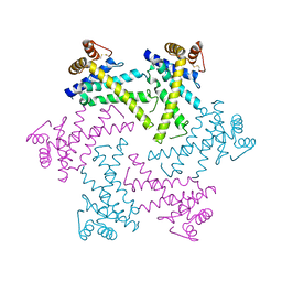
1WDW
 | | Structural basis of mutual activation of the tryptophan synthase a2b2 complex from a hyperthermophile, Pyrococcus furiosus | | 分子名称: | PYRIDOXAL-5'-PHOSPHATE, Tryptophan synthase alpha chain, Tryptophan synthase beta chain 1 | | 著者 | Lee, S.J, Ogasahara, K, Ma, J, Nishio, K, Ishida, M, Yamagata, Y, Tsukihara, T, Yutani, K, RIKEN Structural Genomics/Proteomics Initiative (RSGI) | | 登録日 | 2004-05-19 | | 公開日 | 2005-07-12 | | 最終更新日 | 2023-10-25 | | 実験手法 | X-RAY DIFFRACTION (3 Å) | | 主引用文献 | Conformational Changes in the Tryptophan Synthase from a Hyperthermophile upon alpha(2)beta(2) Complex Formation: Crystal Structure of the Complex
Biochemistry, 44, 2005
|
|
3VOH
 | | CcCel6A catalytic domain complexed with cellobiose | | 分子名称: | Cellobiohydrolase, beta-D-glucopyranose, beta-D-glucopyranose-(1-4)-beta-D-glucopyranose, ... | | 著者 | Tamura, M, Miyazaki, T, Tanaka, Y, Yoshida, M, Nishikawa, A, Tonozuka, T. | | 登録日 | 2012-01-24 | | 公開日 | 2012-03-21 | | 最終更新日 | 2023-11-08 | | 実験手法 | X-RAY DIFFRACTION (2.4 Å) | | 主引用文献 | Comparison of the structural changes in two cellobiohydrolases, CcCel6A and CcCel6C, from Coprinopsis cinerea - a tweezer-like motion in the structure of CcCel6C
Febs J., 279, 2012
|
|
3VOJ
 | | CcCel6A catalytic domain mutant D164A | | 分子名称: | Cellobiohydrolase | | 著者 | Tamura, M, Miyazaki, T, Tanaka, Y, Yoshida, M, Nishikawa, A, Tonozuka, T. | | 登録日 | 2012-01-24 | | 公開日 | 2012-03-21 | | 最終更新日 | 2023-11-08 | | 実験手法 | X-RAY DIFFRACTION (2.29 Å) | | 主引用文献 | Comparison of the structural changes in two cellobiohydrolases, CcCel6A and CcCel6C, from Coprinopsis cinerea - a tweezer-like motion in the structure of CcCel6C
Febs J., 279, 2012
|
|
3VOG
 | | Catalytic domain of the cellobiohydrolase, CcCel6A, from Coprinopsis cinerea | | 分子名称: | 4-(2-HYDROXYETHYL)-1-PIPERAZINE ETHANESULFONIC ACID, Cellobiohydrolase | | 著者 | Tamura, M, Miyazaki, T, Tanaka, Y, Yoshida, M, Nishikawa, A, Tonozuka, T. | | 登録日 | 2012-01-24 | | 公開日 | 2012-03-21 | | 最終更新日 | 2023-11-08 | | 実験手法 | X-RAY DIFFRACTION (1.45 Å) | | 主引用文献 | Comparison of the structural changes in two cellobiohydrolases, CcCel6A and CcCel6C, from Coprinopsis cinerea - a tweezer-like motion in the structure of CcCel6C
Febs J., 279, 2012
|
|
3VOI
 | | CcCel6A catalytic domain complexed with p-nitrophenyl beta-D-cellotrioside | | 分子名称: | 4-nitrophenyl beta-D-glucopyranosyl-(1->4)-beta-D-glucopyranosyl-(1->4)-beta-D-glucopyranoside, Cellobiohydrolase, MAGNESIUM ION | | 著者 | Tamura, M, Miyazaki, T, Tanaka, Y, Yoshida, M, Nishikawa, A, Tonozuka, T. | | 登録日 | 2012-01-24 | | 公開日 | 2012-03-21 | | 最終更新日 | 2023-11-08 | | 実験手法 | X-RAY DIFFRACTION (2 Å) | | 主引用文献 | Comparison of the structural changes in two cellobiohydrolases, CcCel6A and CcCel6C, from Coprinopsis cinerea - a tweezer-like motion in the structure of CcCel6C
Febs J., 279, 2012
|
|
3VOF
 | | Cellobiohydrolase mutant, CcCel6C D102A, in the closed form | | 分子名称: | Cellobiohydrolase, beta-D-glucopyranose | | 著者 | Tamura, M, Miyazaki, T, Tanaka, Y, Yoshida, M, Nishikawa, A, Tonozuka, T. | | 登録日 | 2012-01-23 | | 公開日 | 2012-03-21 | | 最終更新日 | 2023-11-08 | | 実験手法 | X-RAY DIFFRACTION (1.6 Å) | | 主引用文献 | Comparison of the structural changes in two cellobiohydrolases, CcCel6A and CcCel6C, from Coprinopsis cinerea - a tweezer-like motion in the structure of CcCel6C
Febs J., 279, 2012
|
|
3VND
 | | Crystal structure of tryptophan synthase alpha-subunit from the psychrophile Shewanella frigidimarina K14-2 | | 分子名称: | 3,6,9,12,15,18,21-HEPTAOXATRICOSANE-1,23-DIOL, SULFATE ION, Tryptophan synthase alpha chain | | 著者 | Mitsuya, D, Tanaka, S, Matsumura, H, Takano, K, Urano, N, Ishida, M. | | 登録日 | 2012-01-12 | | 公開日 | 2013-01-16 | | 最終更新日 | 2023-11-08 | | 実験手法 | X-RAY DIFFRACTION (2.6 Å) | | 主引用文献 | Strategy for cold adaptation of the tryptophan synthase alpha subunit from the psychrophile Shewanella frigidimarina K14-2: crystal structure and physicochemical properties
J.Biochem., 155, 2014
|
|
2YQG
 | |
2YST
 | |
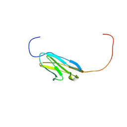
2DKU
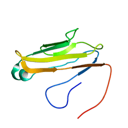
 | | Solution structure of the third Ig-like domain of human KIAA1556 protein | | 分子名称: | KIAA1556 protein | | 著者 | Nagashima, K, Kurosaki, C, Yoshida, M, Hayashi, F, Yokoyama, S, RIKEN Structural Genomics/Proteomics Initiative (RSGI) | | 登録日 | 2006-04-14 | | 公開日 | 2006-10-14 | | 最終更新日 | 2024-05-29 | | 実験手法 | SOLUTION NMR | | 主引用文献 | Solution structure of the third Ig-like domain of human KIAA1556 protein
To be Published
|
|
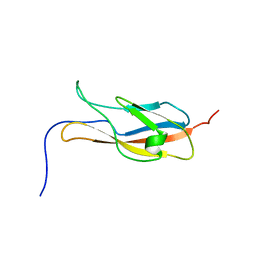
2YR3
 | | Solution structure of the fourth Ig-like domain from myosin light chain kinase, smooth muscle | | 分子名称: | Myosin light chain kinase, smooth muscle | | 著者 | Qin, X.R, Kurosaki, C, Yoshida, M, Hayahsi, F, Yokoyama, S, RIKEN Structural Genomics/Proteomics Initiative (RSGI) | | 登録日 | 2007-04-02 | | 公開日 | 2007-10-02 | | 最終更新日 | 2024-05-29 | | 実験手法 | SOLUTION NMR | | 主引用文献 | Solution structure of the fourth Ig-like domain from myosin light chain kinase, smooth muscle
To be Published
|
|
2YRJ
 | | Solution structure of the C2H2-type zinc finger domain (781-813) from zinc finger protein 473 | | 分子名称: | ZINC ION, Zinc finger protein 473 | | 著者 | Qin, X.R, Izumi, K, Yoshida, M, Hayashi, F, Yokoyama, S, RIKEN Structural Genomics/Proteomics Initiative (RSGI) | | 登録日 | 2007-04-02 | | 公開日 | 2007-10-02 | | 最終更新日 | 2024-05-29 | | 実験手法 | SOLUTION NMR | | 主引用文献 | Solution structure of the C2H2-type zinc finger domain (781-813) from zinc finger protein 473
To be Published
|
|
2EDL
 | |


