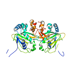
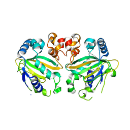
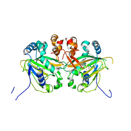
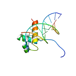
7W6F
 
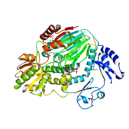
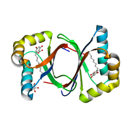 | | Polyketide cyclase OAC-F24I mutant from Cannabis sativa in complex with 6-nonylresorcylic acid | | 分子名称: | 2-nonyl-4,6-bis(oxidanyl)benzoic acid, GLYCEROL, Olivetolic acid cyclase | | 著者 | Nakashima, Y, Lee, Y.E, Morita, H. | | 登録日 | 2021-12-01 | | 公開日 | 2022-02-02 | | 最終更新日 | 2023-11-29 | | 実験手法 | X-RAY DIFFRACTION (1.58 Å) | | 主引用文献 | Dual Engineering of Olivetolic Acid Cyclase and Tetraketide Synthase to Generate Longer Alkyl-Chain Olivetolic Acid Analogs.
Org.Lett., 24, 2022
|
|
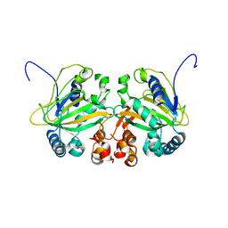
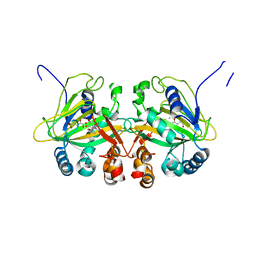
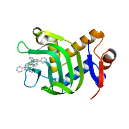
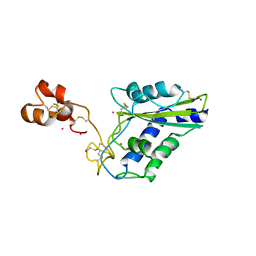
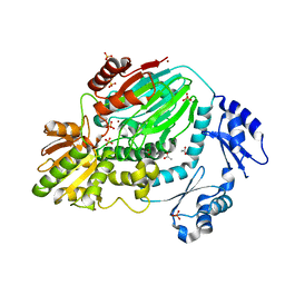
7W6G
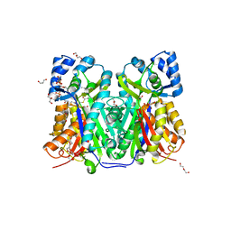 | | TKS-L190G mutant from Cannabis sativa in complex with lauroyl-CoA | | 分子名称: | 3,5,7-trioxododecanoyl-CoA synthase, DI(HYDROXYETHYL)ETHER, GLYCEROL, ... | | 著者 | Nakashima, Y, Lee, Y.E, Morita, H. | | 登録日 | 2021-12-01 | | 公開日 | 2022-02-02 | | 最終更新日 | 2023-11-29 | | 実験手法 | X-RAY DIFFRACTION (2.1 Å) | | 主引用文献 | Dual Engineering of Olivetolic Acid Cyclase and Tetraketide Synthase to Generate Longer Alkyl-Chain Olivetolic Acid Analogs.
Org.Lett., 24, 2022
|
|
7W6E
 | |
7W6D
 | |
5YBQ
 | |
5YBM
 | |
5YBR
 | |
5YBO
 | |
5YBL
 | | Fe(II)/(alpha)ketoglutarate-dependent dioxygenase AusE | | 分子名称: | 2-OXOGLUTARIC ACID, MANGANESE (II) ION, Multifunctional dioxygenase ausE | | 著者 | Nakashima, Y, Senda, M. | | 登録日 | 2017-09-05 | | 公開日 | 2018-01-24 | | 最終更新日 | 2024-03-27 | | 実験手法 | X-RAY DIFFRACTION (2.108 Å) | | 主引用文献 | Structure function and engineering of multifunctional non-heme iron dependent oxygenases in fungal meroterpenoid biosynthesis.
Nat Commun, 9, 2018
|
|
5YBT
 | |
5YBS
 | |
5YBN
 | |
5YBP
 | |
7VM1
 | | Crystal Structure of HasAp Capturing Iron Tetra(4-pyridyl)porphyrin | | 分子名称: | Fe-Tetra(4-pyridyl)porphyrin, GLYCEROL, Heme acquisition protein HasAp | | 著者 | Shisaka, Y, Ueda, G, Sakakibara, E, Sugimoto, H, Shoji, O. | | 登録日 | 2021-10-06 | | 公開日 | 2022-08-17 | | 最終更新日 | 2023-11-29 | | 実験手法 | X-RAY DIFFRACTION (2 Å) | | 主引用文献 | Tetraphenylporphyrin Enters the Ring: First Example of a Complex between Highly Bulky Porphyrins and a Protein.
Chembiochem, 23, 2022
|
|
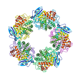
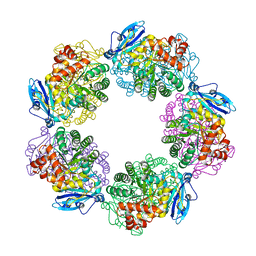
5XSX
 | | Crystal structure of an archaeal chitinase in the substrate-complex form (P212121) | | 分子名称: | 2-acetamido-2-deoxy-beta-D-glucopyranose-(1-4)-2-acetamido-2-deoxy-beta-D-glucopyranose-(1-4)-2-acetamido-2-deoxy-beta-D-glucopyranose-(1-4)-2-acetamido-2-deoxy-beta-D-glucopyranose-(1-4)-2-acetamido-2-deoxy-beta-D-glucopyranose, Chitinase, GLYCEROL, ... | | 著者 | Nishitani, Y, Miki, K. | | 登録日 | 2017-06-15 | | 公開日 | 2018-05-02 | | 最終更新日 | 2023-11-22 | | 実験手法 | X-RAY DIFFRACTION (2.642 Å) | | 主引用文献 | Crystal structures of an archaeal chitinase ChiD and its ligand complexes.
Glycobiology, 28, 2018
|
|
5XSV
 | |
1VFC
 | |
1WVR
 | | Crystal Structure of a CRISP family Ca-channel blocker derived from snake venom | | 分子名称: | CADMIUM ION, Triflin | | 著者 | Shikamoto, Y, Suto, K, Yamazaki, Y, Morita, T, Mizuno, H. | | 登録日 | 2004-12-24 | | 公開日 | 2005-07-05 | | 最終更新日 | 2017-10-11 | | 実験手法 | X-RAY DIFFRACTION (2.4 Å) | | 主引用文献 | Crystal structure of a CRISP family Ca2+ -channel blocker derived from snake venom.
J.Mol.Biol., 350, 2005
|
|
3A65
 | | Crystal structure of 6-aminohexanoate-dimer hydrolase S112A/G181D/H266N mutant with substrate | | 分子名称: | 2-(N-MORPHOLINO)-ETHANESULFONIC ACID, 6-AMINOHEXANOIC ACID, 6-aminohexanoate-dimer hydrolase, ... | | 著者 | Kawashima, Y, Shibata, N, Higuchi, Y, Takeo, M, Negoro, S. | | 登録日 | 2009-08-21 | | 公開日 | 2010-09-01 | | 最終更新日 | 2023-11-15 | | 実験手法 | X-RAY DIFFRACTION (1.7 Å) | | 主引用文献 | Enzymatic Synthesis of Nylon-6 Units in Organic Sol Contained Low-Water: Structural Requirement of 6-Aminohexanoate-Dimer Hydrolase for Efficient Amid Synthesis
To be Published
|
|
3A66
 | | Crystal structure of 6-aminohexanoate-dimer hydrolase S112A/G181D/H266N/D370Y mutant with substrate | | 分子名称: | 2-(N-MORPHOLINO)-ETHANESULFONIC ACID, 6-AMINOHEXANOATE-DIMER HYDROLASE, 6-AMINOHEXANOIC ACID, ... | | 著者 | Kawashima, Y, Shibata, N, Higuchi, Y, Takeo, M, Negoro, S. | | 登録日 | 2009-08-21 | | 公開日 | 2010-09-01 | | 最終更新日 | 2023-11-15 | | 実験手法 | X-RAY DIFFRACTION (1.6 Å) | | 主引用文献 | Enzymatic Synthesis of Nylon-6 Units in Organic Sol Contained Low-Water: Structural Requirement of 6-Aminohexanoate-Dimer Hydrolase for Efficient Amid Synthesis
To be Published
|
|
2Z7E
 | | Crystal structure of Aquifex aeolicus IscU with bound [2Fe-2S] cluster | | 分子名称: | FE2/S2 (INORGANIC) CLUSTER, NifU-like protein, SULFATE ION | | 著者 | Shimomura, Y, Wada, K, Takahashi, Y, Fukuyama, K. | | 登録日 | 2007-08-20 | | 公開日 | 2008-08-19 | | 最終更新日 | 2021-11-10 | | 実験手法 | X-RAY DIFFRACTION (2.3 Å) | | 主引用文献 | The asymmetric trimeric architecture of [2Fe-2S] IscU: implications for its scaffolding during iron-sulfur cluster biosynthesis
J.Mol.Biol., 383, 2008
|
|
3A12
 | | Crystal structure of Type III Rubisco complexed with 2-CABP | | 分子名称: | 2-CARBOXYARABINITOL-1,5-DIPHOSPHATE, MAGNESIUM ION, Ribulose bisphosphate carboxylase | | 著者 | Nishitani, Y, Fujihashi, M, Doi, T, Yoshida, S, Atomi, H, Imanaka, T, Miki, K. | | 登録日 | 2009-03-25 | | 公開日 | 2010-04-07 | | 最終更新日 | 2023-11-15 | | 実験手法 | X-RAY DIFFRACTION (2.3 Å) | | 主引用文献 | Structure-based catalytic optimization of a type III Rubisco from a hyperthermophile
J.Biol.Chem., 285, 2010
|
|
3A13
 | | Crystal structure of Type III Rubisco SP4 mutant complexed with 2-CABP and activated with Ca | | 分子名称: | 2-CARBOXYARABINITOL-1,5-DIPHOSPHATE, CALCIUM ION, MAGNESIUM ION, ... | | 著者 | Nishitani, Y, Fujihashi, M, Doi, T, Yoshida, S, Atomi, H, Imanaka, T, Miki, K. | | 登録日 | 2009-03-25 | | 公開日 | 2010-04-07 | | 最終更新日 | 2023-11-15 | | 実験手法 | X-RAY DIFFRACTION (2.34 Å) | | 主引用文献 | Structure-based optimization of a Type III Rubisco from a hyperthermophile
To be Published
|
|
3AF6
 | | The crystal structure of an archaeal CPSF subunit, PH1404 from Pyrococcus horikoshii complexed with RNA-analog | | 分子名称: | 5'-R(*(SSU)P*(SSU)P*(SSU)P*(SSU)P*(SSU)P*(SSU))-3', Putative uncharacterized protein PH1404, SULFATE ION, ... | | 著者 | Nishida, Y, Ishikawa, H, Nakagawa, N, Masui, R, Kuramitsu, S. | | 登録日 | 2010-02-24 | | 公開日 | 2010-04-21 | | 最終更新日 | 2023-11-01 | | 実験手法 | X-RAY DIFFRACTION (2.6 Å) | | 主引用文献 | Crystal structure of an archaeal cleavage and polyadenylation specificity factor subunit from Pyrococcus horikoshii
Proteins, 78, 2010
|
|
3AF5
 | | The crystal structure of an archaeal CPSF subunit, PH1404 from Pyrococcus horikoshii | | 分子名称: | ACETIC ACID, Putative uncharacterized protein PH1404, SULFATE ION, ... | | 著者 | Nishida, Y, Ishikawa, H, Nakagawa, N, Masui, R, Kuramitsu, S. | | 登録日 | 2010-02-23 | | 公開日 | 2010-04-21 | | 最終更新日 | 2024-03-13 | | 実験手法 | X-RAY DIFFRACTION (2.6 Å) | | 主引用文献 | Crystal structure of an archaeal cleavage and polyadenylation specificity factor subunit from Pyrococcus horikoshii
Proteins, 78, 2010
|
|


