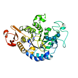
7W6F
 
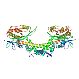
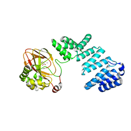
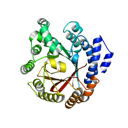
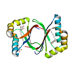 | | Polyketide cyclase OAC-F24I mutant from Cannabis sativa in complex with 6-nonylresorcylic acid | | 分子名称: | 2-nonyl-4,6-bis(oxidanyl)benzoic acid, GLYCEROL, Olivetolic acid cyclase | | 著者 | Nakashima, Y, Lee, Y.E, Morita, H. | | 登録日 | 2021-12-01 | | 公開日 | 2022-02-02 | | 最終更新日 | 2023-11-29 | | 実験手法 | X-RAY DIFFRACTION (1.58 Å) | | 主引用文献 | Dual Engineering of Olivetolic Acid Cyclase and Tetraketide Synthase to Generate Longer Alkyl-Chain Olivetolic Acid Analogs.
Org.Lett., 24, 2022
|
|
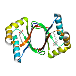
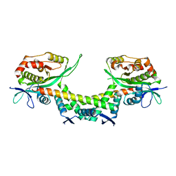
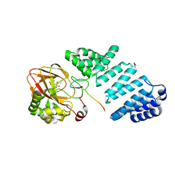
7W6E
 | |
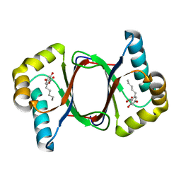
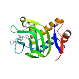
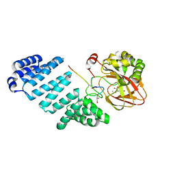
7W6D
 | |
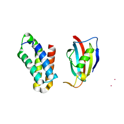
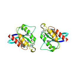
7BON
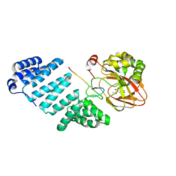
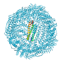 | | Crystal structure of recombinant horse spleen apo-R52C/E56C/R59C/E63C-Fr | | 分子名称: | 1,2-ETHANEDIOL, CADMIUM ION, CHLORIDE ION, ... | | 著者 | Hishikawa, Y, Maity, B, Ito, N, Abe, S, Lu, D, Ueno, T. | | 登録日 | 2020-03-19 | | 公開日 | 2021-01-27 | | 最終更新日 | 2023-11-29 | | 実験手法 | X-RAY DIFFRACTION (1.48 Å) | | 主引用文献 | Design of Multinuclear Gold Binding Site at the Two-fold Symmetric Interface of the Ferritin Cage
Chem Lett., 49, 2021
|
|
5ETY
 | | Crystal Structure of human Tankyrase-1 bound to K-756 | | 分子名称: | 3-[[1-(6,7-dimethoxyquinazolin-4-yl)piperidin-4-yl]methyl]-1,4-dihydroquinazolin-2-one, Tankyrase-1, ZINC ION | | 著者 | Takahashi, Y, Miyagi, H, Suzuki, M, Saito, J. | | 登録日 | 2015-11-18 | | 公開日 | 2016-06-22 | | 最終更新日 | 2024-03-20 | | 実験手法 | X-RAY DIFFRACTION (2.9 Å) | | 主引用文献 | The Discovery and Characterization of K-756, a Novel Wnt/ beta-Catenin Pathway Inhibitor Targeting Tankyrase
Mol.Cancer Ther., 15, 2016
|
|
6AJN
 | |
6AJM
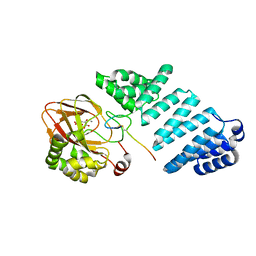
 | | Crystal structure of apo AtaTR | | 分子名称: | DUF1778 domain-containing protein, N-acetyltransferase, TRIETHYLENE GLYCOL | | 著者 | Yashiro, Y, Yamashita, S, Tomita, K. | | 登録日 | 2018-08-28 | | 公開日 | 2019-01-02 | | 最終更新日 | 2023-11-22 | | 実験手法 | X-RAY DIFFRACTION (2.604 Å) | | 主引用文献 | Crystal Structure of the Enterohemorrhagic Escherichia coli AtaT-AtaR Toxin-Antitoxin Complex.
Structure, 27, 2019
|
|
7VM1
 | | Crystal Structure of HasAp Capturing Iron Tetra(4-pyridyl)porphyrin | | 分子名称: | Fe-Tetra(4-pyridyl)porphyrin, GLYCEROL, Heme acquisition protein HasAp | | 著者 | Shisaka, Y, Ueda, G, Sakakibara, E, Sugimoto, H, Shoji, O. | | 登録日 | 2021-10-06 | | 公開日 | 2022-08-17 | | 最終更新日 | 2023-11-29 | | 実験手法 | X-RAY DIFFRACTION (2 Å) | | 主引用文献 | Tetraphenylporphyrin Enters the Ring: First Example of a Complex between Highly Bulky Porphyrins and a Protein.
Chembiochem, 23, 2022
|
|
2RSE
 | | NMR structure of FKBP12-mTOR FRB domain-rapamycin complex structure determined based on PCS | | 分子名称: | Peptidyl-prolyl cis-trans isomerase FKBP1A, Serine/threonine-protein kinase mTOR, TERBIUM(III) ION | | 著者 | Kobashigawa, Y, Ushio, M, Saio, T, Inagaki, F. | | 登録日 | 2012-01-25 | | 公開日 | 2012-05-30 | | 最終更新日 | 2024-05-15 | | 実験手法 | SOLUTION NMR | | 主引用文献 | Convenient method for resolving degeneracies due to symmetry of the magnetic susceptibility tensor and its application to pseudo contact shift-based protein-protein complex structure determination.
J.Biomol.Nmr, 53, 2012
|
|
1QPK
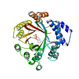
 | | MUTANT (D193G) MALTOTETRAOSE-FORMING EXO-AMYLASE IN COMPLEX WITH MALTOTETRAOSE | | 分子名称: | CALCIUM ION, PROTEIN (MALTOTETRAOSE-FORMING AMYLASE), alpha-D-glucopyranose-(1-4)-alpha-D-glucopyranose-(1-4)-alpha-D-glucopyranose-(1-4)-alpha-D-glucopyranose | | 著者 | Yoshioka, Y, Hasegawa, K, Matsuura, Y, Katsube, Y, Kubota, M. | | 登録日 | 1999-05-26 | | 公開日 | 1999-11-17 | | 最終更新日 | 2021-11-03 | | 実験手法 | X-RAY DIFFRACTION (2 Å) | | 主引用文献 | Roles of catalytic residues in alpha-amylases as evidenced by the structures of the product-complexed mutants of a maltotetraose-forming amylase.
Protein Eng., 12, 1999
|
|
7YBC
 | |
7YB8
 | |
5EZ5
 | |
7YBA
 | |
7YB9
 | |
7YBB
 | |
5XSO
 | | Crystal structure of full-length FixJ from B. japonicum crystallized in space group C2221 | | 分子名称: | FORMIC ACID, GLYCEROL, Response regulator FixJ | | 著者 | Nishizono, Y, Hisano, T, Sawai, H, Shiro, Y, Nakamura, H, Wright, G.S.A, Saeki, A, Hikima, T, Yamamoto, M, Antonyuk, S.V, Hasnain, S.S. | | 登録日 | 2017-06-14 | | 公開日 | 2018-05-23 | | 最終更新日 | 2024-03-27 | | 実験手法 | X-RAY DIFFRACTION (1.778 Å) | | 主引用文献 | Architecture of the complete oxygen-sensing FixL-FixJ two-component signal transduction system.
Sci Signal, 11, 2018
|
|
5XT2
 | | Crystal structures of full-length FixJ from B. japonicum crystallized in space group P212121 | | 分子名称: | FORMIC ACID, GLYCEROL, MAGNESIUM ION, ... | | 著者 | Nishizono, Y, Hisano, T, Shiro, Y, Sawai, H, Wright, G.S.A, Saeki, A, Hikima, T, Nakamura, H, Yamamoto, M, Antonyuk, S.V, Hasnain, S.S. | | 登録日 | 2017-06-16 | | 公開日 | 2018-05-23 | | 最終更新日 | 2024-03-27 | | 実験手法 | X-RAY DIFFRACTION (2.652 Å) | | 主引用文献 | Architecture of the complete oxygen-sensing FixL-FixJ two-component signal transduction system.
Sci Signal, 11, 2018
|
|
7Y3V
 | | Crystal structure of CdpNPT in complex with harmane | | 分子名称: | 1-methyl-9H-pyrido[3,4-b]indole, Cyclic dipeptide N-prenyltransferase, PHOSPHATE ION | | 著者 | Nakashima, Y, Morita, H. | | 登録日 | 2022-06-13 | | 公開日 | 2023-04-05 | | 最終更新日 | 2023-11-29 | | 実験手法 | X-RAY DIFFRACTION (2.43 Å) | | 主引用文献 | Catalytic potential of a fungal indole prenyltransferase toward beta-carbolines, harmine and harman, and their prenylation effects on antibacterial activity.
J.Biosci.Bioeng., 134, 2022
|
|
7XVJ
 | |
7WLR
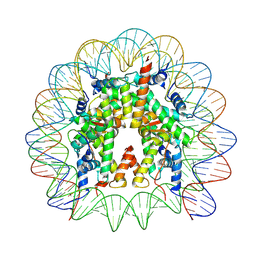 | | Cryo-EM structure of the nucleosome containing Komagataella pastoris histones | | 分子名称: | DNA (145-MER), Histone H2A, Histone H2B, ... | | 著者 | Fukushima, Y, Hatazawa, S, Hirai, S, Kujirai, T, Takizawa, Y, Kurumizaka, H. | | 登録日 | 2022-01-13 | | 公開日 | 2022-07-13 | | 最終更新日 | 2024-06-26 | | 実験手法 | ELECTRON MICROSCOPY (3.54 Å) | | 主引用文献 | Structural and biochemical analyses of the nucleosome containing Komagataella pastoris histones.
J.Biochem., 172, 2022
|
|
7KHE
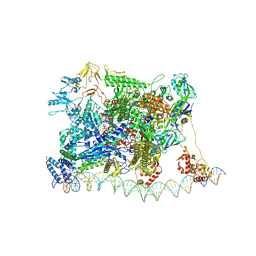
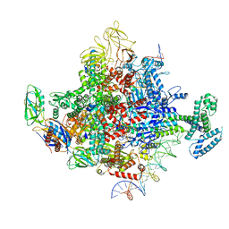 | | Escherichia coli RNA polymerase and rrnBP1 promoter pre-open complex with DksA/ppGpp | | 分子名称: | CHAPSO, DNA (46-MER), DNA (54-MER), ... | | 著者 | Shin, Y, Qayyum, M.Z, Murakami, K.S. | | 登録日 | 2020-10-21 | | 公開日 | 2020-12-09 | | 最終更新日 | 2024-03-06 | | 実験手法 | ELECTRON MICROSCOPY (3.58 Å) | | 主引用文献 | Structural basis of ribosomal RNA transcription regulation.
Nat Commun, 12, 2021
|
|
7KHB
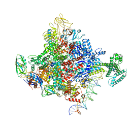
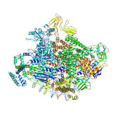 | | Escherichia coli RNA polymerase and rrnBP1 promoter open complex | | 分子名称: | CHAPSO, DNA (60-MER), DNA (64-MER), ... | | 著者 | Shin, Y, Qayyum, M.Z, Murakami, K.S. | | 登録日 | 2020-10-20 | | 公開日 | 2020-10-28 | | 最終更新日 | 2024-03-06 | | 実験手法 | ELECTRON MICROSCOPY (3.53 Å) | | 主引用文献 | Structural basis of ribosomal RNA transcription regulation.
Nat Commun, 12, 2021
|
|
7KHC
 | | Escherichia coli RNA polymerase and rrnBP1 promoter closed complex | | 分子名称: | CHAPSO, DNA (18 MER), DNA (63-MER), ... | | 著者 | Shin, Y, Qayyum, M.Z, Murakami, K.S. | | 登録日 | 2020-10-20 | | 公開日 | 2020-10-28 | | 最終更新日 | 2024-03-06 | | 実験手法 | ELECTRON MICROSCOPY (4.14 Å) | | 主引用文献 | Structural basis of ribosomal RNA transcription regulation.
Nat Commun, 12, 2021
|
|
7KHI
 | | Escherichia coli RNA polymerase and rrnBP1 promoter complex with DksA/ppGpp | | 分子名称: | CHAPSO, DNA (28-MER), DNA (36-MER), ... | | 著者 | Shin, Y, Qayyum, M.Z, Murakami, K.S. | | 登録日 | 2020-10-21 | | 公開日 | 2020-10-28 | | 最終更新日 | 2024-03-06 | | 実験手法 | ELECTRON MICROSCOPY (3.62 Å) | | 主引用文献 | Structural basis of ribosomal RNA transcription regulation.
Nat Commun, 12, 2021
|
|


