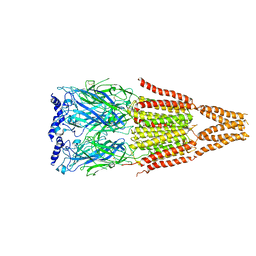
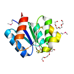
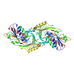
3WV5
 
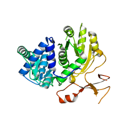 | | Complex structure of VinN with 3-methylaspartate | | 分子名称: | (2S,3S)-3-methyl-aspartic acid, Non-ribosomal peptide synthetase | | 著者 | Miyanaga, A, Cieslak, J, Shinohara, Y, Kudo, F, Eguchi, T. | | 登録日 | 2014-05-15 | | 公開日 | 2014-10-01 | | 最終更新日 | 2023-11-08 | | 実験手法 | X-RAY DIFFRACTION (2.2 Å) | | 主引用文献 | The crystal structure of the adenylation enzyme VinN reveals a unique beta-amino acid recognition mechanism
J.Biol.Chem., 289, 2014
|
|
3WV4
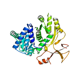 | | Crystal structure of VinN | | 分子名称: | Non-ribosomal peptide synthetase | | 著者 | Miyanaga, A, Cieslak, J, Shinohara, Y, Kudo, F, Eguchi, T. | | 登録日 | 2014-05-15 | | 公開日 | 2014-10-01 | | 最終更新日 | 2023-11-08 | | 実験手法 | X-RAY DIFFRACTION (2.15 Å) | | 主引用文献 | The crystal structure of the adenylation enzyme VinN reveals a unique beta-amino acid recognition mechanism
J.Biol.Chem., 289, 2014
|
|
3WVN
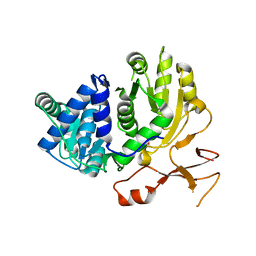 | | Complex structure of VinN with L-aspartate | | 分子名称: | ASPARTIC ACID, Non-ribosomal peptide synthetase | | 著者 | Miyanaga, A, Cieslak, J, Shinohara, Y, Kudo, F, Eguchi, T. | | 登録日 | 2014-05-30 | | 公開日 | 2014-10-01 | | 最終更新日 | 2023-11-08 | | 実験手法 | X-RAY DIFFRACTION (2.2 Å) | | 主引用文献 | The crystal structure of the adenylation enzyme VinN reveals a unique beta-amino acid recognition mechanism
J.Biol.Chem., 289, 2014
|
|
3WD7
 | | Type III polyketide synthase | | 分子名称: | COENZYME A, NICKEL (II) ION, SULFATE ION, ... | | 著者 | Mori, T, Shimokawa, Y, Matsui, T, Kato, R, Sugio, S, Morita, H, Abe, I. | | 登録日 | 2013-06-10 | | 公開日 | 2013-09-04 | | 最終更新日 | 2023-11-08 | | 実験手法 | X-RAY DIFFRACTION (2.35 Å) | | 主引用文献 | Cloning, characterization, and crystal structure analysis of novel type III polyketide synthases from Citrus microcarpa
To be Published
|
|
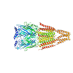
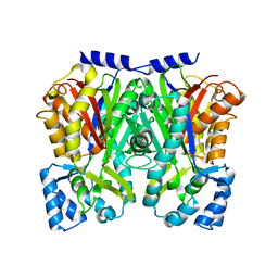
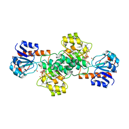
3AQB
 | | M. luteus B-P 26 heterodimeric hexaprenyl diphosphate synthase in complex with magnesium | | 分子名称: | CHLORIDE ION, Component A of hexaprenyl diphosphate synthase, Component B of hexaprenyl diphosphate synthase, ... | | 著者 | Sasaki, D, Fujihashi, M, Okuyama, N, Kobayashi, Y, Noike, M, Koyama, T, Miki, K. | | 登録日 | 2010-10-28 | | 公開日 | 2010-11-10 | | 最終更新日 | 2024-03-13 | | 実験手法 | X-RAY DIFFRACTION (2.4 Å) | | 主引用文献 | Crystal structure of heterodimeric hexaprenyl diphosphate synthase from Micrococcus luteus B-P 26 reveals that the small subunit is directly involved in the product chain length regulation.
J.Biol.Chem., 286, 2011
|
|
4AQ5
 | |
4AQ9
 | |
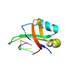
1UHU
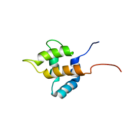 | | Solution structure of the retroviral Gag MA-like domain of RIKEN cDNA 3110009E22 | | 分子名称: | product of RIKEN cDNA 3110009E22 | | 著者 | Suzuki, S, Hatanaka, H, Inoue, M, Kigawa, T, Terada, T, Shirouzu, M, Hayashizaki, Y, Yokoyama, S, RIKEN Structural Genomics/Proteomics Initiative (RSGI) | | 登録日 | 2003-07-10 | | 公開日 | 2004-07-27 | | 最終更新日 | 2023-12-27 | | 実験手法 | SOLUTION NMR | | 主引用文献 | Solution structure of the retroviral Gag MA-like domain of RIKEN cDNA 3110009E22
To be Published
|
|
3WD8
 | | TypeIII polyketide synthases | | 分子名称: | GLYCEROL, Type III polyketide synthase quinolone synthase | | 著者 | Mori, T, Shimokawa, Y, Matsui, T, Morita, H, Abe, I. | | 登録日 | 2013-06-10 | | 公開日 | 2013-09-04 | | 最終更新日 | 2023-11-08 | | 実験手法 | X-RAY DIFFRACTION (2.463 Å) | | 主引用文献 | Cloning, characterization, and crystal structure analysis of novel type III polyketide synthases from Citrus microcarpa
To be Published
|
|
5YXM
 | | Crystal structure of Chlamydomonas Outer Arm Dynein Light Chain 1 | | 分子名称: | Dynein light chain 1, axonemal, PHOSPHATE ION | | 著者 | Toda, A, Tanaka, H, Nishikawa, Y, Yagi, T, Kurisu, G. | | 登録日 | 2017-12-06 | | 公開日 | 2018-03-14 | | 最終更新日 | 2023-11-22 | | 実験手法 | X-RAY DIFFRACTION (1.545 Å) | | 主引用文献 | Structural atlas of dynein motors at atomic resolution.
Biophys Rev, 10, 2018
|
|
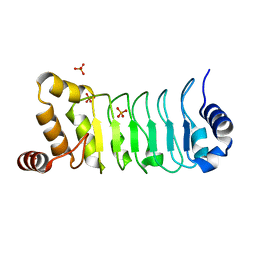
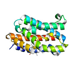
3A0U
 | | Crystal structure of response regulator protein TrrA (TM1360) from Thermotoga maritima in complex with Mg(2+)-BeF (wild type) | | 分子名称: | BERYLLIUM TRIFLUORIDE ION, MAGNESIUM ION, Response regulator, ... | | 著者 | Yamada, S, Sugimoto, H, Kobayashi, M, Ohno, A, Nakamura, H, Shiro, Y. | | 登録日 | 2009-03-24 | | 公開日 | 2009-10-20 | | 最終更新日 | 2023-11-01 | | 実験手法 | X-RAY DIFFRACTION (1.66 Å) | | 主引用文献 | Structure of PAS-linked histidine kinase and the response regulator complex
Structure, 17, 2009
|
|
3A10
 | | Crystal structure of response regulator protein TrrA (TM1360) from Thermotoga maritima in complex with Mg(2+)-BeF (SeMet, L89M) | | 分子名称: | BERYLLIUM TRIFLUORIDE ION, MAGNESIUM ION, Response regulator, ... | | 著者 | Yamada, S, Sugimoto, H, Kobayashi, M, Ohno, A, Nakamura, H, Shiro, Y. | | 登録日 | 2009-03-25 | | 公開日 | 2009-10-20 | | 最終更新日 | 2021-11-10 | | 実験手法 | X-RAY DIFFRACTION (1.63 Å) | | 主引用文献 | Structure of PAS-linked histidine kinase and the response regulator complex
Structure, 17, 2009
|
|
3AJ3
 | | Crystal structure of selenomethionine substituted 4-pyridoxolactonase from Mesorhizobium loti | | 分子名称: | 4-pyridoxolactonase, PHOSPHATE ION, ZINC ION | | 著者 | Kobayashi, J, Yoshikane, Y, Baba, S, Mikami, B, Yagi, T. | | 登録日 | 2010-05-21 | | 公開日 | 2011-05-25 | | 最終更新日 | 2024-03-20 | | 実験手法 | X-RAY DIFFRACTION (1.577 Å) | | 主引用文献 | Structure of 4-pyridoxolactonase from Mesorhizobium loti.
Acta Crystallogr.,Sect.F, 70, 2014
|
|
3X38
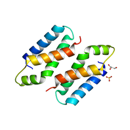 | | Crystal structure of the C-terminal domain of Sld7 | | 分子名称: | GLYCEROL, Mitochondrial morphogenesis protein SLD7, SULFATE ION | | 著者 | Itou, H, Araki, H, Shirakihara, Y. | | 登録日 | 2015-01-16 | | 公開日 | 2015-08-19 | | 最終更新日 | 2024-03-20 | | 実験手法 | X-RAY DIFFRACTION (1.801 Å) | | 主引用文献 | The quaternary structure of the eukaryotic DNA replication proteins Sld7 and Sld3.
Acta Crystallogr.,Sect.D, 71, 2015
|
|
3AQC
 | | M. luteus B-P 26 heterodimeric hexaprenyl diphosphate synthase in complex with magnesium and FPP analogue | | 分子名称: | (2E,6E)-7,11-dimethyldodeca-2,6,10-trien-1-yl trihydrogen diphosphate, CHLORIDE ION, Component A of hexaprenyl diphosphate synthase, ... | | 著者 | Sasaki, D, Fujihashi, M, Okuyama, N, Kobayashi, Y, Noike, M, Koyama, T, Miki, K. | | 登録日 | 2010-10-28 | | 公開日 | 2010-11-10 | | 最終更新日 | 2024-03-13 | | 実験手法 | X-RAY DIFFRACTION (2.61 Å) | | 主引用文献 | Crystal structure of heterodimeric hexaprenyl diphosphate synthase from Micrococcus luteus B-P 26 reveals that the small subunit is directly involved in the product chain length regulation.
J.Biol.Chem., 286, 2011
|
|
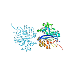
1VE9
 | | Porcine kidney D-amino acid oxidase | | 分子名称: | BENZOIC ACID, D-amino acid oxidase, FLAVIN-ADENINE DINUCLEOTIDE | | 著者 | Mizutani, H, Miyahara, I, Hirotsu, K, Nishina, Y, Shiga, K, Setoyama, C, Miura, R. | | 登録日 | 2004-03-29 | | 公開日 | 2004-04-13 | | 最終更新日 | 2023-12-27 | | 実験手法 | X-RAY DIFFRACTION (2.5 Å) | | 主引用文献 | Three-dimensional structure of porcine kidney D-amino acid oxidase at 3.0 A resolution.
J.Biochem., 120, 1996
|
|
1UFM
 | | Solution structure of the PCI domain | | 分子名称: | COP9 complex subunit 4 | | 著者 | Suzuki, S, Hatanaka, H, Kigawa, T, Shirouzu, M, Hayashizaki, Y, The RIKEN Genome Exploration Research Group Phase I & II Teams, Yokoyama, S, RIKEN Structural Genomics/Proteomics Initiative (RSGI) | | 登録日 | 2003-06-02 | | 公開日 | 2004-06-29 | | 最終更新日 | 2023-12-27 | | 実験手法 | SOLUTION NMR | | 主引用文献 | Solution structure of the PCI domain
To be Published
|
|
2ZOX
 | | Crystal Structure of the Covalent Intermediate of Human Cytosolic beta-Glucosidase | | 分子名称: | 4-nitrophenyl alpha-D-glucopyranoside, Cytosolic beta-glucosidase, GLYCEROL, ... | | 著者 | Noguchi, J, Hayashi, Y, Baba, Y, Okino, N, Kimura, M, Ito, M, Kakuta, Y. | | 登録日 | 2008-06-17 | | 公開日 | 2008-09-30 | | 最終更新日 | 2023-11-01 | | 実験手法 | X-RAY DIFFRACTION (1.9 Å) | | 主引用文献 | Crystal structure of the covalent intermediate of human cytosolic beta-glucosidase
Biochem.Biophys.Res.Commun., 374, 2008
|
|
1VD4
 | | Solution structure of the zinc finger domain of TFIIE alpha | | 分子名称: | Transcription initiation factor IIE, alpha subunit, ZINC ION | | 著者 | Okuda, M, Tanaka, A, Arai, Y, Satoh, M, Okamura, H, Nagadoi, A, Hanaoka, F, Ohkuma, Y, Nishimura, Y. | | 登録日 | 2004-03-18 | | 公開日 | 2004-10-05 | | 最終更新日 | 2023-12-27 | | 実験手法 | SOLUTION NMR | | 主引用文献 | A novel zinc finger structure in the large subunit of human general transcription factor TFIIE.
J.Biol.Chem., 279, 2004
|
|
2E5E
 | | Solution Structure of Variable-type Domain of Human Receptor for Advanced Glycation Endproducts | | 分子名称: | Advanced glycosylation end product-specific receptor | | 著者 | Matsumoto, S, Yoshida, T, Yasumatsu, I, Yamamoto, H, Kobayashi, Y, Ohkubo, T. | | 登録日 | 2006-12-20 | | 公開日 | 2007-12-25 | | 最終更新日 | 2022-03-09 | | 実験手法 | SOLUTION NMR | | 主引用文献 | Solution Structure of Variable-type Domain of Human Receptor for Advanced Glycation Endproducts
to be published
|
|
1X3U
 | | Solution structure of the C-terminal transcriptional activator domain of FixJ from Sinorhizobium melilot | | 分子名称: | Transcriptional regulatory protein fixJ | | 著者 | Kurashima-Ito, K, Kasai, Y, Hosono, K, Tamura, K, Oue, S, Isogai, M, Ito, Y, Nakamura, H, Shiro, Y. | | 登録日 | 2005-05-10 | | 公開日 | 2006-05-02 | | 最終更新日 | 2024-05-29 | | 実験手法 | SOLUTION NMR | | 主引用文献 | Solution structure of the C-terminal transcriptional activator domain of FixJ from Sinorhizobium meliloti and its recognition of the fixK promoter
Biochemistry, 44, 2005
|
|
1WTB
 | | Complex structure of the C-terminal RNA-binding domain of hnRNP D (AUF1) with telomere DNA | | 分子名称: | 5'-D(P*TP*AP*GP*G)-3', Heterogeneous nuclear ribonucleoprotein D0 | | 著者 | Enokizono, Y, Konishi, Y, Nagata, K, Ouhashi, K, Uesugi, S, Ishikawa, F, Katahira, M. | | 登録日 | 2004-11-22 | | 公開日 | 2005-04-05 | | 最終更新日 | 2024-05-29 | | 実験手法 | SOLUTION NMR | | 主引用文献 | Structure of hnRNP D complexed with single-stranded telomere DNA and unfolding of the quadruplex by heterogeneous nuclear ribonucleoprotein D
J.Biol.Chem., 280, 2005
|
|
1X0F
 | | Complex structure of the C-terminal RNA-binding domain of hnRNP D(AUF1) with telomeric DNA | | 分子名称: | 5'-D(P*TP*AP*GP*G)-3', Heterogeneous nuclear ribonucleoprotein D0 | | 著者 | Enokizono, Y, Konishi, Y, Nagata, K, Ouhashi, K, Uesugi, S, Ishikawa, F, Katahira, M. | | 登録日 | 2005-03-22 | | 公開日 | 2005-04-05 | | 最終更新日 | 2024-05-29 | | 実験手法 | SOLUTION NMR | | 主引用文献 | Structure of hnRNP D complexed with single-stranded telomere DNA and unfolding of the quadruplex by heterogeneous nuclear ribonucleoprotein D.
J.Biol.Chem., 280, 2005
|
|
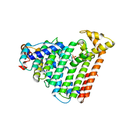
1UBB
 | | Crystal structure of rat HO-1 in complex with ferrous heme | | 分子名称: | Heme oxygenase 1, PROTOPORPHYRIN IX CONTAINING FE | | 著者 | Sugishima, M, Sakamoto, H, Higashimoto, Y, Noguchi, M, Fukuyama, K. | | 登録日 | 2003-04-03 | | 公開日 | 2003-09-02 | | 最終更新日 | 2023-10-25 | | 実験手法 | X-RAY DIFFRACTION (2.3 Å) | | 主引用文献 | Crystal Structures of Ferrous and CO-, CN(-)-, and NO-Bound Forms of Rat Heme Oxygenase-1 (HO-1) in Complex with Heme: Structural Implications for Discrimination between CO and O(2) in HO-1.
Biochemistry, 42, 2003
|
|
2DBR
 | | Crystal Structure of Glyoxylate Reductase (PH0597) from Pyrococcus horikoshii OT3, Complexed with NADP (P1) | | 分子名称: | Glyoxylate reductase, NADP NICOTINAMIDE-ADENINE-DINUCLEOTIDE PHOSPHATE, SULFATE ION | | 著者 | Yoshikawa, S, Arai, R, Kinoshita, Y, Uchikubo-Kamo, T, Akasaka, R, Terada, T, Shirouzu, M, Yokoyama, S, RIKEN Structural Genomics/Proteomics Initiative (RSGI) | | 登録日 | 2005-12-16 | | 公開日 | 2006-06-16 | | 最終更新日 | 2023-10-25 | | 実験手法 | X-RAY DIFFRACTION (2.61 Å) | | 主引用文献 | Structure of archaeal glyoxylate reductase from Pyrococcus horikoshii OT3 complexed with nicotinamide adenine dinucleotide phosphate.
Acta Crystallogr.,Sect.D, 63, 2007
|
|


