Loading
PDBj
MenuPDBj@FacebookPDBj@X(formerly Twitter)PDBj@BlueSkyPDBj@YouTubewwPDB FoundationwwPDB
RCSB PDBPDBeBMRBAdv. SearchSearch help
PDB: 6 results

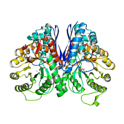
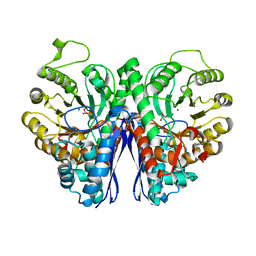
2XH7
DownloadVisualize
BU of 2xh7 by Molmil
Engineering the enolase active site pocket: Crystal structure of the D321A mutant of yeast enolase 1
Descriptor: 2-PHOSPHOGLYCERIC ACID, ENOLASE 1, MAGNESIUM ION
Authors:Schreier, B, Hocker, B.
Deposit date:2010-06-09
Release date:2010-08-25
Last modified:2023-12-20
Method:X-RAY DIFFRACTION (1.8 Å)
Cite:Engineering the Enolase Magnesium II Binding Site -Implications for its Evolution.
Biochemistry, 49, 2010
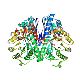
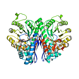
2XGZ
DownloadVisualize
BU of 2xgz by Molmil
Engineering the enolase active site pocket: Crystal structure of the S39N D321R mutant of yeast enolase 1
Descriptor: ENOLASE 1, MAGNESIUM ION, PHOSPHOENOLPYRUVATE
Authors:Schreier, B, Hocker, B.
Deposit date:2010-06-08
Release date:2010-08-25
Last modified:2023-12-20
Method:X-RAY DIFFRACTION (1.8 Å)
Cite:Engineering the Enolase Magnesium II Binding Site -Implications for its Evolution.
Biochemistry, 49, 2010
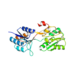
2WRZ
DownloadVisualize
BU of 2wrz by Molmil
Crystal structure of an arabinose binding protein with designed serotonin binding site in open, ligand-free state
Descriptor: L-ARABINOSE-BINDING PERIPLASMIC PROTEIN
Authors:Schreier, B, Stumpp, C, Wiesner, S, Hocker, B.
Deposit date:2009-09-03
Release date:2009-10-13
Last modified:2023-12-20
Method:X-RAY DIFFRACTION (2.2 Å)
Cite:The Computational Design of Ligand Binding is not a Solved Problem
Proc.Natl.Acad.Sci.USA, 106, 2009
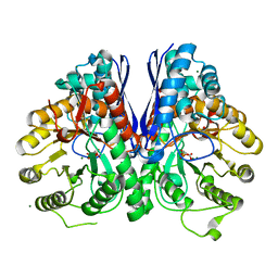
2XH2
DownloadVisualize
BU of 2xh2 by Molmil
Engineering the enolase active site pocket: Crystal structure of the S39N D321A mutant of yeast enolase 1
Descriptor: 2-PHOSPHOGLYCERIC ACID, ENOLASE 1, MAGNESIUM ION
Authors:Schreier, B, Hocker, B.
Deposit date:2010-06-08
Release date:2010-08-25
Last modified:2023-12-20
Method:X-RAY DIFFRACTION (1.8 Å)
Cite:Engineering the Enolase Magnesium II Binding Site -Implications for its Evolution.
Biochemistry, 49, 2010
2XH0
DownloadVisualize
BU of 2xh0 by Molmil
Engineering the enolase active site pocket: Crystal structure of the S39N Q167K D321R mutant of yeast enolase 1
Descriptor: ENOLASE 1, MAGNESIUM ION, PHOSPHOENOLPYRUVATE
Authors:Schreier, B, Hocker, B.
Deposit date:2010-06-08
Release date:2010-08-25
Last modified:2023-12-20
Method:X-RAY DIFFRACTION (1.7 Å)
Cite:Engineering the Enolase Magnesium II Binding Site -Implications for its Evolution.
Biochemistry, 49, 2010
2XH4
DownloadVisualize
BU of 2xh4 by Molmil
Engineering the enolase active site pocket: Crystal structure of the S39A D321A mutant of yeast enolase 1
Descriptor: 2-PHOSPHOGLYCERIC ACID, ENOLASE 1, MAGNESIUM ION
Authors:Schreier, B, Hocker, B.
Deposit date:2010-06-09
Release date:2010-08-25
Last modified:2023-12-20
Method:X-RAY DIFFRACTION (1.7 Å)
Cite:Engineering the Enolase Magnesium II Binding Site -Implications for its Evolution.
Biochemistry, 49, 2010

227561

PDB entries from 2024-11-20

PDB statisticsPDBj update infoContact PDBjnumon