Loading
PDBj
MenuPDBj@FacebookPDBj@TwitterPDBj@YouTubewwPDB FoundationwwPDB
RCSB PDBPDBeBMRBAdv. SearchSearch help
PDB: 424 results

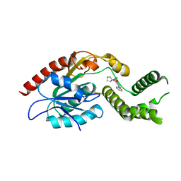
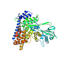
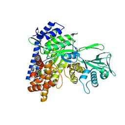
5CJN
DownloadVisualize
BU of 5cjn by Molmil
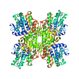
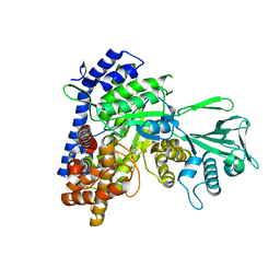
Crystal structure of Mycobacterium tuberculosis malate synthase in complex with 3-(3-oxo-3,4-dihydroquinoxalin-2-yl)acrylate
Descriptor: 3-(3-oxo-3,4-dihydroquinoxalin-2-yl)propanoic acid, MAGNESIUM ION, Malate synthase G
Authors:Huang, H.-L, Sacchettini, J.C.
Deposit date:2015-07-14
Release date:2016-08-10
Last modified:2023-11-15
Method:X-RAY DIFFRACTION (2.19 Å)
Cite:Mycobacterium tuberculosis Malate Synthase Structures with Fragments Reveal a Portal for Substrate/Product Exchange.
J. Biol. Chem., 291, 2016
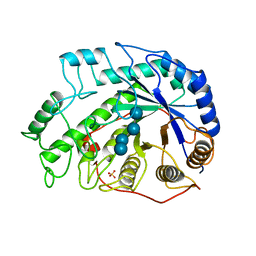
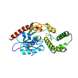
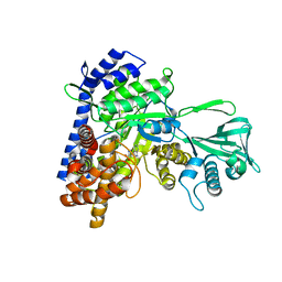
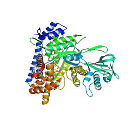
1BYB
DownloadVisualize
BU of 1byb by Molmil
CRYSTAL STRUCTURES OF SOYBEAN BETA-AMYLASE REACTED WITH BETA-MALTOSE AND MALTAL: ACTIVE SITE COMPONENTS AND THEIR APPARENT ROLE IN CATALYSIS
Descriptor: BETA-AMYLASE, SULFATE ION, alpha-D-glucopyranose-(1-4)-alpha-D-glucopyranose-(1-4)-alpha-D-glucopyranose-(1-4)-alpha-D-glucopyranose
Authors:Mikami, B, Degano, M, Hehre, E.J, Sacchettini, J.C.
Deposit date:1994-01-25
Release date:1994-07-31
Last modified:2024-02-07
Method:X-RAY DIFFRACTION (1.9 Å)
Cite:Crystal structures of soybean beta-amylase reacted with beta-maltose and maltal: active site components and their apparent roles in catalysis.
Biochemistry, 33, 1994
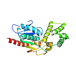
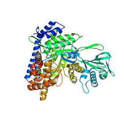
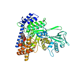
1BYC
DownloadVisualize
BU of 1byc by Molmil
CRYSTAL STRUCTURES OF SOYBEAN BETA-AMYLASE REACTED WITH BETA-MALTOSE AND MALTAL: ACTIVE SITE COMPONENTS AND THEIR APPARENT ROLE IN CATALYSIS
Descriptor: BETA-AMYLASE, SULFATE ION, alpha-D-glucopyranose-(1-4)-alpha-D-glucopyranose-(1-4)-alpha-D-glucopyranose-(1-4)-beta-D-glucopyranose
Authors:Mikami, B, Degano, M, Hehre, E.J, Sacchettini, J.C.
Deposit date:1994-01-25
Release date:1994-07-31
Last modified:2024-02-07
Method:X-RAY DIFFRACTION (2.2 Å)
Cite:Crystal structures of soybean beta-amylase reacted with beta-maltose and maltal: active site components and their apparent roles in catalysis.
Biochemistry, 33, 1994
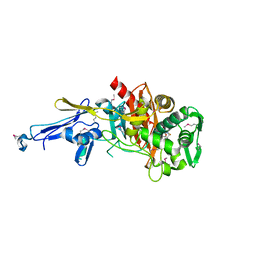
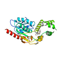
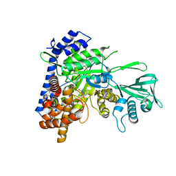
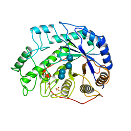
4N1X
DownloadVisualize
BU of 4n1x by Molmil
Structure of a putative peptidoglycan glycosyltransferase from Atopobium parvulum in complex with penicillin G
Descriptor: OPEN FORM - PENICILLIN G, Peptidoglycan glycosyltransferase
Authors:Filippova, E.V, Minasov, G, Shuvalova, L, Kiryukhina, O, Babnigg, G, Rubin, E, Sacchettini, J, Joachimiak, A, Anderson, W.F, Midwest Center for Structural Genomics (MCSG), Structures of Mtb Proteins Conferring Susceptibility to Known Mtb Inhibitors (MTBI)
Deposit date:2013-10-04
Release date:2013-10-30
Last modified:2023-12-06
Method:X-RAY DIFFRACTION (2 Å)
Cite:Structure of a putative peptidoglycan glycosyltransferase from Atopobium parvulum in complex with penicillin G
To be Published
2Z9I
DownloadVisualize
BU of 2z9i by Molmil
Crystal structure of RV0983 from Mycobacterium tuberculosis- Proteolytically active form
Descriptor: GATV, PROBABLE SERINE PROTEASE PEPD, SVEQV
Authors:Palaninathan, S.K, Mohamedmohaideen, N.N, Sacchettini, J.C.
Deposit date:2007-09-20
Release date:2008-06-10
Last modified:2011-07-13
Method:X-RAY DIFFRACTION (2 Å)
Cite:Structure and function of the virulence-associated high-temperature requirement A of Mycobacterium tuberculosis
Biochemistry, 47, 2008
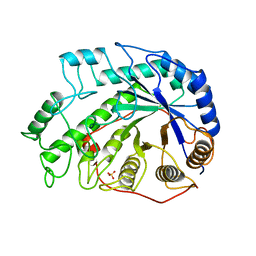
1BYD
DownloadVisualize
BU of 1byd by Molmil
CRYSTAL STRUCTURES OF SOYBEAN BETA-AMYLASE REACTED WITH BETA-MALTOSE AND MALTAL: ACTIVE SITE COMPONENTS AND THEIR APPARENT ROLE IN CATALYSIS
Descriptor: BETA-AMYLASE, SULFATE ION, alpha-D-glucopyranose-(1-4)-2-deoxy-beta-D-arabino-hexopyranose
Authors:Mikami, B, Degano, M, Hehre, E.J, Sacchettini, J.C.
Deposit date:1994-01-25
Release date:1994-07-31
Last modified:2024-02-07
Method:X-RAY DIFFRACTION (2.2 Å)
Cite:Crystal structures of soybean beta-amylase reacted with beta-maltose and maltal: active site components and their apparent roles in catalysis.
Biochemistry, 33, 1994
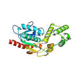
5V3Z
DownloadVisualize
BU of 5v3z by Molmil
Crystal Structure of the D1607N mutant form of Thioesterase domain of Mtb Pks13
Descriptor: PENTAETHYLENE GLYCOL, Polyketide synthase Pks13 (Termination polyketide synthase), SULFATE ION
Authors:Aggarwal, A, Sacchettini, J.C.
Deposit date:2017-03-08
Release date:2017-07-05
Last modified:2023-10-04
Method:X-RAY DIFFRACTION (1.881 Å)
Cite:Development of a Novel Lead that Targets M. tuberculosis Polyketide Synthase 13.
Cell, 170, 2017
5V42
DownloadVisualize
BU of 5v42 by Molmil
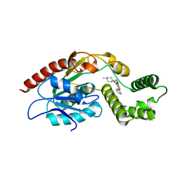
Crystal Structure of Mtb Pks13 Thioesterase domain in complex with inhibitor TAM3
Descriptor: Polyketide synthase Pks13 (Termination polyketide synthase), ethyl 5-hydroxy-2-phenyl-4-(pyrrolidin-1-ylmethyl)-1-benzofuran-3-carboxylate
Authors:Aggarwal, A, Sacchettini, J.C.
Deposit date:2017-03-08
Release date:2017-07-05
Last modified:2023-10-04
Method:X-RAY DIFFRACTION (1.987 Å)
Cite:Development of a Novel Lead that Targets M. tuberculosis Polyketide Synthase 13.
Cell, 170, 2017
5V3Y
DownloadVisualize
BU of 5v3y by Molmil
Crystal Structure of Mtb Pks13 Thioesterase domain in complex with inhibitor TAM16
Descriptor: 2-(4-hydroxyphenyl)-~{N}-methyl-5-oxidanyl-4-(piperidin-1-ylmethyl)-1-benzofuran-3-carboxamide, Polyketide synthase Pks13 (Termination polyketide synthase), SULFATE ION
Authors:Aggarwal, A, Sacchettini, J.C.
Deposit date:2017-03-08
Release date:2017-07-05
Last modified:2023-10-04
Method:X-RAY DIFFRACTION (1.98 Å)
Cite:Development of a Novel Lead that Targets M. tuberculosis Polyketide Synthase 13.
Cell, 170, 2017
5V3W
DownloadVisualize
BU of 5v3w by Molmil
Crystal Structure of the Apo form of Thioesterase domain of Mtb Pks13
Descriptor: (2R)-2-{[(2R)-2-{[(2S)-2-{[(2R)-2-hydroxypropyl]oxy}propyl]oxy}propyl]oxy}propan-1-ol, Polyketide synthase Pks13 (Termination polyketide synthase)
Authors:Aggarwal, A, Sacchettini, J.C.
Deposit date:2017-03-08
Release date:2017-07-05
Last modified:2023-10-04
Method:X-RAY DIFFRACTION (1.723 Å)
Cite:Development of a Novel Lead that Targets M. tuberculosis Polyketide Synthase 13.
Cell, 170, 2017
5V41
DownloadVisualize
BU of 5v41 by Molmil
Crystal Structure of Mtb Pks13 Thioesterase domain in complex with inhibitor TAM5
Descriptor: Polyketide synthase Pks13 (Termination polyketide synthase), ethyl 5-hydroxy-4-(morpholin-4-ylmethyl)-2-phenyl-1-benzofuran-3-carboxylate
Authors:Aggarwal, A, Sacchettini, J.C.
Deposit date:2017-03-08
Release date:2017-07-05
Last modified:2023-10-04
Method:X-RAY DIFFRACTION (2.051 Å)
Cite:Development of a Novel Lead that Targets M. tuberculosis Polyketide Synthase 13.
Cell, 170, 2017
5V3X
DownloadVisualize
BU of 5v3x by Molmil
Crystal Structure of Mtb Pks13 Thioesterase domain in complex with inhibitor TAM1
Descriptor: Polyketide synthase Pks13 (Termination polyketide synthase), ethyl 5-hydroxy-4-[(4-methylpiperidin-1-yl)methyl]-2-phenyl-1-benzofuran-3-carboxylate
Authors:Aggarwal, A, Sacchettini, J.C.
Deposit date:2017-03-08
Release date:2017-07-05
Last modified:2023-10-04
Method:X-RAY DIFFRACTION (1.936 Å)
Cite:Development of a Novel Lead that Targets M. tuberculosis Polyketide Synthase 13.
Cell, 170, 2017
3BV0
DownloadVisualize
BU of 3bv0 by Molmil
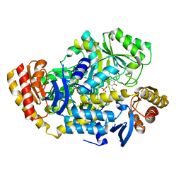
Crystal Structure of PLP Bound 7,8-Diaminopelargonic Acid Synthase in Mycobacterium Tuberculosis
Descriptor: Adenosylmethionine-8-amino-7-oxononanoate aminotransferase, PYRIDOXAL-5'-PHOSPHATE
Authors:Dey, S, Sacchettini, J.C.
Deposit date:2008-01-04
Release date:2008-02-05
Last modified:2023-08-30
Method:X-RAY DIFFRACTION (2.21 Å)
Cite:Structural characterization of the Mycobacterium tuberculosis biotin biosynthesis enzymes 7,8-diaminopelargonic acid synthase and dethiobiotin synthetase .
Biochemistry, 49, 2010
2ZIZ
DownloadVisualize
BU of 2ziz by Molmil
Crystal structure of Mycobacterium tuberculosis S-adenosyl-L-homocysteine hydrolase in ternary complex with NAD and 3-deazaadenosine
Descriptor: 3-DEAZA-ADENOSINE, Adenosylhomocysteinase, NICOTINAMIDE-ADENINE-DINUCLEOTIDE
Authors:Reddy, M.C.M, Gokulan, K, Shetty, N.D, Owen, J.L, Ioerger, T.R, Sacchettini, J.C.
Deposit date:2008-02-29
Release date:2008-09-16
Last modified:2023-08-30
Method:X-RAY DIFFRACTION (2.2 Å)
Cite:Crystal structures of Mycobacterium tuberculosis S-adenosyl-L-homocysteine hydrolase in ternary complex with substrate and inhibitors.
Protein Sci., 17, 2008
6OYY
DownloadVisualize
BU of 6oyy by Molmil
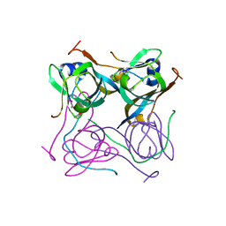
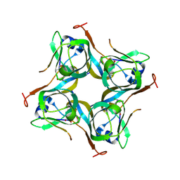
Crystal structure of Mtb aspartate decarboxylase, pyrazinoic acid complex
Descriptor: Aspartate 1-decarboxylase alpha chain, Aspartate 1-decarboxylase beta chain, PYRAZINE-2-CARBOXYLIC ACID
Authors:Sun, Q, Li, X, Sacchettini, J.C, TB Structural Genomics Consortium (TBSGC)
Deposit date:2019-05-15
Release date:2020-02-05
Last modified:2023-11-15
Method:X-RAY DIFFRACTION (2.7 Å)
Cite:The molecular basis of pyrazinamide activity on Mycobacterium tuberculosis PanD.
Nat Commun, 11, 2020
6P02
DownloadVisualize
BU of 6p02 by Molmil
Crystal structure of Mtb aspartate decarboxylase, 6-Chlorine pyrazinoic acid complex
Descriptor: 6-chloropyrazine-2-carboxylic acid, Aspartate 1-decarboxylase alpha chain, Aspartate 1-decarboxylase beta chain
Authors:Sun, Q, Li, X, Sacchettini, J.C, TB Structural Genomics Consortium (TBSGC)
Deposit date:2019-05-16
Release date:2020-02-05
Last modified:2023-11-15
Method:X-RAY DIFFRACTION (2.25 Å)
Cite:The molecular basis of pyrazinamide activity on Mycobacterium tuberculosis PanD.
Nat Commun, 11, 2020
5CAH
DownloadVisualize
BU of 5cah by Molmil
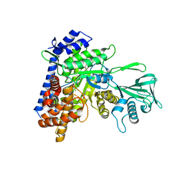
Crystal structure of Mycobacterium tuberculosis malate synthase in complex with 6H-thieno[2,3-b]pyrrole-5-carboxylic acid
Descriptor: 6H-thieno[2,3-b]pyrrole-5-carboxylic acid, MAGNESIUM ION, Malate synthase G
Authors:Huang, H.-L, Sacchettini, J.C.
Deposit date:2015-06-29
Release date:2016-08-10
Last modified:2023-09-27
Method:X-RAY DIFFRACTION (2.303 Å)
Cite:Mycobacterium tuberculosis Malate Synthase Structures with Fragments Reveal a Portal for Substrate/Product Exchange.
J. Biol. Chem., 291, 2016
5C7V
DownloadVisualize
BU of 5c7v by Molmil
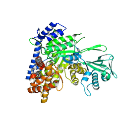
Crystal structure of Mycobacterium tuberculosis malate synthase in complex with 1H-pyrrole-2-carboxylic acid
Descriptor: MAGNESIUM ION, Malate synthase G, PYRROLE-2-CARBOXYLATE
Authors:Huang, H.-L, Sacchettini, J.C.
Deposit date:2015-06-25
Release date:2016-08-10
Last modified:2023-09-27
Method:X-RAY DIFFRACTION (2.504 Å)
Cite:Mycobacterium tuberculosis Malate Synthase Structures with Fragments Reveal a Portal for Substrate/Product Exchange.
J. Biol. Chem., 291, 2016
5CC5
DownloadVisualize
BU of 5cc5 by Molmil
Crystal structure of Mycobacterium tuberculosis malate synthase in complex with 1H-indole-3-carboxylic acid
Descriptor: 1H-INDOLE-3-CARBOXYLIC ACID, GLYOXYLIC ACID, MAGNESIUM ION, ...
Authors:Huang, H.-L, Sacchettini, J.C.
Deposit date:2015-07-01
Release date:2016-08-10
Last modified:2023-09-27
Method:X-RAY DIFFRACTION (2.14 Å)
Cite:Crystal structures of fragment-bound malate synthase from Mycobacterium tuberculosis provide insights into mechanism and potential inhibitor designs
To Be Published
5C9X
DownloadVisualize
BU of 5c9x by Molmil
Crystal structure of Mycobacterium tuberculosis malate synthase in complex with 2,4-dichloro-5-fluorobenzoic acid
Descriptor: 2,4-dichloro-5-fluorobenzoic acid, MAGNESIUM ION, Malate synthase G
Authors:Huang, H.-L, Sacchettini, J.C.
Deposit date:2015-06-29
Release date:2016-08-10
Last modified:2023-09-27
Method:X-RAY DIFFRACTION (2.1 Å)
Cite:Mycobacterium tuberculosis Malate Synthase Structures with Fragments Reveal a Portal for Substrate/Product Exchange.
J. Biol. Chem., 291, 2016
5CBB
DownloadVisualize
BU of 5cbb by Molmil
Crystal structure of Mycobacterium tuberculosis malate synthase in complex with 5-(3H-indol-3-ylidene)-2,5-dihydro-1H-pyrazole-3-carboxylate
Descriptor: (5E)-5-(3H-indol-3-ylidene)-2,5-dihydro-1H-pyrazole-3-carboxylic acid, MAGNESIUM ION, Malate synthase G
Authors:Huang, H.-L, Sacchettini, J.C.
Deposit date:2015-06-30
Release date:2016-08-10
Last modified:2023-09-27
Method:X-RAY DIFFRACTION (2.012 Å)
Cite:Mycobacterium tuberculosis Malate Synthase Structures with Fragments Reveal a Portal for Substrate/Product Exchange.
J. Biol. Chem., 291, 2016
5CC3
DownloadVisualize
BU of 5cc3 by Molmil
Crystal structure of Mycobacterium tuberculosis malate synthase in complex with 6-bromo-1H-indole-2-carboxylic acid
Descriptor: 6-bromanyl-1H-indole-2-carboxylic acid, MAGNESIUM ION, Malate synthase G
Authors:Huang, H.-L, Sacchettini, J.C.
Deposit date:2015-07-01
Release date:2016-08-10
Last modified:2023-09-27
Method:X-RAY DIFFRACTION (2.204 Å)
Cite:Crystal structures of fragment-bound malate synthase from Mycobacterium tuberculosis provide insights into mechanism and potential inhibitor designs
To Be Published
5C9W
DownloadVisualize
BU of 5c9w by Molmil
Crystal structure of Mycobacterium tuberculosis malate synthase in complex with (Z)-N-(2-bromophenyl)-2-(hydroxyimino)acetamide
Descriptor: (2E)-N-(2-bromophenyl)-2-(hydroxyimino)acetamide, MAGNESIUM ION, Malate synthase G
Authors:Huang, H.-L, Sacchettini, J.C.
Deposit date:2015-06-29
Release date:2016-08-10
Last modified:2023-09-27
Method:X-RAY DIFFRACTION (2.0928 Å)
Cite:Mycobacterium tuberculosis Malate Synthase Structures with Fragments Reveal a Portal for Substrate/Product Exchange.
J. Biol. Chem., 291, 2016
5CC7
DownloadVisualize
BU of 5cc7 by Molmil
Crystal structure of Mycobacterium tuberculosis malate synthase in complex with 1H-indole-6-carboxylic acid
Descriptor: 1H-indole-6-carboxylic acid, MAGNESIUM ION, Malate synthase G
Authors:Huang, H.-L, Sacchettini, J.C.
Deposit date:2015-07-01
Release date:2016-08-10
Last modified:2023-09-27
Method:X-RAY DIFFRACTION (2.12 Å)
Cite:Crystal structures of fragment-bound malate synthase from Mycobacterium tuberculosis provide insights into mechanism and potential inhibitor designs
To Be Published
5CEW
DownloadVisualize
BU of 5cew by Molmil
Crystal structure of Mycobacterium tuberculosis malate synthase in complex with 2-(pyridin-4-yl)thiazolidine-4-carboxylic acid
Descriptor: (2S,4S)-2-(pyridin-4-yl)-1,3-thiazolidine-4-carboxylic acid, MAGNESIUM ION, Malate synthase G
Authors:Huang, H.-L, Sacchettini, J.C.
Deposit date:2015-07-07
Release date:2016-08-10
Last modified:2023-09-27
Method:X-RAY DIFFRACTION (2.03 Å)
Cite:Mycobacterium tuberculosis Malate Synthase Structures with Fragments Reveal a Portal for Substrate/Product Exchange.
J. Biol. Chem., 291, 2016

224004

數據於2024-08-21公開中

PDB statisticsPDBj update infoContact PDBjnumon