Loading
PDBj
MenuPDBj@FacebookPDBj@X(formerly Twitter)PDBj@BlueSkyPDBj@YouTubewwPDB FoundationwwPDBDonate
RCSB PDBPDBeBMRBAdv. SearchSearch help
PDB: 426 results

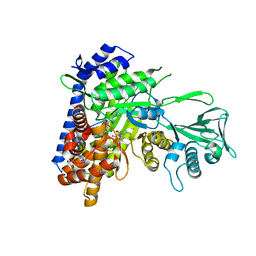
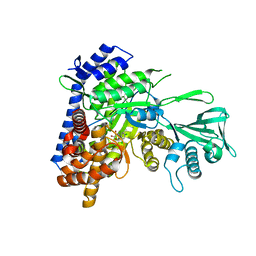
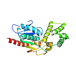
5V3Y
DownloadVisualize
BU of 5v3y by Molmil
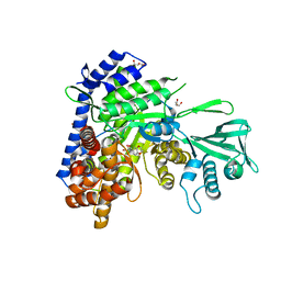
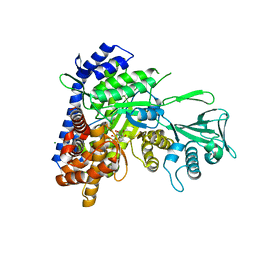
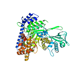
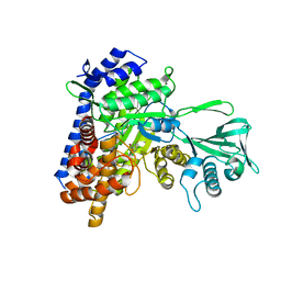
Crystal Structure of Mtb Pks13 Thioesterase domain in complex with inhibitor TAM16
Descriptor: 2-(4-hydroxyphenyl)-~{N}-methyl-5-oxidanyl-4-(piperidin-1-ylmethyl)-1-benzofuran-3-carboxamide, Polyketide synthase Pks13 (Termination polyketide synthase), SULFATE ION
Authors:Aggarwal, A, Sacchettini, J.C.
Deposit date:2017-03-08
Release date:2017-07-05
Last modified:2023-10-04
Method:X-RAY DIFFRACTION (1.98 Å)
Cite:Development of a Novel Lead that Targets M. tuberculosis Polyketide Synthase 13.
Cell, 170, 2017
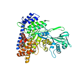
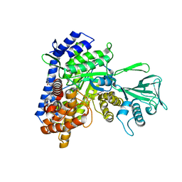
5V3W
DownloadVisualize
BU of 5v3w by Molmil
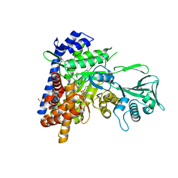
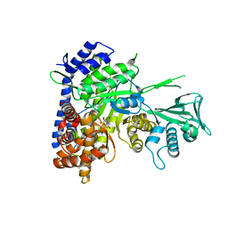
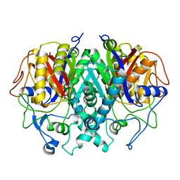
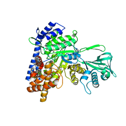
Crystal Structure of the Apo form of Thioesterase domain of Mtb Pks13
Descriptor: (2R)-2-{[(2R)-2-{[(2S)-2-{[(2R)-2-hydroxypropyl]oxy}propyl]oxy}propyl]oxy}propan-1-ol, Polyketide synthase Pks13 (Termination polyketide synthase)
Authors:Aggarwal, A, Sacchettini, J.C.
Deposit date:2017-03-08
Release date:2017-07-05
Last modified:2023-10-04
Method:X-RAY DIFFRACTION (1.723 Å)
Cite:Development of a Novel Lead that Targets M. tuberculosis Polyketide Synthase 13.
Cell, 170, 2017
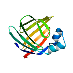
1KZX
DownloadVisualize
BU of 1kzx by Molmil
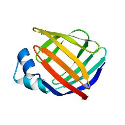
Solution structure of human intestinal fatty acid binding protein with a naturally-occurring single amino acid substitution (A54T)
Descriptor: INTESTINAL FATTY ACID-BINDING PROTEIN (T54)
Authors:Zhang, F, Luecke, C, Baier, L.J, Sacchettini, J.C, Hamilton, J.A.
Deposit date:2002-02-08
Release date:2003-07-01
Last modified:2024-05-22
Method:SOLUTION NMR
Cite:Solution structure of human intestinal fatty acid binding protein with a naturally-occurring single amino acid substitution (A54T) that is associated with altered lipid metabolism
Biochemistry, 42, 2003
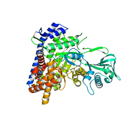
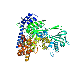
6AU9
DownloadVisualize
BU of 6au9 by Molmil
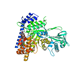
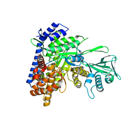
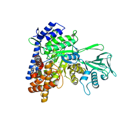
Crystal structure of Mycobacterium tuberculosis malate synthase in complex with dioxine-phenyldiketoacid
Descriptor: (2Z)-4-(2,3-dihydro-1,4-benzodioxin-6-yl)-2-hydroxy-4-oxobut-2-enoic acid, DI(HYDROXYETHYL)ETHER, MAGNESIUM ION, ...
Authors:Krieger, I.V, Sacchettini, J.C, TB Structural Genomics Consortium (TBSGC)
Deposit date:2017-08-31
Release date:2018-09-05
Last modified:2023-10-04
Method:X-RAY DIFFRACTION (2.1 Å)
Cite:Anion-pi Interactions in Computer-Aided Drug Design: Modeling the Inhibition of Malate Synthase by Phenyl-Diketo Acids.
J Chem Inf Model, 58, 2018
6APZ
DownloadVisualize
BU of 6apz by Molmil
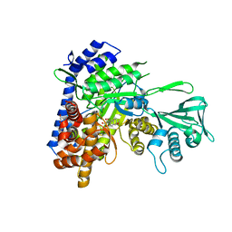
Crystal structure of Mycobacterium tuberculosis malate synthase in complex with 3-hydroxy-phenyldiketoacid
Descriptor: (2Z)-2-hydroxy-4-(3-hydroxyphenyl)-4-oxobut-2-enoic acid, DI(HYDROXYETHYL)ETHER, MAGNESIUM ION, ...
Authors:Krieger, I.V, Sacchettini, J.C, TB Structural Genomics Consortium (TBSGC)
Deposit date:2017-08-18
Release date:2018-08-22
Last modified:2023-10-04
Method:X-RAY DIFFRACTION (2.254 Å)
Cite:Anion-pi Interactions in Computer-Aided Drug Design: Modeling the Inhibition of Malate Synthase by Phenyl-Diketo Acids.
J Chem Inf Model, 58, 2018
6ASU
DownloadVisualize
BU of 6asu by Molmil
Crystal structure of Mycobacterium tuberculosis malate synthase in complex with 4-methyl-phenyldiketoacid
Descriptor: (2Z)-2-hydroxy-4-(4-methylphenyl)-4-oxobut-2-enoic acid, MAGNESIUM ION, Malate synthase G, ...
Authors:Krieger, I.V, Sacchettini, J.C, TB Structural Genomics Consortium (TBSGC)
Deposit date:2017-08-25
Release date:2018-08-29
Last modified:2023-10-04
Method:X-RAY DIFFRACTION (2.316 Å)
Cite:Anion-pi Interactions in Computer-Aided Drug Design: Modeling the Inhibition of Malate Synthase by Phenyl-Diketo Acids.
J Chem Inf Model, 58, 2018
6AS6
DownloadVisualize
BU of 6as6 by Molmil
Crystal structure of Mycobacterium tuberculosis malate synthase in complex with 3-Prop-6-Me-phenyldiketoacid
Descriptor: (2Z)-4-(5-cyclopropyl-2-methylphenyl)-2-hydroxy-4-oxobut-2-enoic acid, DI(HYDROXYETHYL)ETHER, MAGNESIUM ION, ...
Authors:Krieger, I.V, Sacchettini, J.C, TB Structural Genomics Consortium (TBSGC)
Deposit date:2017-08-23
Release date:2018-08-29
Last modified:2023-10-04
Method:X-RAY DIFFRACTION (1.4 Å)
Cite:Anion-pi Interactions in Computer-Aided Drug Design: Modeling the Inhibition of Malate Synthase by Phenyl-Diketo Acids.
J Chem Inf Model, 58, 2018
1KZW
DownloadVisualize
BU of 1kzw by Molmil
Solution structure of Human Intestinal Fatty acid binding protein
Descriptor: INTESTINAL FATTY ACID-BINDING PROTEIN (A54)
Authors:Zhang, F, Luecke, C, Baier, L.J, Sacchettini, J.C, Hamilton, J.A.
Deposit date:2002-02-08
Release date:2003-07-01
Last modified:2024-05-22
Method:SOLUTION NMR
Cite:Solution structure of human intestinal fatty acid binding protein with a naturally-occurring single amino acid substitution (A54T) that is associated with altered lipid metabolism
Biochemistry, 42, 2003
6BA7
DownloadVisualize
BU of 6ba7 by Molmil
Crystal structure of Mycobacterium tuberculosis malate synthase in complex with 2-Cl-4-OH-phenyldiketoacid
Descriptor: (2Z)-4-(2-chloro-4-hydroxyphenyl)-2-hydroxy-4-oxobut-2-enoic acid, MAGNESIUM ION, Malate synthase G
Authors:Krieger, I.V, Sacchettini, J.C, TB Structural Genomics Consortium (TBSGC)
Deposit date:2017-10-12
Release date:2018-09-05
Last modified:2023-10-04
Method:X-RAY DIFFRACTION (2.496 Å)
Cite:Anion-pi Interactions in Computer-Aided Drug Design: Modeling the Inhibition of Malate Synthase by Phenyl-Diketo Acids.
J Chem Inf Model, 58, 2018
6AXB
DownloadVisualize
BU of 6axb by Molmil
Crystal structure of Mycobacterium tuberculosis malate synthase in complex with 2-naphthyldiketoacid
Descriptor: (2Z)-2-hydroxy-4-(naphthalen-2-yl)-4-oxobut-2-enoic acid, DI(HYDROXYETHYL)ETHER, MAGNESIUM ION, ...
Authors:Krieger, I.V, Sacchettini, J.C, TB Structural Genomics Consortium (TBSGC)
Deposit date:2017-09-06
Release date:2018-09-05
Last modified:2023-10-04
Method:X-RAY DIFFRACTION (1.8 Å)
Cite:Anion-pi Interactions in Computer-Aided Drug Design: Modeling the Inhibition of Malate Synthase by Phenyl-Diketo Acids.
J Chem Inf Model, 58, 2018
6BU1
DownloadVisualize
BU of 6bu1 by Molmil
Crystal structure of Mycobacterium tuberculosis malate synthase in complex with 2-Br-3-OH-phenyldiketoacid
Descriptor: (2Z)-4-(2-bromo-3-hydroxyphenyl)-2-hydroxy-4-oxobut-2-enoic acid, MAGNESIUM ION, Malate synthase G
Authors:Krieger, I.V, Sacchettini, J.C, TB Structural Genomics Consortium (TBSGC)
Deposit date:2017-12-08
Release date:2018-09-05
Last modified:2023-10-04
Method:X-RAY DIFFRACTION (1.584 Å)
Cite:Anion-pi Interactions in Computer-Aided Drug Design: Modeling the Inhibition of Malate Synthase by Phenyl-Diketo Acids.
J Chem Inf Model, 58, 2018
6C6O
DownloadVisualize
BU of 6c6o by Molmil
Crystal structure of Mycobacterium tuberculosis malate synthase in complex with 2-Br-4-OH-phenyldiketoacid
Descriptor: (2Z)-4-(2-bromo-4-hydroxyphenyl)-2-hydroxy-4-oxobut-2-enoic acid, MAGNESIUM ION, Malate synthase G
Authors:Krieger, I.V, Sacchettini, J.C, TB Structural Genomics Consortium (TBSGC)
Deposit date:2018-01-19
Release date:2018-09-05
Last modified:2023-10-04
Method:X-RAY DIFFRACTION (2.3 Å)
Cite:Anion-pi Interactions in Computer-Aided Drug Design: Modeling the Inhibition of Malate Synthase by Phenyl-Diketo Acids.
J Chem Inf Model, 58, 2018
6C8P
DownloadVisualize
BU of 6c8p by Molmil
Crystal structure of Mycobacterium tuberculosis malate synthase in complex with 2-F-phenyldiketoacid
Descriptor: (2Z)-4-(2-fluorophenyl)-2-hydroxy-4-oxobut-2-enoic acid, DI(HYDROXYETHYL)ETHER, MAGNESIUM ION, ...
Authors:Krieger, I.V, Sacchettini, J.C, TB Structural Genomics Consortium (TBSGC)
Deposit date:2018-01-25
Release date:2018-09-05
Last modified:2023-10-04
Method:X-RAY DIFFRACTION (1.635 Å)
Cite:Anion-pi Interactions in Computer-Aided Drug Design: Modeling the Inhibition of Malate Synthase by Phenyl-Diketo Acids.
J Chem Inf Model, 58, 2018
6C7B
DownloadVisualize
BU of 6c7b by Molmil
Crystal structure of Mycobacterium tuberculosis malate synthase in complex with Methoxynaphthyldiketoacid
Descriptor: (2Z)-2-hydroxy-4-(6-methoxynaphthalen-2-yl)-4-oxobut-2-enoic acid, DI(HYDROXYETHYL)ETHER, MAGNESIUM ION, ...
Authors:Krieger, I.V, Sacchettini, J.C, TB Structural Genomics Consortium (TBSGC)
Deposit date:2018-01-22
Release date:2018-09-05
Last modified:2023-10-04
Method:X-RAY DIFFRACTION (2.133 Å)
Cite:Anion-pi Interactions in Computer-Aided Drug Design: Modeling the Inhibition of Malate Synthase by Phenyl-Diketo Acids.
J Chem Inf Model, 58, 2018
6C2X
DownloadVisualize
BU of 6c2x by Molmil
Crystal structure of Mycobacterium tuberculosis malate synthase in complex with 2-Br-6-Me-phenyldiketoacid
Descriptor: (2S)-4-(2-bromo-6-methylphenyl)-2-hydroxy-4-oxobutanoic acid, MAGNESIUM ION, Malate synthase G
Authors:Krieger, I.V, Sacchettini, J.C, TB Structural Genomics Consortium (TBSGC)
Deposit date:2018-01-09
Release date:2018-09-05
Last modified:2023-10-04
Method:X-RAY DIFFRACTION (2.6 Å)
Cite:Anion-pi Interactions in Computer-Aided Drug Design: Modeling the Inhibition of Malate Synthase by Phenyl-Diketo Acids.
J Chem Inf Model, 58, 2018
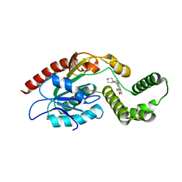
2GP6
DownloadVisualize
BU of 2gp6 by Molmil
X-ray crystal structure of Mycobacterium tuberculosis beta-ketoacyl acyl carrier protein synthase II (mtKasB)
Descriptor: 3-oxoacyl-[acyl-carrier-protein] synthase 2
Authors:Sridharan, S, Sacchettini, J, TB Structural Genomics Consortium (TBSGC)
Deposit date:2006-04-17
Release date:2006-07-18
Last modified:2024-02-14
Method:X-RAY DIFFRACTION (2.4 Å)
Cite:X-Ray Crystal Structure of Mycobacterium tuberculosis beta-Ketoacyl Acyl Carrier Protein Synthase II (mtKasB).
J.Mol.Biol., 366, 2007
6D8I
DownloadVisualize
BU of 6d8i by Molmil
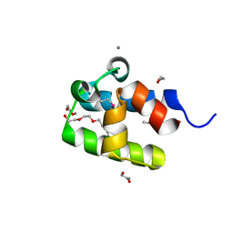
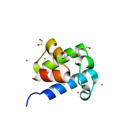
Mycobacterium tuberculosis polyketide synthase 13 N-terminal acyl carrier protein domain
Descriptor: 1,2-ETHANEDIOL, CALCIUM ION, Polyketide synthase, ...
Authors:Tang, S, Sacchettini, J.
Deposit date:2018-04-26
Release date:2018-06-06
Last modified:2024-11-13
Method:X-RAY DIFFRACTION (1.65 Å)
Cite:Mycobacterium tuberculosis polyketide synthase 13 N-terminal acyl carrier protein domain
To be Published
6DNM
DownloadVisualize
BU of 6dnm by Molmil
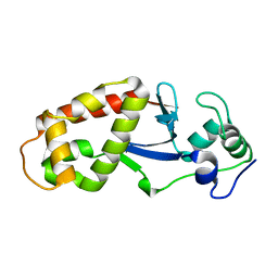
The crystal structure of SatS c-terminal domain
Descriptor: Export chaperone SatS
Authors:Hughes, R.C, Sacchettini, J.C, TB Structural Genomics Consortium (TBSGC)
Deposit date:2018-06-07
Release date:2019-01-23
Last modified:2024-03-13
Method:X-RAY DIFFRACTION (1.397 Å)
Cite:Mycobacterium tuberculosisSatS is a chaperone for the SecA2 protein export pathway.
Elife, 8, 2019
6D8J
DownloadVisualize
BU of 6d8j by Molmil
Mycobacterium tuberculosis polyketide synthase 13 N-terminal acyl carrier protein domain
Descriptor: 1,2-ETHANEDIOL, CALCIUM ION, Polyketide synthase, ...
Authors:Tang, S, Sacchettini, J.
Deposit date:2018-04-26
Release date:2018-06-06
Last modified:2023-10-04
Method:X-RAY DIFFRACTION (1.63 Å)
Cite:Mycobacterium tuberculosis polyketide synthase 13 N-terminal acyl carrier protein domain
To be Published
5C9X
DownloadVisualize
BU of 5c9x by Molmil
Crystal structure of Mycobacterium tuberculosis malate synthase in complex with 2,4-dichloro-5-fluorobenzoic acid
Descriptor: 2,4-dichloro-5-fluorobenzoic acid, MAGNESIUM ION, Malate synthase G
Authors:Huang, H.-L, Sacchettini, J.C.
Deposit date:2015-06-29
Release date:2016-08-10
Last modified:2023-09-27
Method:X-RAY DIFFRACTION (2.1 Å)
Cite:Mycobacterium tuberculosis Malate Synthase Structures with Fragments Reveal a Portal for Substrate/Product Exchange.
J. Biol. Chem., 291, 2016
5CBB
DownloadVisualize
BU of 5cbb by Molmil
Crystal structure of Mycobacterium tuberculosis malate synthase in complex with 5-(3H-indol-3-ylidene)-2,5-dihydro-1H-pyrazole-3-carboxylate
Descriptor: (5E)-5-(3H-indol-3-ylidene)-2,5-dihydro-1H-pyrazole-3-carboxylic acid, MAGNESIUM ION, Malate synthase G
Authors:Huang, H.-L, Sacchettini, J.C.
Deposit date:2015-06-30
Release date:2016-08-10
Last modified:2023-09-27
Method:X-RAY DIFFRACTION (2.012 Å)
Cite:Mycobacterium tuberculosis Malate Synthase Structures with Fragments Reveal a Portal for Substrate/Product Exchange.
J. Biol. Chem., 291, 2016
5CJN
DownloadVisualize
BU of 5cjn by Molmil
Crystal structure of Mycobacterium tuberculosis malate synthase in complex with 3-(3-oxo-3,4-dihydroquinoxalin-2-yl)acrylate
Descriptor: 3-(3-oxo-3,4-dihydroquinoxalin-2-yl)propanoic acid, MAGNESIUM ION, Malate synthase G
Authors:Huang, H.-L, Sacchettini, J.C.
Deposit date:2015-07-14
Release date:2016-08-10
Last modified:2024-11-13
Method:X-RAY DIFFRACTION (2.19 Å)
Cite:Mycobacterium tuberculosis Malate Synthase Structures with Fragments Reveal a Portal for Substrate/Product Exchange.
J. Biol. Chem., 291, 2016
5C9U
DownloadVisualize
BU of 5c9u by Molmil
Crystal structure of Mycobacterium tuberculosis malate synthase in complex with 2-(2-(2,4-dichlorophenyl)hydrazinyl)-2-oxoacetic acid
Descriptor: MAGNESIUM ION, Malate synthase G, [2-(2,4-dichlorophenyl)hydrazinyl](oxo)acetic acid
Authors:Huang, H.-L, Sacchettini, J.C.
Deposit date:2015-06-29
Release date:2016-08-10
Last modified:2023-09-27
Method:X-RAY DIFFRACTION (1.95 Å)
Cite:Mycobacterium tuberculosis Malate Synthase Structures with Fragments Reveal a Portal for Substrate/Product Exchange.
J. Biol. Chem., 291, 2016
5CBI
DownloadVisualize
BU of 5cbi by Molmil
Crystal structure of Mycobacterium tuberculosis malate synthase in complex with 5-chloro-2-hydroxybenzonitrile
Descriptor: 5-chloro-2-hydroxybenzonitrile, MAGNESIUM ION, Malate synthase G
Authors:Huang, H.-L, Sacchettini, J.C.
Deposit date:2015-06-30
Release date:2016-08-10
Last modified:2023-09-27
Method:X-RAY DIFFRACTION (1.988 Å)
Cite:Mycobacterium tuberculosis Malate Synthase Structures with Fragments Reveal a Portal for Substrate/Product Exchange.
J. Biol. Chem., 291, 2016
5CC6
DownloadVisualize
BU of 5cc6 by Molmil
Crystal structure of Mycobacterium tuberculosis malate synthase in complex with 1H-indole-5-carboxylic acid
Descriptor: 1H-indole-5-carboxylic acid, MAGNESIUM ION, Malate synthase G
Authors:Huang, H.-L, Sacchettini, J.C.
Deposit date:2015-07-01
Release date:2016-08-10
Last modified:2023-09-27
Method:X-RAY DIFFRACTION (2.1 Å)
Cite:Crystal structures of fragment-bound malate synthase from Mycobacterium tuberculosis provide insights into mechanism and potential inhibitor designs
To Be Published

234136

PDB entries from 2025-04-02

PDB statisticsPDBj update infoContact PDBjnumon