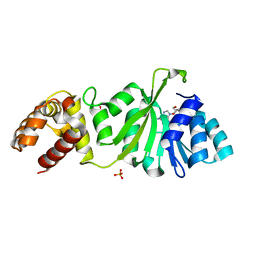
1WTS
 
 | |
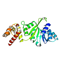
1WTT
 | |
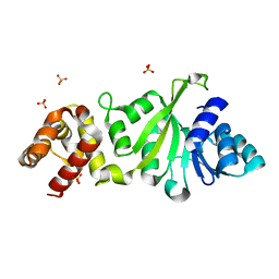
1JUR
 | |
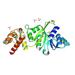
3TPZ
 | |
4FAK
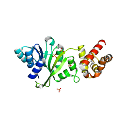
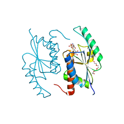 | | Crystal Structure of OrfX in Complex with S-Adenosylmethionine | | 分子名称: | PHOSPHATE ION, Ribosomal RNA large subunit methyltransferase H, S-ADENOSYLMETHIONINE, ... | | 著者 | Safo, M.K, Musayev, F.N, Boundy, S, Archer, G.L, Rife, J.P, O'Farrell, H.C. | | 登録日 | 2012-05-22 | | 公開日 | 2012-06-06 | | 最終更新日 | 2023-09-13 | | 実験手法 | X-RAY DIFFRACTION (1.7 Å) | | 主引用文献 | Characterization of the Staphylococcus aureus rRNA Methyltransferase Encoded by orfX, the Gene Containing the Staphylococcal Chromosome Cassette mec (SCCmec) Insertion Site.
J.Biol.Chem., 288, 2013
|
|
4ADV
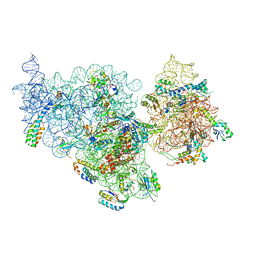 | | Structure of the E. coli methyltransferase KsgA bound to the E. coli 30S ribosomal subunit | | 分子名称: | 16S RIBOSOMAL RNA, 30S RIBOSOMAL PROTEIN S10, 30S RIBOSOMAL PROTEIN S11, ... | | 著者 | Boehringer, D, O'Farrell, H.C, Rife, J.P, Ban, N. | | 登録日 | 2012-01-03 | | 公開日 | 2012-02-15 | | 最終更新日 | 2024-05-08 | | 実験手法 | ELECTRON MICROSCOPY (13.5 Å) | | 主引用文献 | Structural Insights Into Methyltransferase Ksga Function in 30S Ribosomal Subunit Biogenesis
J.Biol.Chem., 287, 2012
|
|
1QYR
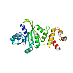 | | 2.1 Angstrom Crystal structure of KsgA: A Universally Conserved Adenosine Dimethyltransferase | | 分子名称: | High level Kasugamycin resistance protein | | 著者 | O'Farrell, H.C, Scarsdale, J.N, Wright, H.T, Rife, J.P. | | 登録日 | 2003-09-11 | | 公開日 | 2004-06-29 | | 最終更新日 | 2024-02-14 | | 実験手法 | X-RAY DIFFRACTION (2.1 Å) | | 主引用文献 | Crystal structure of KsgA, a universally conserved rRNA adenine dimethyltransferase in Escherichia coli
J.Mol.Biol., 339, 2004
|
|
3GRR
 | |
3GRV
 | |
3GRY
 | |
3FYD
 | |
3GRU
 | |
3FYC
 | |
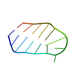
1KD3
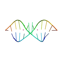 | | The Crystal Structure of r(GGUCACAGCCC)2, Thallium form | | 分子名称: | 5'-R(*GP*GP*UP*CP*AP*CP*AP*GP*CP*CP*C)-3', THALLIUM (I) ION | | 著者 | Kacer, V, Scaringe, S.A, Scarsdale, J.N, Rife, J.P. | | 登録日 | 2001-11-12 | | 公開日 | 2003-03-04 | | 最終更新日 | 2024-02-07 | | 実験手法 | X-RAY DIFFRACTION (1.8 Å) | | 主引用文献 | Crystal structures of r(GGUCACAGCCC)2.
Acta Crystallogr.,Sect.D, 59, 2003
|
|
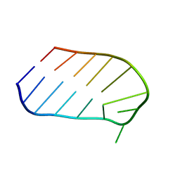
1KD4
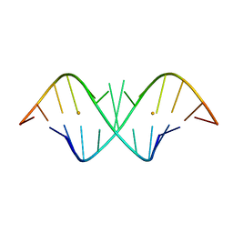 | | The Crystal Structure of r(GGUCACAGCCC)2, Barium form | | 分子名称: | 5'-R(*GP*GP*UP*CP*AP*CP*AP*GP*CP*CP*C)-3', BARIUM ION | | 著者 | Kacer, V, Scaringe, S.A, Scarsdale, J.N, Rife, J.P. | | 登録日 | 2001-11-12 | | 公開日 | 2003-03-04 | | 最終更新日 | 2024-02-07 | | 実験手法 | X-RAY DIFFRACTION (1.85 Å) | | 主引用文献 | Crystal structures of r(GGUCACAGCCC)2.
Acta Crystallogr.,Sect.D, 59, 2003
|
|
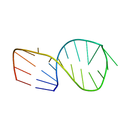
1KD5
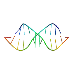 | | The Crystal Structure of r(GGUCACAGCCC)2 metal free form | | 分子名称: | 5'-R(*GP*GP*UP*CP*AP*CP*AP*GP*CP*CP*C)-3' | | 著者 | Kacer, V, Scaringe, S.A, Scarsdale, J.N, Rife, J.P. | | 登録日 | 2001-11-12 | | 公開日 | 2003-03-04 | | 最終更新日 | 2024-02-07 | | 実験手法 | X-RAY DIFFRACTION (1.58 Å) | | 主引用文献 | Crystal structures of r(GGUCACAGCCC)2.
Acta Crystallogr.,Sect.D, 59, 2003
|
|


