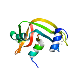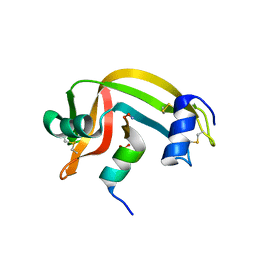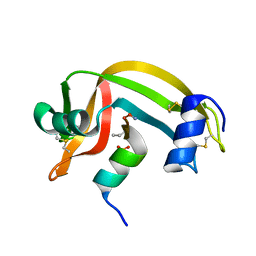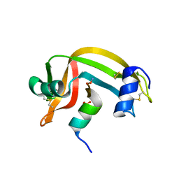
Loading
PDBj
MenuPDBj@FacebookPDBj@TwitterPDBj@YouTubewwPDB FoundationwwPDB
RCSB PDBPDBeBMRBAdv. SearchSearch help
PDB: 45 results

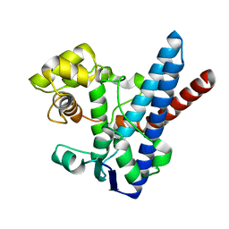
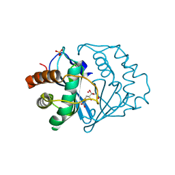
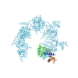
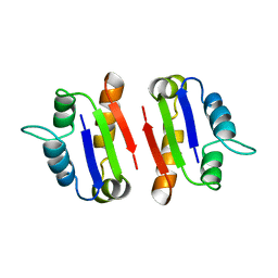
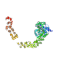
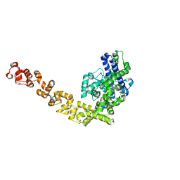
5HM5
DownloadVisualize
BU of 5hm5 by Molmil
Crystal structure of Topo-97, an N-terminal 97kDa fragment of topoisomerase V
Descriptor: Topoisomerase V
Authors:Rajan, R, Osterman, A, Mondragon, A.
Deposit date:2016-01-15
Release date:2016-03-09
Last modified:2024-10-30
Method:X-RAY DIFFRACTION (2.4 Å)
Cite:Methanopyrus kandleri topoisomerase V contains three distinct AP lyase active sites in addition to the topoisomerase active site.
Nucleic Acids Res., 44, 2016
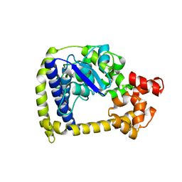
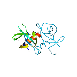
4GFJ
DownloadVisualize
BU of 4gfj by Molmil
Crystal structure of Topo-78, an N-terminal 78kDa fragment of topoisomerase V
Descriptor: GLYCEROL, Topoisomerase V, ZINC ION
Authors:Rajan, R, Prasad, R, Taneja, B, Wilson, S.H, Mondragon, A.
Deposit date:2012-08-03
Release date:2012-12-05
Last modified:2023-09-13
Method:X-RAY DIFFRACTION (2.91 Å)
Cite:Identification of one of the apurinic/apyrimidinic lyase active sites of topoisomerase V by structural and functional studies.
Nucleic Acids Res., 41, 2013
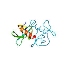
3M7G
DownloadVisualize
BU of 3m7g by Molmil
Structure of topoisomerase domain of topoisomerase V protein
Descriptor: Topoisomerase V
Authors:Rajan, R, Taneja, B, Mondragon, A.
Deposit date:2010-03-16
Release date:2010-08-04
Last modified:2023-09-06
Method:X-RAY DIFFRACTION (2.4 Å)
Cite:Structures of minimal catalytic fragments of topoisomerase v reveals conformational changes relevant for DNA binding.
Structure, 18, 2010
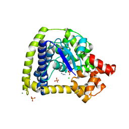
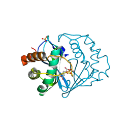
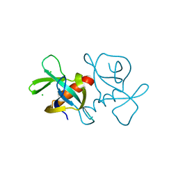
3M6K
DownloadVisualize
BU of 3m6k by Molmil
Crystal Structure of N-terminal 44 kDa fragment of topoisomerase V in the presence of guanidium hydrochloride
Descriptor: PHOSPHATE ION, Topoisomerase V
Authors:Rajan, R, Taneja, B, Mondragon, A.
Deposit date:2010-03-15
Release date:2010-08-04
Last modified:2011-07-13
Method:X-RAY DIFFRACTION (2.6 Å)
Cite:Structures of minimal catalytic fragments of topoisomerase v reveals conformational changes relevant for DNA binding.
Structure, 18, 2010
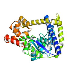
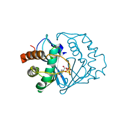
3M6Z
DownloadVisualize
BU of 3m6z by Molmil
Crystal structure of an N-terminal 44 kDa fragment of topoisomerase V in the presence of guanidium hydrochloride
Descriptor: CHLORIDE ION, GUANIDINE, MAGNESIUM ION, ...
Authors:Rajan, R, Taneja, B, Mondragon, A.
Deposit date:2010-03-16
Release date:2010-08-04
Last modified:2024-11-06
Method:X-RAY DIFFRACTION (1.4 Å)
Cite:Structures of minimal catalytic fragments of topoisomerase v reveals conformational changes relevant for DNA binding.
Structure, 18, 2010
3M7D
DownloadVisualize
BU of 3m7d by Molmil
Crystal structure of an N-terminal 44 kDA fragment of topoisomerase V in the presence of dioxane
Descriptor: Topoisomerase V
Authors:Rajan, R, Taneja, B, Mondragon, A.
Deposit date:2010-03-16
Release date:2010-08-04
Last modified:2023-09-06
Method:X-RAY DIFFRACTION (1.815 Å)
Cite:Structures of minimal catalytic fragments of topoisomerase v reveals conformational changes relevant for DNA binding.
Structure, 18, 2010
1YCL
DownloadVisualize
BU of 1ycl by Molmil
Crystal Structure of B. subtilis LuxS in Complex with a Catalytic 2-Ketone Intermediate
Descriptor: (S)-2-AMINO-4-[(2S,3R)-2,3,5-TRIHYDROXY-4-OXO-PENTYL]MERCAPTO-BUTYRIC ACID, COBALT (II) ION, S-ribosylhomocysteinase, ...
Authors:Rajan, R, Zhu, J, Hu, X, Pei, D, Bell, C.E.
Deposit date:2004-12-22
Release date:2005-03-15
Last modified:2024-03-13
Method:X-RAY DIFFRACTION (1.8 Å)
Cite:Crystal Structure of S-Ribosylhomocysteinase (LuxS) in Complex with a Catalytic 2-Ketone Intermediate.
Biochemistry, 44, 2005
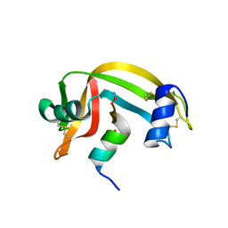
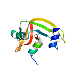
1RBE
DownloadVisualize
BU of 1rbe by Molmil
CRYSTALLOGRAPHIC STRUCTURES OF RIBONUCLEASE S VARIANTS WITH NONPOLAR SUBSTITUTION AT POSITION 13: PACKING AND CAVITIES
Descriptor: RIBONUCLEASE S (S-PEPTIDE), RIBONUCLEASE S (S-PROTEIN), SULFATE ION
Authors:Varadarajan, R, Richards, F.M.
Deposit date:1992-06-12
Release date:1993-10-31
Last modified:2017-11-29
Method:X-RAY DIFFRACTION (1.75 Å)
Cite:Crystallographic structures of ribonuclease S variants with nonpolar substitution at position 13: packing and cavities.
Biochemistry, 31, 1992
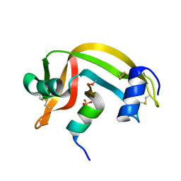
1RBF
DownloadVisualize
BU of 1rbf by Molmil
CRYSTALLOGRAPHIC STRUCTURES OF RIBONUCLEASE S VARIANTS WITH NONPOLAR SUBSTITUTION AT POSITION 13: PACKING AND CAVITIES
Descriptor: RIBONUCLEASE S (S-PEPTIDE), RIBONUCLEASE S (S-PROTEIN), SULFATE ION
Authors:Varadarajan, R, Richards, F.M.
Deposit date:1992-06-12
Release date:1993-10-31
Last modified:2024-10-09
Method:X-RAY DIFFRACTION (1.8 Å)
Cite:Crystallographic structures of ribonuclease S variants with nonpolar substitution at position 13: packing and cavities.
Biochemistry, 31, 1992
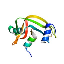
1RBI
DownloadVisualize
BU of 1rbi by Molmil
CRYSTALLOGRAPHIC STRUCTURES OF RIBONUCLEASE S VARIANTS WITH NONPOLAR SUBSTITUTION AT POSITION 13: PACKING AND CAVITIES
Descriptor: RIBONUCLEASE S (S-PEPTIDE), RIBONUCLEASE S (S-PROTEIN), SULFATE ION
Authors:Varadarajan, R, Richards, F.M.
Deposit date:1992-06-12
Release date:1993-10-31
Last modified:2017-11-29
Method:X-RAY DIFFRACTION (1.8 Å)
Cite:Crystallographic structures of ribonuclease S variants with nonpolar substitution at position 13: packing and cavities.
Biochemistry, 31, 1992
1RBC
DownloadVisualize
BU of 1rbc by Molmil
CRYSTALLOGRAPHIC STRUCTURES OF RIBONUCLEASE S VARIANTS WITH NONPOLAR SUBSTITUTION AT POSITION 13: PACKING AND CAVITIES
Descriptor: RIBONUCLEASE S (S-PEPTIDE), RIBONUCLEASE S (S-PROTEIN), SULFATE ION
Authors:Varadarajan, R, Richards, F.M.
Deposit date:1992-06-12
Release date:1993-10-31
Last modified:2024-11-06
Method:X-RAY DIFFRACTION (2 Å)
Cite:Crystallographic structures of ribonuclease S variants with nonpolar substitution at position 13: packing and cavities.
Biochemistry, 31, 1992
1RBG
DownloadVisualize
BU of 1rbg by Molmil
CRYSTALLOGRAPHIC STRUCTURES OF RIBONUCLEASE S VARIANTS WITH NONPOLAR SUBSTITUTION AT POSITION 13: PACKING AND CAVITIES
Descriptor: RIBONUCLEASE S (S-PEPTIDE), RIBONUCLEASE S (S-PROTEIN), SULFATE ION
Authors:Varadarajan, R, Richards, F.M.
Deposit date:1992-06-12
Release date:1993-10-31
Last modified:2024-10-23
Method:X-RAY DIFFRACTION (1.8 Å)
Cite:Crystallographic structures of ribonuclease S variants with nonpolar substitution at position 13: packing and cavities.
Biochemistry, 31, 1992
1RBD
DownloadVisualize
BU of 1rbd by Molmil
CRYSTALLOGRAPHIC STRUCTURES OF RIBONUCLEASE S VARIANTS WITH NONPOLAR SUBSTITUTION AT POSITION 13: PACKING AND CAVITIES
Descriptor: RIBONUCLEASE S (S-PEPTIDE), RIBONUCLEASE S (S-PROTEIN), SULFATE ION
Authors:Varadarajan, R, Richards, F.M.
Deposit date:1992-06-12
Release date:1993-10-31
Last modified:2017-11-29
Method:X-RAY DIFFRACTION (1.7 Å)
Cite:Crystallographic structures of ribonuclease S variants with nonpolar substitution at position 13: packing and cavities.
Biochemistry, 31, 1992
1RBH
DownloadVisualize
BU of 1rbh by Molmil
CRYSTALLOGRAPHIC STRUCTURES OF RIBONUCLEASE S VARIANTS WITH NONPOLAR SUBSTITUTION AT POSITION 13: PACKING AND CAVITIES
Descriptor: RIBONUCLEASE S (S-PEPTIDE), RIBONUCLEASE S (S-PROTEIN), SULFATE ION
Authors:Varadarajan, R, Richards, F.M.
Deposit date:1992-06-12
Release date:1993-10-31
Last modified:2024-10-23
Method:X-RAY DIFFRACTION (1.7 Å)
Cite:Crystallographic structures of ribonuclease S variants with nonpolar substitution at position 13: packing and cavities.
Biochemistry, 31, 1992
1XP8
DownloadVisualize
BU of 1xp8 by Molmil
Deinococcus radiodurans RecA in complex with ATP-gamma-S
Descriptor: PHOSPHOTHIOPHOSPHORIC ACID-ADENYLATE ESTER, RecA protein
Authors:Bell, C.E, Rajan, R.
Deposit date:2004-10-08
Release date:2004-12-21
Last modified:2023-08-23
Method:X-RAY DIFFRACTION (2.5 Å)
Cite:Crystal structure of RecA from Deinococcus radiodurans: insights into the structural basis of extreme radioresistance.
J.Mol.Biol., 344, 2004
2FQT
DownloadVisualize
BU of 2fqt by Molmil
Crystal structure of B.subtilis LuxS in complex with (2S)-2-Amino-4-[(2R,3S)-2,3-dihydroxy-3-N-hydroxycarbamoyl-propylmercapto]butyric acid
Descriptor: (2S)-2-AMINO-4-[(2R,3S)-2,3-DIHYDROXY-3-N-HYDROXYCARBAMOYL-PROPYLMERCAPTO]BUTYRIC ACID, COBALT (II) ION, S-ribosylhomocysteine lyase, ...
Authors:Shen, G, Rajan, R, Zhu, J, Bell, C.E, Pei, D.
Deposit date:2006-01-18
Release date:2006-05-30
Last modified:2023-08-30
Method:X-RAY DIFFRACTION (1.79 Å)
Cite:Design and Synthesis of Substrate Analogue Inhibitors of S-Ribosylhomocysteinase (LuxS)
J.Med.Chem., 49, 2006
2FQO
DownloadVisualize
BU of 2fqo by Molmil
Crystal structure of B. subtilis LuxS in complex with (2S)-2-Amino-4-[(2R,3R)-2,3-dihydroxy-3-N- hydroxycarbamoyl-propylmercapto]butyric acid
Descriptor: (2S)-2-AMINO-4-[(2R,3R)-2,3-DIHYDROXY-3-N-HYDROXYCARBAMOYL-PROPYLMERCAPTO]BUTYRIC ACID, COBALT (II) ION, S-ribosylhomocysteine lyase, ...
Authors:Shen, G, Rajan, R, Zhu, J, Bell, C.E, Pei, D.
Deposit date:2006-01-18
Release date:2006-05-30
Last modified:2023-08-30
Method:X-RAY DIFFRACTION (1.87 Å)
Cite:Design and Synthesis of Substrate and Intermediate Analogue Inhibitors of S-Ribosylhomocysteinase
J.Med.Chem., 49, 2006
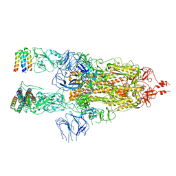
7X7N
DownloadVisualize
BU of 7x7n by Molmil
3D model of the 3-RBD up single trimeric spike protein of SARS-CoV2 in the presence of synthetic peptide SIH-5.
Descriptor: 2-acetamido-2-deoxy-beta-D-glucopyranose, Spike glycoprotein, Synthetic peptide SIH-5
Authors:Khatri, B, Pramanick, I, Malladi, S.K, Rajmani, R.S, Kumar, S, Ghosh, P, Sengupta, N, Rahisuddin, R, Kumaran, S, Ringe, R.P, Varadarajan, R, Dutta, S, Chatterjee, J.
Deposit date:2022-03-10
Release date:2022-04-27
Last modified:2022-11-16
Method:ELECTRON MICROSCOPY (4.47 Å)
Cite:A dimeric proteomimetic prevents SARS-CoV-2 infection by dimerizing the spike protein.
Nat.Chem.Biol., 18, 2022
1A19
DownloadVisualize
BU of 1a19 by Molmil
BARSTAR (FREE), C82A MUTANT
Descriptor: BARSTAR
Authors:Ratnaparkhi, G.S, Varadarajan, R.
Deposit date:1997-12-25
Release date:1998-04-08
Last modified:2024-05-22
Method:X-RAY DIFFRACTION (2.76 Å)
Cite:Discrepancies between the NMR and X-ray structures of uncomplexed barstar: analysis suggests that packing densities of protein structures determined by NMR are unreliable.
Biochemistry, 37, 1998
7EPI
DownloadVisualize
BU of 7epi by Molmil
Crystal structure of E.coli CcdB mutant S60E
Descriptor: CHLORIDE ION, Toxin CcdB
Authors:Manjunath, K, Goyal, P, Varadarajan, R.
Deposit date:2021-04-26
Release date:2022-06-08
Last modified:2023-11-29
Method:X-RAY DIFFRACTION (1.931 Å)
Cite:Mechanistic insights into global suppressors of protein folding defects.
Plos Genet., 18, 2022
7EPG
DownloadVisualize
BU of 7epg by Molmil
Crystal structure of E.coli CcdB mutant S12G
Descriptor: CHLORIDE ION, Toxin CcdB
Authors:Manjunath, K, Varadarajan, R.
Deposit date:2021-04-26
Release date:2022-06-08
Last modified:2023-11-29
Method:X-RAY DIFFRACTION (1.631 Å)
Cite:Mechanistic insights into global suppressors of protein folding defects.
Plos Genet., 18, 2022
7EPJ
DownloadVisualize
BU of 7epj by Molmil
Crystal structure of E.coli CcdB mutant V46L
Descriptor: CHLORIDE ION, Toxin CcdB
Authors:Manjunath, K, Goyal, P, Varadarajan, R.
Deposit date:2021-04-26
Release date:2022-06-08
Last modified:2023-11-29
Method:X-RAY DIFFRACTION (1.354 Å)
Cite:Mechanistic insights into global suppressors of protein folding defects.
Plos Genet., 18, 2022
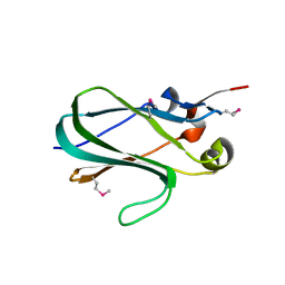
5FCE
DownloadVisualize
BU of 5fce by Molmil
The crystal structure of the ligand binding region of Serine-glutamate repeat protein A (SgrA) of Enterococcus faecium
Descriptor: LPXTG family cell surface protein Fms2
Authors:Ponnuraj, K, Nagarajan, R.
Deposit date:2015-12-15
Release date:2016-07-06
Last modified:2024-11-06
Method:X-RAY DIFFRACTION (1.798 Å)
Cite:The crystal structure of the ligand-binding region of serine-glutamate repeat containing protein A (SgrA) of Enterococcus faecium reveals a new protein fold: functional characterization and insights into its adhesion function.
FEBS J., 283, 2016
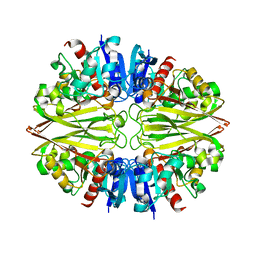
6IEP
DownloadVisualize
BU of 6iep by Molmil
GAPDH of Streptococcus agalactiae
Descriptor: Glyceraldehyde-3-phosphate dehydrogenase
Authors:Ponnuraj, K, Nagarajan, R.
Deposit date:2018-09-15
Release date:2019-01-02
Last modified:2023-11-22
Method:X-RAY DIFFRACTION (2.6 Å)
Cite:Crystal structure of GAPDH of Streptococcus agalactiae and characterization of its interaction with extracellular matrix molecules
Microb. Pathog., 127, 2018
1D5D
DownloadVisualize
BU of 1d5d by Molmil
The role of phenylalanine 8 in the stabilization of the s protein-s peptide interaction: packing and cavities
Descriptor: RNASE S, S PEPTIDE, SULFATE ION
Authors:Ratnaparkhi, G.S, Varadarajan, R.
Deposit date:1999-10-07
Release date:1999-10-20
Last modified:2018-03-14
Method:X-RAY DIFFRACTION (2.25 Å)
Cite:Thermodynamic and structural studies of cavity formation in proteins suggest that loss of packing interactions rather than the hydrophobic effect dominates the observed energetics.
Biochemistry, 39, 2000


12>

227111

數據於2024-11-06公開中

PDB statisticsPDBj update infoContact PDBjnumon