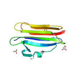
5D9R
 
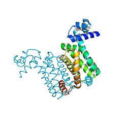
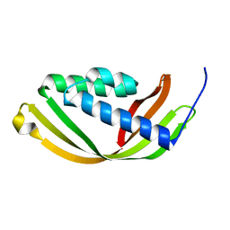 | | Crystal structure of a conserved domain in the intermembrane space region of the plastid division protein ARC6 | | 分子名称: | Protein ACCUMULATION AND REPLICATION OF CHLOROPLASTS 6, chloroplastic | | 著者 | Radhakrishnan, A, Kumar, N, Su, C.-C, Chou, T.-H, Yu, E. | | 登録日 | 2015-08-18 | | 公開日 | 2015-11-11 | | 最終更新日 | 2024-03-06 | | 実験手法 | X-RAY DIFFRACTION (2.052 Å) | | 主引用文献 | Crystal structure of a conserved domain in the intermembrane space region of the plastid division protein ARC6.
Protein Sci., 25, 2016
|
|
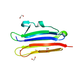
6LNR
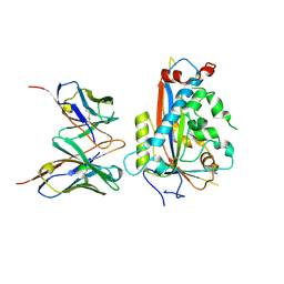
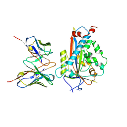
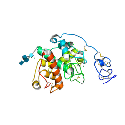 | | Structure of intact chitinase with hevein domain from the plant Simarouba glauca, known for its traditional anti-inflammatory efficacy | | 分子名称: | 2-acetamido-2-deoxy-beta-D-glucopyranose-(1-4)-2-acetamido-2-deoxy-beta-D-glucopyranose, 2-acetamido-2-deoxy-beta-D-glucopyranose-(1-4)-2-acetamido-2-deoxy-beta-D-glucopyranose-(1-4)-[2-acetamido-2-deoxy-beta-D-glucopyranose-(3-3)]2-acetamido-2-deoxy-beta-D-glucopyranose, CHLORIDE ION, ... | | 著者 | KanalElamparithi, B, Ramya, K.S, Ankur, T, Radha, A, Gunasekaran, K. | | 登録日 | 2020-01-01 | | 公開日 | 2020-12-23 | | 最終更新日 | 2023-11-29 | | 実験手法 | X-RAY DIFFRACTION (1.66 Å) | | 主引用文献 | Structure of intact chitinase with hevein domain from the plant Simarouba glauca, known for its traditional anti-inflammatory efficacy.
Int.J.Biol.Macromol., 161, 2020
|
|
1PLY
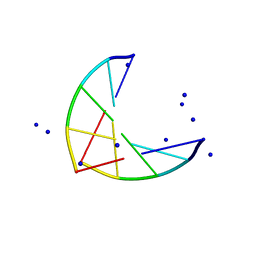 | | SODIUM IONS AND WATER MOLECULES IN THE STRUCTURE OF POLY D(A)(DOT)POLY D(T) | | 分子名称: | DNA (5'-D(P*AP*AP*AP*AP*A)-3'), DNA (5'-D(P*TP*TP*TP*TP*T)-3'), SODIUM ION | | 著者 | Chandrasekaran, R, Radha, A, Park, H.-S. | | 登録日 | 1995-02-28 | | 公開日 | 1995-06-03 | | 最終更新日 | 2024-02-14 | | 実験手法 | FIBER DIFFRACTION (3.2 Å) | | 主引用文献 | Sodium ions and water molecules in the structure of poly(dA).poly(dT).
Acta Crystallogr.,Sect.D, 51, 1995
|
|
6BHP
 | |
7LKF
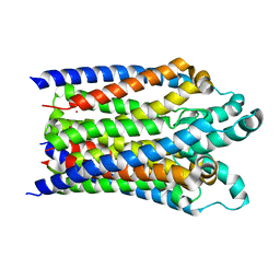
 | | WT Chicken Scap L1-L7 / Fab 4G10 complex focused refinement | | 分子名称: | 2-acetamido-2-deoxy-beta-D-glucopyranose-(1-4)-2-acetamido-2-deoxy-beta-D-glucopyranose, 4G10 heavy chain, 4G10 light chain, ... | | 著者 | Kober, D.L, Radhakrishnan, A, Goldstein, J.L, Brown, M.S, Clark, L.D, Bai, X.-C, Rosenbaum, D.M. | | 登録日 | 2021-02-02 | | 公開日 | 2021-06-30 | | 最終更新日 | 2021-07-28 | | 実験手法 | ELECTRON MICROSCOPY (2.9 Å) | | 主引用文献 | Scap structures highlight key role for rotation of intertwined luminal loops in cholesterol sensing.
Cell, 184, 2021
|
|
7LKH
 | | Chicken Scap D435V L1-L7 domain / Fab complex focused map | | 分子名称: | 2-acetamido-2-deoxy-beta-D-glucopyranose-(1-4)-2-acetamido-2-deoxy-beta-D-glucopyranose, 4G10 Fab heavy chain, 4G10 Fab kappa chain, ... | | 著者 | Kober, D.L, Radhakrishnan, A, Goldstein, J.L, Brown, M.S, Clark, L.D, Bai, X.-C, Rosenbaum, D.M. | | 登録日 | 2021-02-02 | | 公開日 | 2021-06-30 | | 最終更新日 | 2024-10-16 | | 実験手法 | ELECTRON MICROSCOPY (3.5 Å) | | 主引用文献 | Scap structures highlight key role for rotation of intertwined luminal loops in cholesterol sensing.
Cell, 184, 2021
|
|
4NB5
 | |
4NN1
 | |
6MYI
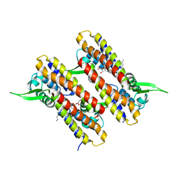
 | | Pleurotus ostreatus OstreolysinA | | 分子名称: | 1,2-ETHANEDIOL, Ostreolysin A6, SODIUM ION | | 著者 | Tomchick, D.R, Radhakrishnan, A, Endapally, S. | | 登録日 | 2018-11-01 | | 公開日 | 2019-02-13 | | 最終更新日 | 2024-03-13 | | 実験手法 | X-RAY DIFFRACTION (1.15 Å) | | 主引用文献 | Molecular Discrimination between Two Conformations of Sphingomyelin in Plasma Membranes.
Cell, 176, 2019
|
|
6MYK
 | | Pleurotus ostreatus OstreolysinA mutant E69A with Bis-Tris | | 分子名称: | 1,2-ETHANEDIOL, 2-[BIS-(2-HYDROXY-ETHYL)-AMINO]-2-HYDROXYMETHYL-PROPANE-1,3-DIOL, Ostreolysin A6, ... | | 著者 | Tomchick, D.R, Radhakrishnan, A, Endapally, S. | | 登録日 | 2018-11-01 | | 公開日 | 2019-02-13 | | 最終更新日 | 2023-10-11 | | 実験手法 | X-RAY DIFFRACTION (1.8 Å) | | 主引用文献 | Molecular Discrimination between Two Conformations of Sphingomyelin in Plasma Membranes.
Cell, 176, 2019
|
|
6MYJ
 | | Pleurotus ostreatus OstreolysinA plus sphingomyelin | | 分子名称: | 1,2-ETHANEDIOL, N-[(2S)-1-hydroxypropan-2-yl]butanamide, Ostreolysin A6, ... | | 著者 | Tomchick, D.R, Radhakrishnan, A, Endapally, S. | | 登録日 | 2018-11-01 | | 公開日 | 2019-02-13 | | 最終更新日 | 2023-10-11 | | 実験手法 | X-RAY DIFFRACTION (1.33 Å) | | 主引用文献 | Molecular Discrimination between Two Conformations of Sphingomyelin in Plasma Membranes.
Cell, 176, 2019
|
|


