8JU7
 
 | |
8OIW
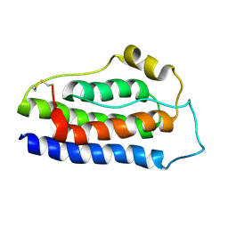
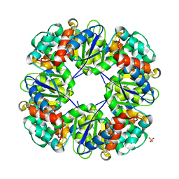
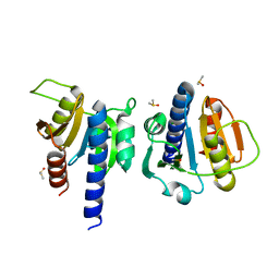
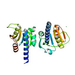
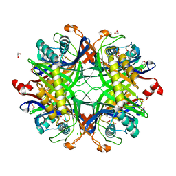 | | Crystal structure of the cysteine-rich Gallus gallus urate oxidase in complex with the 8-azaxanthine inhibitor under oxidising conditions (space group P 21 21 21) | | 分子名称: | 1,2-ETHANEDIOL, 8-AZAXANTHINE, CHLORIDE ION, ... | | 著者 | Di Palma, M, Chegkazi, M, Bui, S, Mori, G, Percudani, R, Steiner, R.A. | | 登録日 | 2023-03-23 | | 公開日 | 2024-01-17 | | 実験手法 | X-RAY DIFFRACTION (1.89 Å) | | 主引用文献 | Cysteine Enrichment Mediates Co-Option of Uricase in Reptilian Skin and Transition to Uricotelism.
Mol.Biol.Evol., 40, 2023
|
|
8OPV
 | |
6Z8V
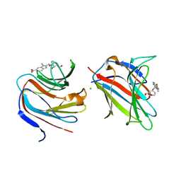
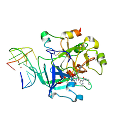 | | X-ray structure of the complex between human alpha thrombin and a thrombin binding aptamer variant (TBA-3L), which contains 1-beta-D-lactopyranosyl residue in the side chain of Thy3 at N3. | | 分子名称: | D-phenylalanyl-N-[(2S,3S)-6-{[amino(iminio)methyl]amino}-1-chloro-2-hydroxyhexan-3-yl]-L-prolinamide, POTASSIUM ION, Prothrombin, ... | | 著者 | Troisi, R, Timofeev, E.N, Sica, F. | | 登録日 | 2020-06-02 | | 公開日 | 2021-01-27 | | 最終更新日 | 2024-01-24 | | 実験手法 | X-RAY DIFFRACTION (1.58 Å) | | 主引用文献 | Expanding the recognition interface of the thrombin-binding aptamer HD1 through modification of residues T3 and T12.
Mol Ther Nucleic Acids, 23, 2021
|
|
8K6Z
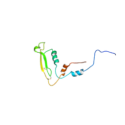
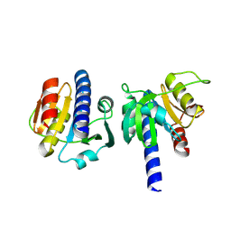
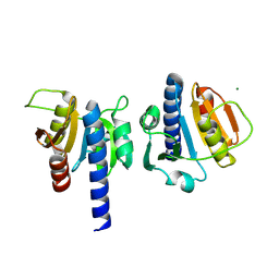
 | | NMR structure of human leptin | | 分子名称: | Leptin | | 著者 | Fan, X, Qin, R, Yuan, W, Fan, J, Huang, W, Lin, Z. | | 登録日 | 2023-07-26 | | 公開日 | 2024-02-07 | | 最終更新日 | 2024-05-15 | | 実験手法 | SOLUTION NMR | | 主引用文献 | The solution structure of human leptin reveals a conformational plasticity important for receptor recognition.
Structure, 32, 2024
|
|
8OIL
 | |
6ZJM
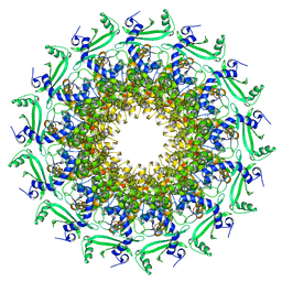
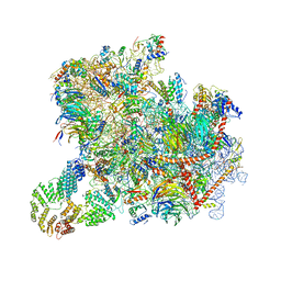
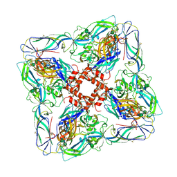 | | Atomic model of Andes virus glycoprotein spike tetramer generated by fitting into a Tula virus reconstruction | | 分子名称: | 2-acetamido-2-deoxy-beta-D-glucopyranose, 2-acetamido-2-deoxy-beta-D-glucopyranose-(1-4)-2-acetamido-2-deoxy-beta-D-glucopyranose, Envelope polyprotein,Envelope polyprotein,Envelope polyprotein,Envelope polyprotein,Envelope polyprotein,Envelope polyprotein,Envelope polyprotein,Envelope polyprotein,Envelope polyprotein, ... | | 著者 | Stass, R, Huiskonen, J.T, Rey, F, Guardado-Calvo, P. | | 登録日 | 2020-06-29 | | 公開日 | 2020-10-14 | | 最終更新日 | 2020-10-28 | | 実験手法 | ELECTRON MICROSCOPY (11.4 Å) | | 主引用文献 | The Hantavirus Surface Glycoprotein Lattice and Its Fusion Control Mechanism.
Cell, 183, 2020
|
|
8OGY
 | | PanDDA analysis group deposition -- CdaA in complex with fragment F2X-Entry D04 | | 分子名称: | Cyclic di-AMP synthase CdaA, MAGNESIUM ION, methyl 4-fluoro-L-phenylalaninate | | 著者 | Garbers, T.B, Neumann, P, Wollenhaupt, J, Weiss, M.S, Ficner, R. | | 登録日 | 2023-03-20 | | 公開日 | 2024-03-27 | | 実験手法 | X-RAY DIFFRACTION (1.23 Å) | | 主引用文献 | PanDDA analysis group deposition -- CdaA in complex with fragment F2X-Entry D04
To Be Published
|
|
5GQY
 | |
8OF6
 | |
6TJP
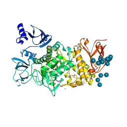
 | | Crystal structure of T7 bacteriophage portal protein, 13mer, closed valve - P212121 | | 分子名称: | Portal protein | | 著者 | Fabrega-Ferrer, M, Cuervo, A, Fernandez, F.J, Machon, C, Perez-Luque, R, Pous, J, Vega, M.C, Carrascosa, J.L, Coll, M. | | 登録日 | 2019-11-26 | | 公開日 | 2020-12-16 | | 最終更新日 | 2024-05-01 | | 実験手法 | X-RAY DIFFRACTION (3.74 Å) | | 主引用文献 | Using a partial atomic model from medium-resolution cryo-EM to solve a large crystal structure.
Acta Crystallogr D Struct Biol, 77, 2021
|
|
5GR1
 | |
8OHB
 | | PanDDA analysis group deposition -- CdaA in complex with fragment F2X-Entry E08 | | 分子名称: | 5-(2-methoxyethyl)-1,3,4-oxadiazol-2-amine, Cyclic di-AMP synthase CdaA, MAGNESIUM ION | | 著者 | Garbers, T.B, Neumann, P, Wollenhaupt, J, Weiss, M.S, Ficner, R. | | 登録日 | 2023-03-21 | | 公開日 | 2024-04-03 | | 実験手法 | X-RAY DIFFRACTION (1.11 Å) | | 主引用文献 | PanDDA analysis group deposition -- CdaA in complex with fragment F2X-Entry E08
To Be Published
|
|
6ZCE
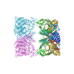
 | | Structure of a yeast ABCE1-bound 43S pre-initiation complex | | 分子名称: | 18S ribosomal RNA (1719-MER), 40S ribosomal protein S0-A, 40S ribosomal protein S1-A, ... | | 著者 | Kratzat, H, Mackens-Kiani, T, Cheng, J, Berninghausen, O, Becker, T, Beckmann, R. | | 登録日 | 2020-06-10 | | 公開日 | 2020-10-07 | | 最終更新日 | 2021-01-13 | | 実験手法 | ELECTRON MICROSCOPY (5.3 Å) | | 主引用文献 | A structural inventory of native ribosomal ABCE1-43S pre-initiation complexes.
Embo J., 40, 2021
|
|
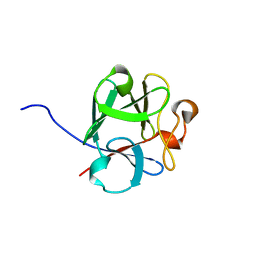
7P11
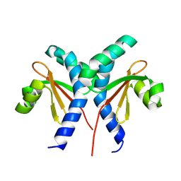
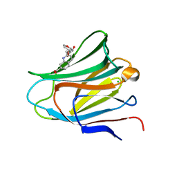 | | Galectin-8 N-terminal carbohydrate recognition domain in complex with quinoline D-galactal ligand | | 分子名称: | 2-[[(2~{R},3~{R},4~{R})-2-(hydroxymethyl)-3-oxidanyl-3,4-dihydro-2~{H}-pyran-4-yl]oxymethyl]quinoline-7-carboxylic acid, CHLORIDE ION, Galectin-8, ... | | 著者 | Hassan, M, Hakansson, M, Nilsson, J.U, Kovacic, R. | | 登録日 | 2021-07-01 | | 公開日 | 2021-12-15 | | 最終更新日 | 2024-01-31 | | 実験手法 | X-RAY DIFFRACTION (2.1 Å) | | 主引用文献 | Structure-Guided Design of d-Galactal Derivatives with High Affinity and Selectivity for the Galectin-8 N-Terminal Domain.
Acs Med.Chem.Lett., 12, 2021
|
|
8OHL
 | | PanDDA analysis group deposition -- CdaA in complex with fragment F2X-Entry H09 | | 分子名称: | (2~{R})-2-fluoranyl-2-(4-fluoranyl-1,2,4$l^{4}-triazacyclopenta-2,4-dien-1-yl)-1-phenyl-ethanone, Cyclic di-AMP synthase CdaA, MAGNESIUM ION | | 著者 | Garbers, T.B, Neumann, P, Wollenhaupt, J, Weiss, M.S, Ficner, R. | | 登録日 | 2023-03-21 | | 公開日 | 2024-04-03 | | 実験手法 | X-RAY DIFFRACTION (1.29 Å) | | 主引用文献 | PanDDA analysis group deposition -- CdaA in complex with fragment F2X-Entry H09
To Be Published
|
|
8OJY
 | |
8OM6
 | |
8OGK
 | |
8OGR
 | | PanDDA analysis group deposition -- CdaA in complex with fragment F2X-Entry B07 | | 分子名称: | Cyclic di-AMP synthase CdaA, MAGNESIUM ION, N-[3-(diethylamino)phenyl]ethanamide | | 著者 | Garbers, T.B, Neumann, P, Wollenhaupt, J, Weiss, M.S, Ficner, R. | | 登録日 | 2023-03-20 | | 公開日 | 2024-03-27 | | 実験手法 | X-RAY DIFFRACTION (1.35 Å) | | 主引用文献 | PanDDA analysis group deposition -- CdaA in complex with fragment F2X-Entry B07
To Be Published
|
|
8OGV
 | | PanDDA analysis group deposition -- CdaA in complex with fragment F2X-Entry C08 | | 分子名称: | (3S)-3-hydroxy-2-methyl-2,3-dihydro-1H-isoindol-1-one, Cyclic di-AMP synthase CdaA, MAGNESIUM ION | | 著者 | Garbers, T.B, Neumann, P, Wollenhaupt, J, Weiss, M.S, Ficner, R. | | 登録日 | 2023-03-20 | | 公開日 | 2024-03-27 | | 実験手法 | X-RAY DIFFRACTION (1.25 Å) | | 主引用文献 | PanDDA analysis group deposition -- CdaA in complex with fragment F2X-Entry C08
To Be Published
|
|
8OGZ
 | | PanDDA analysis group deposition -- CdaA in complex with fragment F2X-Entry D06 | | 分子名称: | (3-methoxyphenyl)-pyrrol-1-yl-methanone, Cyclic di-AMP synthase CdaA, MAGNESIUM ION | | 著者 | Garbers, T.B, Neumann, P, Wollenhaupt, J, Weiss, M.S, Ficner, R. | | 登録日 | 2023-03-20 | | 公開日 | 2024-03-27 | | 実験手法 | X-RAY DIFFRACTION (1.14 Å) | | 主引用文献 | PanDDA analysis group deposition -- CdaA in complex with fragment F2X-Entry D06
To Be Published
|
|
8K1R
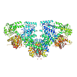
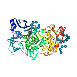
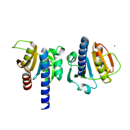
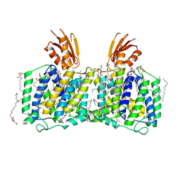 | | YeeE(TsuA)-YeeD(TsuB) complex for thiosulfate uptake | | 分子名称: | (2R)-2,3-dihydroxypropyl (9Z)-octadec-9-enoate, Spirochaeta thermophila YeeE(TsuA)-YeeD(TsuB),UPF0033 domain-containing protein, SirA-like domain-containing protein (chimera) | | 著者 | Ikei, M, Miyazaki, R, Monden, K, Naito, Y, Takeuchi, A, Takahashi, Y.S, Tanaka, Y, Ichikawa, M, Tsukazaki, T. | | 登録日 | 2023-07-11 | | 公開日 | 2024-03-27 | | 実験手法 | X-RAY DIFFRACTION (2.6 Å) | | 主引用文献 | Structure and function of YeeE-YeeD complex for sophisticated thiosulfate uptake
To Be Published
|
|
8OHJ
 | | PanDDA analysis group deposition -- CdaA in complex with fragment F2X-Entry G08 | | 分子名称: | Cyclic di-AMP synthase CdaA, MAGNESIUM ION, N-ethyl-2-{[5-(propan-2-yl)-1,3,4-oxadiazol-2-yl]sulfanyl}acetamide | | 著者 | Garbers, T.B, Neumann, P, Wollenhaupt, J, Weiss, M.S, Ficner, R. | | 登録日 | 2023-03-21 | | 公開日 | 2024-04-03 | | 実験手法 | X-RAY DIFFRACTION (1.22 Å) | | 主引用文献 | PanDDA analysis group deposition -- CdaA in complex with fragment F2X-Entry G08
To Be Published
|
|
7P1M
 | | Galectin-8 N-terminal carbohydrate recognition domain in complex with benzimidazole D-galactal ligand | | 分子名称: | 2-[[(2R,3R,4R)-2-(hydroxymethyl)-3-oxidanyl-3,4-dihydro-2H-pyran-4-yl]oxymethyl]-3-methyl-benzimidazole-5-carboxylic acid, CHLORIDE ION, Galectin-8 | | 著者 | Hassan, M, Hakansson, M, Nilsson, J.U, Kovacic, R. | | 登録日 | 2021-07-02 | | 公開日 | 2021-12-15 | | 最終更新日 | 2024-01-31 | | 実験手法 | X-RAY DIFFRACTION (1.52 Å) | | 主引用文献 | Structure-Guided Design of d-Galactal Derivatives with High Affinity and Selectivity for the Galectin-8 N-Terminal Domain.
Acs Med.Chem.Lett., 12, 2021
|
|


