Loading
PDBj
MenuPDBj@FacebookPDBj@TwitterPDBj@YouTubewwPDB FoundationwwPDB
RCSB PDBPDBeBMRBAdv. SearchSearch help
PDB: 43 results

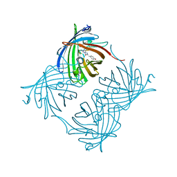
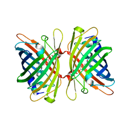
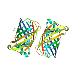
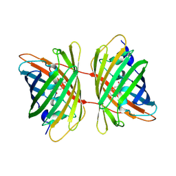
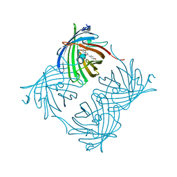
2PXS
DownloadVisualize
BU of 2pxs by Molmil
Crystal Structure of N66D Mutant of Green Fluorescent Protein from Zoanthus sp. at 2.2 A Resolution (Mature State)
Descriptor: GFP-like fluorescent chromoprotein FP506
Authors:Pletnev, S.V, Pletneva, N.V, Tikhonova, T.V, Pletnev, V.Z.
Deposit date:2007-05-14
Release date:2007-09-25
Last modified:2024-04-03
Method:X-RAY DIFFRACTION (2.2 Å)
Cite:Refined crystal structures of red and green fluorescent proteins from the button polyp Zoanthus.
Acta Crystallogr.,Sect.D, 63, 2007
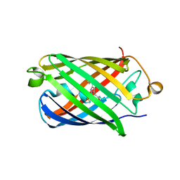
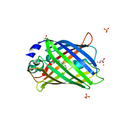
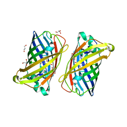
2OJK
DownloadVisualize
BU of 2ojk by Molmil
Crystal Structure of Green Fluorescent Protein from Zoanthus sp at 2.2 A Resolution
Descriptor: GFP-like fluorescent chromoprotein FP506
Authors:Pletneva, N.V, Pletnev, S.V, Tikhonova, T.V, Pletnev, V.Z.
Deposit date:2007-01-12
Release date:2007-09-25
Last modified:2023-11-15
Method:X-RAY DIFFRACTION (2.2 Å)
Cite:Refined crystal structures of red and green fluorescent proteins from the button polyp Zoanthus.
Acta Crystallogr.,Sect.D, 63, 2007
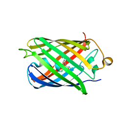
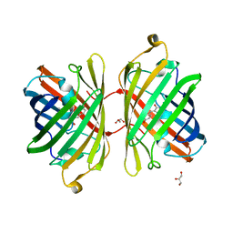
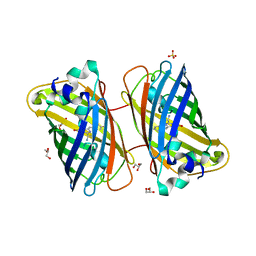
4EDS
DownloadVisualize
BU of 4eds by Molmil
Crystal structure of far-red fluorescent protein eqFP670
Descriptor: far-red fluorescent protein eqFP650
Authors:Pletnev, S, Pletnev, V.Z, Pletneva, N.V.
Deposit date:2012-03-27
Release date:2012-09-05
Last modified:2013-03-27
Method:X-RAY DIFFRACTION (1.6 Å)
Cite:Structural basis for bathochromic shift of fluorescence in far-red fluorescent proteins eqFP650 and eqFP670.
Acta Crystallogr.,Sect.D, 68, 2012
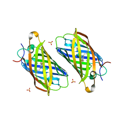
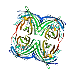
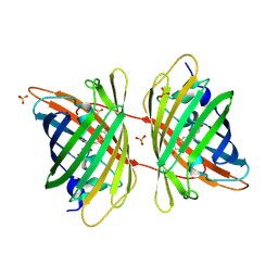
4EDO
DownloadVisualize
BU of 4edo by Molmil
Crystal structure of far-red fluorescent protein eqFP650
Descriptor: far-red fluorescent protein eqFP650
Authors:Pletnev, S, Pletnev, V.Z, Pletneva, N.V.
Deposit date:2012-03-27
Release date:2012-09-05
Last modified:2013-03-27
Method:X-RAY DIFFRACTION (1.8 Å)
Cite:Structural basis for bathochromic shift of fluorescence in far-red fluorescent proteins eqFP650 and eqFP670.
Acta Crystallogr.,Sect.D, 68, 2012
3PJ5
DownloadVisualize
BU of 3pj5 by Molmil
Crystal structure of far-red fluorescent protein Katushka crystallized at pH 5.0
Descriptor: Red fluorescent protein eqFP578, SULFATE ION
Authors:Pletnev, S, Pletneva, N.V, Pletnev, V.Z.
Deposit date:2010-11-08
Release date:2011-05-25
Last modified:2012-02-22
Method:X-RAY DIFFRACTION (1.6 Å)
Cite:Crystallographic study of red fluorescent protein eqFP578 and its far-red variant Katushka reveals opposite pH-induced isomerization of chromophore.
Protein Sci., 20, 2011
4HE4
DownloadVisualize
BU of 4he4 by Molmil
Crystal structure of the yellow fluorescent protein phiYFP (Phialidium sp.)
Descriptor: Yellow fluorescent protein
Authors:Pletneva, N, Pletnev, S, Pletnev, V.Z.
Deposit date:2012-10-03
Release date:2013-05-29
Last modified:2023-12-06
Method:X-RAY DIFFRACTION (2.05 Å)
Cite:Yellow fluorescent protein phiYFPv (Phialidium): structure and structure-based mutagenesis.
Acta Crystallogr.,Sect.D, 69, 2013
4N3D
DownloadVisualize
BU of 4n3d by Molmil
Crystal structure of the dimeric variant EGFP-K162Q in P61 space group
Descriptor: GLYCEROL, Green fluorescent protein, PHOSPHATE ION, ...
Authors:Pletneva, N.V, Pletnev, V.Z, Pletnev, S.V.
Deposit date:2013-10-07
Release date:2014-08-27
Method:X-RAY DIFFRACTION (1.34 Å)
Cite:Three dimensional structure of the dimeric gene-engineered variant of green fluorescent protein egfp-K162Q in P61 crystal space group
Rus.J.Bioorg.Chem., 40, 2014
3PJB
DownloadVisualize
BU of 3pjb by Molmil
Crystal structure of red fluorescent protein eqFP578 crystallized at pH 4.0
Descriptor: GLYCEROL, Red fluorescent protein eqFP578
Authors:Pletnev, S, Pletneva, N.V, Pletnev, V.Z.
Deposit date:2010-11-09
Release date:2011-05-25
Last modified:2018-01-24
Method:X-RAY DIFFRACTION (1.75 Å)
Cite:Crystallographic study of red fluorescent protein eqFP578 and its far-red variant Katushka reveals opposite pH-induced isomerization of chromophore.
Protein Sci., 20, 2011
3PIB
DownloadVisualize
BU of 3pib by Molmil
Crystal structure of red fluorescent protein eqFP578 crystallized at pH 5.5
Descriptor: GLYCEROL, eqFP578 fluorescent protein
Authors:Pletnev, S, Pletneva, N.V, Pletnev, V.Z.
Deposit date:2010-11-05
Release date:2011-05-25
Last modified:2012-02-22
Method:X-RAY DIFFRACTION (1.154 Å)
Cite:Crystallographic study of red fluorescent protein eqFP578 and its far-red variant Katushka reveals opposite pH-induced isomerization of chromophore.
Protein Sci., 20, 2011
3PJ7
DownloadVisualize
BU of 3pj7 by Molmil
Crystal structure of far-red fluorescent protein Katushka crystallized at pH 8.5
Descriptor: Red fluorescent protein eqFP578
Authors:Pletnev, S, Pletneva, N.V, Pletnev, V.Z.
Deposit date:2010-11-08
Release date:2011-05-25
Last modified:2012-02-22
Method:X-RAY DIFFRACTION (1.85 Å)
Cite:Crystallographic study of red fluorescent protein eqFP578 and its far-red variant Katushka reveals opposite pH-induced isomerization of chromophore.
Protein Sci., 20, 2011
3LVD
DownloadVisualize
BU of 3lvd by Molmil
Crystal structure of GFP-like protein aceGFP_G222E (A. coerulescens). UV-photoconverted green form.
Descriptor: GLYCEROL, Green fluorescent protein
Authors:Pletneva, N.V, Pletnev, V.Z, Pletnev, S.V.
Deposit date:2010-02-19
Release date:2010-03-09
Last modified:2023-11-15
Method:X-RAY DIFFRACTION (1.751 Å)
Cite:Structural evidence for a dehydrated intermediate in green fluorescent protein chromophore biosynthesis.
J.Biol.Chem., 285, 2010
3LVC
DownloadVisualize
BU of 3lvc by Molmil
Crystal structure of GFP-like protein aceGFP_G222E (A. coerulescens). Colorless form.
Descriptor: GLYCEROL, Green fluorescent protein
Authors:Pletneva, N.V, Pletnev, V.Z, Pletnev, S.V.
Deposit date:2010-02-19
Release date:2010-03-09
Last modified:2023-11-15
Method:X-RAY DIFFRACTION (1.14 Å)
Cite:Structural evidence for a dehydrated intermediate in green fluorescent protein chromophore biosynthesis.
J.Biol.Chem., 285, 2010
3LVA
DownloadVisualize
BU of 3lva by Molmil
Crystal structure of colorless GFP-like protein from Aequorea coerulescens
Descriptor: GLYCEROL, Green fluorescent protein, SULFATE ION
Authors:Pletneva, N.V, Pletnev, V.Z, Pletnev, S.V.
Deposit date:2010-02-19
Release date:2010-03-09
Last modified:2023-11-15
Method:X-RAY DIFFRACTION (1.5 Å)
Cite:Structural evidence for a dehydrated intermediate in green fluorescent protein chromophore biosynthesis.
J.Biol.Chem., 285, 2010
3GB3
DownloadVisualize
BU of 3gb3 by Molmil
X-ray structure of genetically encoded photosensitizer KillerRed in native form
Descriptor: KillerRed, SULFATE ION
Authors:Pletnev, S, Pletneva, N.V, Pletnev, V.Z.
Deposit date:2009-02-18
Release date:2009-09-08
Last modified:2023-11-15
Method:X-RAY DIFFRACTION (1.75 Å)
Cite:Structural basis for phototoxicity of the genetically encoded photosensitizer KillerRed.
J.Biol.Chem., 284, 2009
3GL4
DownloadVisualize
BU of 3gl4 by Molmil
X-ray structure of photobleached killerred
Descriptor: KillerRed
Authors:Pletnev, S, Pletneva, N.V, Pletnev, V.Z.
Deposit date:2009-03-11
Release date:2009-09-08
Last modified:2023-11-15
Method:X-RAY DIFFRACTION (2.15 Å)
Cite:Structural basis for phototoxicity of the genetically encoded photosensitizer KillerRed.
J.Biol.Chem., 284, 2009
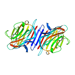
2BQP
DownloadVisualize
BU of 2bqp by Molmil
THE STRUCTURE OF THE PEA LECTIN-D-GLUCOPYRANOSE COMPLEX
Descriptor: CALCIUM ION, MANGANESE (II) ION, PROTEIN (PEA LECTIN), ...
Authors:Ruzeinikov, S.N, Mikhailova Yu, I, Tsygannik, I.N, Pangborn, W, Duax, W, Pletnev, V.Z.
Deposit date:1998-12-08
Release date:1998-12-16
Last modified:2023-08-23
Method:X-RAY DIFFRACTION (1.9 Å)
Cite:The Structure of Pea Lectin-D-Glucopyranose Complex at a 1.9 A Resolution
RUSS.J.BIOORGANIC CHEM., 23, 1997
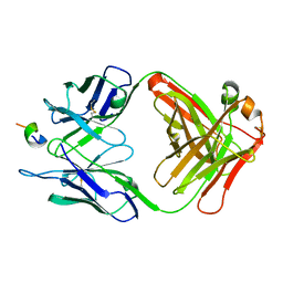
1F90
DownloadVisualize
BU of 1f90 by Molmil
FAB FRAGMENT OF MONOCLONAL ANTIBODY (LNKB-2) AGAINST HUMAN INTERLEUKIN-2 IN COMPLEX WITH ANTIGENIC PEPTIDE
Descriptor: ANTIGENIC NONAPEPTIDE, FAB FRAGMENT OF MONOCLONAL ANTIBODY
Authors:Afonin, P.V, Fokin, A.V, Tsigannik, I.N, Mikhailova, I.Y, Onoprienko, L.V, Mikhaleva, I.I, Ivanov, V.T, Mareeva, T.Y, Nesmeyanov, V.A, Li, N, Duax, W.L, Pletnev, V.Z.
Deposit date:2000-07-06
Release date:2001-07-11
Last modified:2018-04-04
Method:X-RAY DIFFRACTION (2.6 Å)
Cite:Crystal structure of an anti-interleukin-2 monoclonal antibody Fab complexed with an antigenic nonapeptide.
Protein Sci., 10, 2001
1BQP
DownloadVisualize
BU of 1bqp by Molmil
THE STRUCTURE OF THE PEA LECTIN-D-MANNOPYRANOSE COMPLEX
Descriptor: CALCIUM ION, MANGANESE (II) ION, PROTEIN (LECTIN), ...
Authors:Ruzeinikov, S.N, Mikhailova, I.Y, Tsygannik, I.N, Pangborn, W, Duax, W, Pletnev, V.Z.
Deposit date:1998-08-17
Release date:1998-08-26
Last modified:2023-08-09
Method:X-RAY DIFFRACTION (2.1 Å)
Cite:The Structure of the Pea Lectin-D-Mannopyranose Complex at a 2.1 A Resolution
RUSS.J.BIOORGANIC CHEM., 24, 1998
<12


222926

건을2024-07-24부터공개중

PDB statisticsPDBj update infoContact PDBjnumon