Loading
PDBj
メニューPDBj@FacebookPDBj@TwitterPDBj@YouTubewwPDB FoundationwwPDB
RCSB PDBPDBeBMRBAdv. SearchSearch help
PDB: 101 件

7SFA
DownloadVisualize
BU of 7sfa by Molmil
Branchiostoma floridae fluorescent protein LanFP10A2
分子名称: Fluorescent protein lanFP10A2
著者Pletnev, S, Pletneva, N, Pletnev, V.Z, Muslinkina, L.
登録日2021-10-03
公開日2022-03-30
最終更新日2023-11-15
実験手法X-RAY DIFFRACTION (1.65 Å)
主引用文献A novel violet fluorescent protein contains a unique oxidized tyrosine as the simplest chromophore ever reported in fluorescent proteins.
Protein Sci., 31, 2022
7SF9
DownloadVisualize
BU of 7sf9 by Molmil
Branchiostoma Floridae Violet Fluorescent Protein
分子名称: Violet Fluorescent Protein
著者Pletnev, S, Pletneva, N, Pletnev, V.Z, Muslinkina, L.
登録日2021-10-03
公開日2022-03-30
最終更新日2023-10-18
実験手法X-RAY DIFFRACTION (1.8 Å)
主引用文献A novel violet fluorescent protein contains a unique oxidized tyrosine as the simplest chromophore ever reported in fluorescent proteins.
Protein Sci., 31, 2022
3GB3
DownloadVisualize
BU of 3gb3 by Molmil
X-ray structure of genetically encoded photosensitizer KillerRed in native form
分子名称: KillerRed, SULFATE ION
著者Pletnev, S, Pletneva, N.V, Pletnev, V.Z.
登録日2009-02-18
公開日2009-09-08
最終更新日2023-11-15
実験手法X-RAY DIFFRACTION (1.75 Å)
主引用文献Structural basis for phototoxicity of the genetically encoded photosensitizer KillerRed.
J.Biol.Chem., 284, 2009
3GL4
DownloadVisualize
BU of 3gl4 by Molmil
X-ray structure of photobleached killerred
分子名称: KillerRed
著者Pletnev, S, Pletneva, N.V, Pletnev, V.Z.
登録日2009-03-11
公開日2009-09-08
最終更新日2023-11-15
実験手法X-RAY DIFFRACTION (2.15 Å)
主引用文献Structural basis for phototoxicity of the genetically encoded photosensitizer KillerRed.
J.Biol.Chem., 284, 2009
4EDS
DownloadVisualize
BU of 4eds by Molmil
Crystal structure of far-red fluorescent protein eqFP670
分子名称: far-red fluorescent protein eqFP650
著者Pletnev, S, Pletnev, V.Z, Pletneva, N.V.
登録日2012-03-27
公開日2012-09-05
最終更新日2013-03-27
実験手法X-RAY DIFFRACTION (1.6 Å)
主引用文献Structural basis for bathochromic shift of fluorescence in far-red fluorescent proteins eqFP650 and eqFP670.
Acta Crystallogr.,Sect.D, 68, 2012
4EDO
DownloadVisualize
BU of 4edo by Molmil
Crystal structure of far-red fluorescent protein eqFP650
分子名称: far-red fluorescent protein eqFP650
著者Pletnev, S, Pletnev, V.Z, Pletneva, N.V.
登録日2012-03-27
公開日2012-09-05
最終更新日2013-03-27
実験手法X-RAY DIFFRACTION (1.8 Å)
主引用文献Structural basis for bathochromic shift of fluorescence in far-red fluorescent proteins eqFP650 and eqFP670.
Acta Crystallogr.,Sect.D, 68, 2012
6U1A
DownloadVisualize
BU of 6u1a by Molmil
Crystal Structure of Fluorescent Protein FusionRed
分子名称: CALCIUM ION, NICKEL (II) ION, Red fluorescent protein
著者Pletnev, S, Muslinkina, L, Pletneva, N, Pletnev, V.Z.
登録日2019-08-15
公開日2020-04-22
最終更新日2023-11-15
実験手法X-RAY DIFFRACTION (1.09 Å)
主引用文献Two independent routes of post-translational chemistry in fluorescent protein FusionRed.
Int.J.Biol.Macromol., 155, 2020
3PJ5
DownloadVisualize
BU of 3pj5 by Molmil
Crystal structure of far-red fluorescent protein Katushka crystallized at pH 5.0
分子名称: Red fluorescent protein eqFP578, SULFATE ION
著者Pletnev, S, Pletneva, N.V, Pletnev, V.Z.
登録日2010-11-08
公開日2011-05-25
最終更新日2012-02-22
実験手法X-RAY DIFFRACTION (1.6 Å)
主引用文献Crystallographic study of red fluorescent protein eqFP578 and its far-red variant Katushka reveals opposite pH-induced isomerization of chromophore.
Protein Sci., 20, 2011
3PJB
DownloadVisualize
BU of 3pjb by Molmil
Crystal structure of red fluorescent protein eqFP578 crystallized at pH 4.0
分子名称: GLYCEROL, Red fluorescent protein eqFP578
著者Pletnev, S, Pletneva, N.V, Pletnev, V.Z.
登録日2010-11-09
公開日2011-05-25
最終更新日2018-01-24
実験手法X-RAY DIFFRACTION (1.75 Å)
主引用文献Crystallographic study of red fluorescent protein eqFP578 and its far-red variant Katushka reveals opposite pH-induced isomerization of chromophore.
Protein Sci., 20, 2011
3PIB
DownloadVisualize
BU of 3pib by Molmil
Crystal structure of red fluorescent protein eqFP578 crystallized at pH 5.5
分子名称: GLYCEROL, eqFP578 fluorescent protein
著者Pletnev, S, Pletneva, N.V, Pletnev, V.Z.
登録日2010-11-05
公開日2011-05-25
最終更新日2012-02-22
実験手法X-RAY DIFFRACTION (1.154 Å)
主引用文献Crystallographic study of red fluorescent protein eqFP578 and its far-red variant Katushka reveals opposite pH-induced isomerization of chromophore.
Protein Sci., 20, 2011
3PJ7
DownloadVisualize
BU of 3pj7 by Molmil
Crystal structure of far-red fluorescent protein Katushka crystallized at pH 8.5
分子名称: Red fluorescent protein eqFP578
著者Pletnev, S, Pletneva, N.V, Pletnev, V.Z.
登録日2010-11-08
公開日2011-05-25
最終更新日2012-02-22
実験手法X-RAY DIFFRACTION (1.85 Å)
主引用文献Crystallographic study of red fluorescent protein eqFP578 and its far-red variant Katushka reveals opposite pH-induced isomerization of chromophore.
Protein Sci., 20, 2011
4Q7R
DownloadVisualize
BU of 4q7r by Molmil
Crystal structure of large Stokes shift fluorescent protein LSSmOrange
分子名称: ACETATE ION, LSSmOrange, ZINC ION
著者Pletnev, S, Dauter, Z.
登録日2014-04-25
公開日2014-07-09
最終更新日2023-12-06
実験手法X-RAY DIFFRACTION (1.4 Å)
主引用文献Orange Fluorescent Proteins: Structural Studies of LSSmOrange, PSmOrange and PSmOrange2.
Plos One, 9, 2014
3H1R
DownloadVisualize
BU of 3h1r by Molmil
Order-disorder structure of fluorescent protein FP480
分子名称: Fluorescent protein FP480
著者Pletnev, S, Morozova, K.S, Verkhusha, V.V, Dauter, Z.
登録日2009-04-13
公開日2009-09-08
最終更新日2017-11-01
実験手法X-RAY DIFFRACTION (2.41 Å)
主引用文献Rotational order-disorder structure of fluorescent protein FP480
Acta Crystallogr.,Sect.D, 65, 2009
3H1O
DownloadVisualize
BU of 3h1o by Molmil
The Structure of Fluorescent Protein FP480
分子名称: Fluorescent protein FP480, GLYCEROL
著者Pletnev, S, Morozova, K.S, Verkhusha, V.V, Dauter, Z.
登録日2009-04-13
公開日2009-09-08
最終更新日2017-11-01
実験手法X-RAY DIFFRACTION (2 Å)
主引用文献Rotational order-disorder structure of fluorescent protein FP480
Acta Crystallogr.,Sect.D, 65, 2009
6MGH
DownloadVisualize
BU of 6mgh by Molmil
X-ray structure of monomeric near-infrared fluorescent protein miRFP670nano
分子名称: 3-[2-[(~{Z})-[5-[(~{Z})-[(3~{S},4~{R})-3-ethenyl-4-methyl-5-oxidanylidene-pyrrolidin-2-ylidene]methyl]-3-(3-hydroxy-3-oxopropyl)-4-methyl-pyrrol-2-ylidene]methyl]-5-[(~{Z})-(4-ethenyl-3-methyl-5-oxidanylidene-pyrrol-2-ylidene)methyl]-4-methyl-1~{H}-pyrrol-3-yl]propanoic acid, GLYCEROL, ISOPROPYL ALCOHOL, ...
著者Pletnev, S.
登録日2018-09-13
公開日2018-12-19
最終更新日2019-01-30
実験手法X-RAY DIFFRACTION (1.95 Å)
主引用文献Smallest near-infrared fluorescent protein evolved from cyanobacteriochrome as versatile tag for spectral multiplexing.
Nat Commun, 10, 2019
8SXC
DownloadVisualize
BU of 8sxc by Molmil
Crystal Structure of Fluorescent Protein Fusion Red 2
分子名称: Far-red Fluorescent Protein Fusion Red 2, GLYCEROL, HEXAETHYLENE GLYCOL, ...
著者Pletnev, S, Pletneva, N, Pletnev, V.
登録日2023-05-21
公開日2024-02-07
実験手法X-RAY DIFFRACTION (1.5 Å)
主引用文献Crystal Structure of Fluorescent Protein Fusion Red 2
To Be Published
3BXA
DownloadVisualize
BU of 3bxa by Molmil
Monomeric Far-red Fluorescent Protein mKate Crystallized at pH 4.2
分子名称: CITRIC ACID, Far-red fluorescent protein mKate
著者Pletnev, S, Pletneva, N, Pletnev, V.
登録日2008-01-12
公開日2008-07-22
最終更新日2017-10-25
実験手法X-RAY DIFFRACTION (1.75 Å)
主引用文献A Crystallographic Study of Bright Far-Red Fluorescent Protein mKate Reveals pH-induced cis-trans Isomerization of the Chromophore.
J.Biol.Chem., 283, 2008
3BXC
DownloadVisualize
BU of 3bxc by Molmil
Monomeric Far-red Fluorescent Protein mKate Crystallized at pH 9.0
分子名称: Far-red fluorescent protein mKate
著者Pletnev, S, Pletneva, N, Pletnev, V.
登録日2008-01-12
公開日2008-07-22
最終更新日2019-07-24
実験手法X-RAY DIFFRACTION (2.6 Å)
主引用文献A Crystallographic Study of Bright Far-Red Fluorescent Protein mKate Reveals pH-induced cis-trans Isomerization of the Chromophore.
J.Biol.Chem., 283, 2008
3BX9
DownloadVisualize
BU of 3bx9 by Molmil
Monomeric Far-red Fluorescent Protein mKate Crystallized at pH 2.0
分子名称: CITRIC ACID, Far-red fluorescent protein mKate, GLYCEROL
著者Pletnev, S, Pletneva, N, Pletnev, V.
登録日2008-01-11
公開日2008-07-22
最終更新日2023-11-15
実験手法X-RAY DIFFRACTION (1.8 Å)
主引用文献A Crystallographic Study of Bright Far-Red Fluorescent Protein mKate Reveals pH-induced cis-trans Isomerization of the Chromophore.
J.Biol.Chem., 283, 2008
3BXB
DownloadVisualize
BU of 3bxb by Molmil
Monomeric Far-red Fluorescent Protein mKate Crystallized at pH 7.0
分子名称: Far-red fluorescent protein mKate
著者Pletnev, S, Pletneva, N, Pletnev, V.
登録日2008-01-12
公開日2008-07-22
最終更新日2019-07-24
実験手法X-RAY DIFFRACTION (2.6 Å)
主引用文献A Crystallographic Study of Bright Far-Red Fluorescent Protein mKate Reveals pH-induced cis-trans Isomerization of the Chromophore.
J.Biol.Chem., 283, 2008
4XTQ
DownloadVisualize
BU of 4xtq by Molmil
Crystal structure of a mutant (C20S) of a near-infrared fluorescent protein BphP1-FP
分子名称: 3-[2-[(Z)-[5-[(Z)-[(3R,4R)-3-ethenyl-4-methyl-5-oxidanylidene-pyrrolidin-2-ylidene]methyl]-3-(3-hydroxy-3-oxopropyl)-4-methyl-pyrrol-2-ylidene]methyl]-5-[(Z)-(4-ethenyl-3-methyl-5-oxidanylidene-pyrrol-2-ylidene)methyl]-4-methyl-1H-pyrrol-3-yl]propanoic acid, BphP1-FP/C20S, CHLORIDE ION
著者Pletnev, S, Malashkevich, V.N.
登録日2015-01-23
公開日2015-12-09
最終更新日2019-12-04
実験手法X-RAY DIFFRACTION (1.64 Å)
主引用文献Molecular Basis of Spectral Diversity in Near-Infrared Phytochrome-Based Fluorescent Proteins.
Chem.Biol., 22, 2015
4LIS
DownloadVisualize
BU of 4lis by Molmil
Crystal Structure of UDP-galactose-4-epimerase from Aspergillus nidulans
分子名称: GLYCEROL, IODIDE ION, NICOTINAMIDE-ADENINE-DINUCLEOTIDE, ...
著者Dalrymple, S.A, Ko, J, Sheoran, I, Kaminskyj, S.G.W, Sanders, D.A.R.
登録日2013-07-03
公開日2013-10-23
最終更新日2023-09-20
実験手法X-RAY DIFFRACTION (2.8 Å)
主引用文献Elucidation of Substrate Specificity in Aspergillus nidulans UDP-Galactose-4-Epimerase.
Plos One, 8, 2013
8DEF
DownloadVisualize
BU of 8def by Molmil
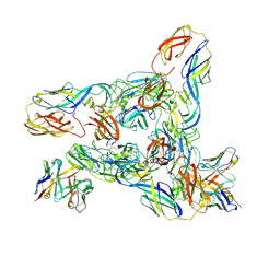
Cryo-EM Structure of Western Equine Encephalitis Virus VLP in complex with SKW24 fab
分子名称: SKW24 Fab heavy chain, SKW24 Fab light chain, Spike glycoprotein E1, ...
著者Pletnev, S, Tsybovsky, Y, Verardi, R, Roedeger, M, Kwong, P.D.
登録日2022-06-20
公開日2023-07-05
最終更新日2024-01-17
実験手法ELECTRON MICROSCOPY (4.2 Å)
主引用文献Vaccine elicitation and structural basis for antibody protection against alphaviruses.
Cell, 186, 2023
4MO2
DownloadVisualize
BU of 4mo2 by Molmil
Crystal Structure of UDP-N-acetylgalactopyranose mutase from Campylobacter jejuni
分子名称: AMMONIUM ION, DIHYDROFLAVINE-ADENINE DINUCLEOTIDE, FLAVIN-ADENINE DINUCLEOTIDE, ...
著者Dalrymple, S.A, Protsko, C, Poulin, M.B, Lowary, T.L, Sanders, D.A.R.
登録日2013-09-11
公開日2014-01-01
最終更新日2024-02-28
実験手法X-RAY DIFFRACTION (2 Å)
主引用文献Specificity of a UDP-GalNAc Pyranose-Furanose Mutase: A Potential Therapeutic Target for Campylobacter jejuni Infections.
Chembiochem, 15, 2014
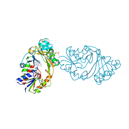
8D9Y
DownloadVisualize
BU of 8d9y by Molmil
Crystal structure of Taipan alpha-neurotoxin in complex with Centi-3FTX-D09 antibody
分子名称: 1-(2-METHOXY-ETHOXY)-2-{2-[2-(2-METHOXY-ETHOXY]-ETHOXY}-ETHANE, 4-(2-HYDROXYETHYL)-1-PIPERAZINE ETHANESULFONIC ACID, Centi-3FTX-D09 Fab heavy chain, ...
著者Pletnev, S, Verardi, R, Tully, E.S, Glanville, J, Kwong, P.D.
登録日2022-06-12
公開日2023-06-14
実験手法X-RAY DIFFRACTION (2.2 Å)
主引用文献Venom protection by antibody from a snakebite hyperimmune subject
To Be Published

222624

件を2024-07-17に公開中

PDB statisticsPDBj update infoContact PDBjnumon