Loading
PDBj
MenuPDBj@FacebookPDBj@X(formerly Twitter)PDBj@BlueSkyPDBj@YouTubewwPDB FoundationwwPDBDonate
RCSB PDBPDBeBMRBAdv. SearchSearch help
PDB: 35 results

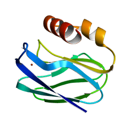
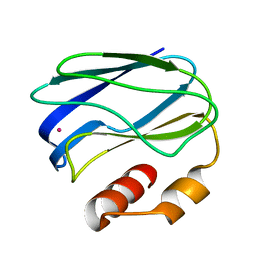
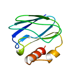
1PZB
DownloadVisualize
BU of 1pzb by Molmil
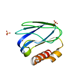
THE CRYSTAL STRUCTURES OF REDUCED PSEUDOAZURIN FROM ALCALIGENES FAECALIS S-6 AT TWO PH VALUES
Descriptor: COPPER (II) ION, PSEUDOAZURIN
Authors:Petratos, K.
Deposit date:1994-08-03
Release date:1994-11-30
Last modified:2024-02-14
Method:X-RAY DIFFRACTION (1.8 Å)
Cite:The crystal structures of reduced pseudoazurin from Alcaligenes faecalis S-6 at two pH values.
FEBS Lett., 347, 1994
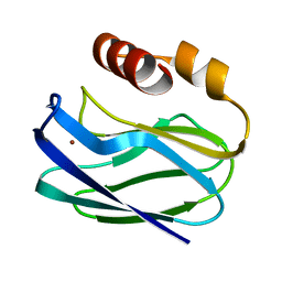
1PZA
DownloadVisualize
BU of 1pza by Molmil
THE CRYSTAL STRUCTURES OF REDUCED PSEUDOAZURIN FROM ALCALIGENES FAECALIS S-6 AT TWO PH VALUES
Descriptor: COPPER (II) ION, PSEUDOAZURIN
Authors:Petratos, K.
Deposit date:1994-09-06
Release date:1994-11-30
Last modified:2024-02-14
Method:X-RAY DIFFRACTION (1.8 Å)
Cite:The crystal structures of reduced pseudoazurin from Alcaligenes faecalis S-6 at two pH values.
FEBS Lett., 347, 1994
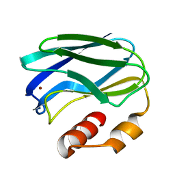
1PZC
DownloadVisualize
BU of 1pzc by Molmil
APO-PSEUDOAZURIN (METAL FREE PROTEIN)
Descriptor: PSEUDOAZURIN
Authors:Petratos, K.
Deposit date:1995-02-22
Release date:1995-09-15
Last modified:2024-02-14
Method:X-RAY DIFFRACTION (1.85 Å)
Cite:The crystal structure of apo-pseudoazurin from Alcaligenes faecalis S-6.
Febs Lett., 368, 1995
1PAZ
DownloadVisualize
BU of 1paz by Molmil
REFINEMENT OF THE STRUCTURE OF PSEUDOAZURIN FROM ALCALIGENES FAECALIS S-6 AT 1.55 ANGSTROMS RESOLUTION
Descriptor: COPPER (II) ION, PSEUDOAZURIN PRECURSOR
Authors:Petratos, K, Dauter, Z, Wilson, K.S.
Deposit date:1988-06-28
Release date:1988-10-09
Last modified:2024-02-14
Method:X-RAY DIFFRACTION (1.55 Å)
Cite:Refinement of the structure of pseudoazurin from Alcaligenes faecalis S-6 at 1.55 A resolution.
Acta Crystallogr.,Sect.B, 44, 1988
5EKA
DownloadVisualize
BU of 5eka by Molmil
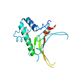
HU DNA-binding protein from Thermus thermophilus
Descriptor: DNA-binding protein HU, GLYCEROL
Authors:Papageorgiou, A, Adam, P, Stavros, P, Nounesis, G, Meijers, R, Petratos, K, Vorgias, C.E.
Deposit date:2015-11-03
Release date:2016-07-06
Last modified:2024-01-10
Method:X-RAY DIFFRACTION (1.69 Å)
Cite:HU histone-like DNA-binding protein from Thermus thermophilus: structural and evolutionary analyses.
Extremophiles, 20, 2016
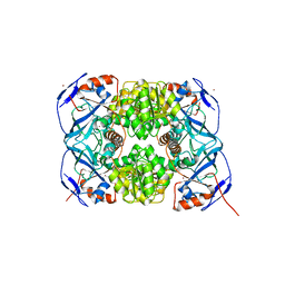
4Z6K
DownloadVisualize
BU of 4z6k by Molmil
Alcohol dehydrogenase from the antarctic psychrophile Moraxella sp. TAE 123
Descriptor: Alcohol dehydrogenase, ZINC ION
Authors:Papanikolau, Y, Bouriotis, V, Petratos, K.
Deposit date:2015-04-05
Release date:2016-04-13
Last modified:2024-05-08
Method:X-RAY DIFFRACTION (1.9 Å)
Cite:Structure and Dynamics of a Thermostable Alcohol Dehydrogenase from the Antarctic Psychrophile Moraxella sp. TAE123
Acs Omega, 2020
6EVH
DownloadVisualize
BU of 6evh by Molmil
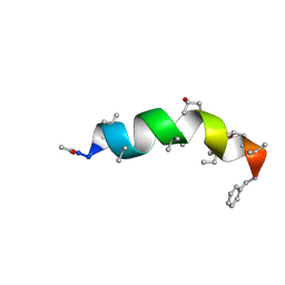
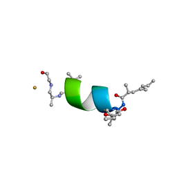
Lipoaminopeptide helioferin A and B from Mycogone rosea
Descriptor: CHLORIDE ION, FLUORIDE ION, Lipoaminopeptide helioferin A and B
Authors:Gessmann, R, Petratos, K.
Deposit date:2017-11-01
Release date:2018-07-18
Last modified:2023-11-15
Method:X-RAY DIFFRACTION (0.9 Å)
Cite:Aminolipopeptide helioferin A and B
Acta Cryst. D, 74, 2018
5MAS
DownloadVisualize
BU of 5mas by Molmil
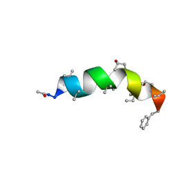
Peptaibol Bergofungin A
Descriptor: Bergofungin A
Authors:Gessmann, R, Petratos, K.
Deposit date:2016-11-04
Release date:2017-02-22
Last modified:2017-03-01
Method:X-RAY DIFFRACTION (0.84 Å)
Cite:A natural, single-residue substitution yields a less active peptaibiotic: the structure of bergofungin A at atomic resolution.
Acta Crystallogr F Struct Biol Commun, 73, 2017
3SBN
DownloadVisualize
BU of 3sbn by Molmil
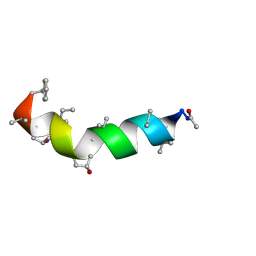
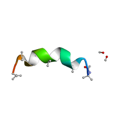
trichovirin I-4A in polar environment at 0.9 Angstroem
Descriptor: ACETONITRILE, METHANOL, Trichovirin I-4A
Authors:Gessmann, R, Axford, D, Petratos, K.
Deposit date:2011-06-06
Release date:2011-12-28
Last modified:2023-11-15
Method:X-RAY DIFFRACTION (0.9 Å)
Cite:Four complete turns of a curved 310-helix at atomic resolution: The crystal structure of the peptaibol trichovirin I-4A in polar environment suggests a transition to alpha-helix for membrane function
Acta Crystallogr.,Sect.D, 68, 2012
4BY8
DownloadVisualize
BU of 4by8 by Molmil
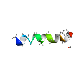
Microheterogeneous Paracelsin-X from Trichoderma reesei
Descriptor: METHANOL, PARACELSIN-X
Authors:Gessmann, R, Petratos, K.
Deposit date:2013-07-18
Release date:2013-08-21
Last modified:2019-04-17
Method:X-RAY DIFFRACTION (0.94 Å)
Cite:Microheterogeneous Paracelsin-X from Trichoderma Reesei
To be Published
1ABA
DownloadVisualize
BU of 1aba by Molmil
THE STRUCTURE OF OXIDIZED BACTERIOPHAGE T4 GLUTAREDOXIN (THIOREDOXIN). REFINEMENT OF NATIVE AND MUTANT PROTEINS
Descriptor: 2-(N-MORPHOLINO)-ETHANESULFONIC ACID, GLUTAREDOXIN
Authors:Eklund, H, Ingelman, M, Soderberg, B.-O, Uhlin, T, Nordlund, P, Nikkola, M, Sonnerstam, U, Joelson, T, Petratos, K.
Deposit date:1992-04-24
Release date:1993-10-31
Last modified:2024-10-23
Method:X-RAY DIFFRACTION (1.45 Å)
Cite:Structure of oxidized bacteriophage T4 glutaredoxin (thioredoxin). Refinement of native and mutant proteins.
J.Mol.Biol., 228, 1992
1AAZ
DownloadVisualize
BU of 1aaz by Molmil
THE STRUCTURE OF OXIDIZED BACTERIOPHAGE T4 GLUTAREDOXIN (THIOREDOXIN)
Descriptor: CADMIUM ION, GLUTAREDOXIN
Authors:Eklund, H, Ingelman, M, Soderberg, B.-O, Uhlin, T, Nordlund, P, Nikkola, M, Sonnerstam, U, Joelson, T, Petratos, K.
Deposit date:1992-04-24
Release date:1993-10-31
Last modified:2024-10-16
Method:X-RAY DIFFRACTION (2 Å)
Cite:Structure of oxidized bacteriophage T4 glutaredoxin (thioredoxin). Refinement of native and mutant proteins.
J.Mol.Biol., 228, 1992
3NYK
DownloadVisualize
BU of 3nyk by Molmil
The structure of cobalt-substituted pseudoazurin from Alcaligenes faecalis
Descriptor: COBALT (II) ION, Pseudoazurin
Authors:Gessmann, R, Petratos, K.
Deposit date:2010-07-15
Release date:2010-10-20
Last modified:2024-02-21
Method:X-RAY DIFFRACTION (1.86 Å)
Cite:The crystal structure of cobalt-substituted pseudoazurin from Alcaligenes faecalis.
Biopolymers, 95, 2011
2RVE
DownloadVisualize
BU of 2rve by Molmil
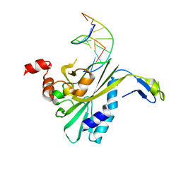
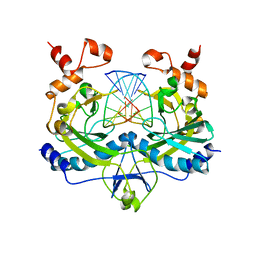
THE CRYSTAL STRUCTURE OF ECORV ENDONUCLEASE AND OF ITS COMPLEXES WITH COGNATE AND NON-COGNATE DNA SEGMENTS
Descriptor: DNA (5'-D(*CP*GP*AP*GP*CP*TP*CP*G)-3'), PROTEIN (ECO RV (E.C.3.1.21.4))
Authors:Winkler, F.K, Banner, D.W, Oefner, C, Tsernoglou, D, Brown, R.S, Heathman, S.P, Bryan, R.K, Martin, P.D, Petratos, K, Wilson, K.S.
Deposit date:1991-03-19
Release date:1992-01-15
Last modified:2024-02-21
Method:X-RAY DIFFRACTION (3 Å)
Cite:The crystal structure of EcoRV endonuclease and of its complexes with cognate and non-cognate DNA fragments.
EMBO J., 12, 1993
4G13
DownloadVisualize
BU of 4g13 by Molmil
Crystal structure of samarosporin I at 100K
Descriptor: SAMAROSPORIN I
Authors:Gessmann, R, Axford, D, Petratos, K.
Deposit date:2012-07-10
Release date:2012-10-03
Last modified:2018-07-18
Method:X-RAY DIFFRACTION (0.8 Å)
Cite:The crystal structure of samarosporin I at atomic resolution.
J.Pept.Sci., 18, 2012
4G14
DownloadVisualize
BU of 4g14 by Molmil
Crystal structure of samarosporin I at 293K
Descriptor: SAMAROSPORIN I
Authors:Gessmann, R, Axford, D, Petratos, K.
Deposit date:2012-07-10
Release date:2012-10-03
Last modified:2023-12-06
Method:X-RAY DIFFRACTION (1.09 Å)
Cite:The crystal structure of samarosporin I at atomic resolution.
J.Pept.Sci., 18, 2012
2B7U
DownloadVisualize
BU of 2b7u by Molmil
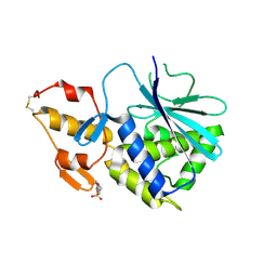
Ribosome inactivating protein type 1 from Charybdis maritima AGG
Descriptor: 2-(N-MORPHOLINO)-ETHANESULFONIC ACID, CHARYBDIN
Authors:Gessmann, R, Petratos, K.
Deposit date:2005-10-05
Release date:2006-06-27
Last modified:2024-10-23
Method:X-RAY DIFFRACTION (1.6 Å)
Cite:Isolation, characterization, sequencing and crystal structure of charybdin, a type 1 ribosome-inactivating protein from Charybdis maritima agg.
Febs J., 273, 2006
4RVE
DownloadVisualize
BU of 4rve by Molmil
THE CRYSTAL STRUCTURE OF ECORV ENDONUCLEASE AND OF ITS COMPLEXES WITH COGNATE AND NON-COGNATE DNA SEGMENTS
Descriptor: DNA (5'-D(*GP*GP*GP*AP*TP*AP*TP*CP*CP*C)-3'), PROTEIN (ECO RV (E.C.3.1.21.4))
Authors:Winkler, F.K, Banner, D.W, Oefner, C, Tsernoglou, D, Brown, R.S, Heathman, S.P, Bryan, R.K, Martin, P.D, Petratos, K, Wilso, K.S.
Deposit date:1993-02-18
Release date:1993-04-15
Last modified:2024-02-28
Method:X-RAY DIFFRACTION (3 Å)
Cite:The crystal structure of EcoRV endonuclease and of its complexes with cognate and non-cognate DNA fragments.
EMBO J., 12, 1993
4RH4
DownloadVisualize
BU of 4rh4 by Molmil
Zinc-substituted pseudoazurin solved by S/Zn-SAD phasing
Descriptor: Pseudoazurin, SULFATE ION, ZINC ION
Authors:Gessmann, R, Petratos, K.
Deposit date:2014-10-01
Release date:2015-01-14
Last modified:2024-02-28
Method:X-RAY DIFFRACTION (1.6 Å)
Cite:Zinc-substituted pseudoazurin solved by S/Zn-SAD phasing.
Acta Crystallogr F Struct Biol Commun, 71, 2015
1FFQ
DownloadVisualize
BU of 1ffq by Molmil
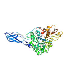
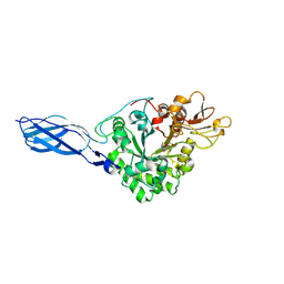
CRYSTAL STRUCTURE OF CHITINASE A COMPLEXED WITH ALLOSAMIDIN
Descriptor: 2-acetamido-2-deoxy-beta-D-allopyranose-(1-4)-2-acetamido-2-deoxy-beta-D-allopyranose, ALLOSAMIZOLINE, CHITINASE A
Authors:Papanikolau, Y, Tavlas, G, Vorgias, C.E, Petratos, K.
Deposit date:2000-07-26
Release date:2003-02-11
Last modified:2024-11-06
Method:X-RAY DIFFRACTION (1.9 Å)
Cite:De novo purification scheme and crystallization conditions yield high-resolution structures of chitinase A and its complex with the inhibitor allosamidin.
Acta Crystallogr.,Sect.D, 59, 2003
1FFR
DownloadVisualize
BU of 1ffr by Molmil
CRYSTAL STRUCTURE OF CHITINASE A MUTANT Y390F COMPLEXED WITH HEXA-N-ACETYLCHITOHEXAOSE (NAG)6
Descriptor: 2-acetamido-2-deoxy-beta-D-glucopyranose-(1-4)-2-acetamido-2-deoxy-beta-D-glucopyranose-(1-4)-2-acetamido-2-deoxy-beta-D-glucopyranose-(1-4)-2-acetamido-2-deoxy-beta-D-glucopyranose-(1-4)-2-acetamido-2-deoxy-beta-D-glucopyranose-(1-4)-2-acetamido-2-deoxy-beta-D-glucopyranose-(1-4)-2-acetamido-2-deoxy-beta-D-glucopyranose, CHITINASE A
Authors:Papanikolau, Y, Prag, G, Tavlas, G, Vorgias, C.E, Oppenheim, A.B, Petratos, K.
Deposit date:2000-07-26
Release date:2001-09-26
Last modified:2024-11-20
Method:X-RAY DIFFRACTION (1.8 Å)
Cite:High resolution structural analyses of mutant chitinase A complexes with substrates provide new insight into the mechanism of catalysis.
Biochemistry, 40, 2001
1EIB
DownloadVisualize
BU of 1eib by Molmil
CRYSTAL STRUCTURE OF CHITINASE A MUTANT D313A COMPLEXED WITH OCTA-N-ACETYLCHITOOCTAOSE (NAG)8.
Descriptor: 2-acetamido-2-deoxy-beta-D-glucopyranose-(1-4)-2-acetamido-2-deoxy-beta-D-glucopyranose-(1-4)-2-acetamido-2-deoxy-beta-D-glucopyranose-(1-4)-2-acetamido-2-deoxy-beta-D-glucopyranose-(1-4)-2-acetamido-2-deoxy-beta-D-glucopyranose-(1-4)-2-acetamido-2-deoxy-beta-D-glucopyranose-(1-4)-2-acetamido-2-deoxy-beta-D-glucopyranose-(1-4)-2-acetamido-2-deoxy-beta-D-glucopyranose, CHITINASE A
Authors:Papanikolau, Y, Prag, G, Tavlas, G, Vorgias, C.E, Oppenheim, A.B, Petratos, K.
Deposit date:2000-02-25
Release date:2001-02-25
Last modified:2024-11-06
Method:X-RAY DIFFRACTION (1.8 Å)
Cite:High resolution structural analyses of mutant chitinase A complexes with substrates provide new insight into the mechanism of catalysis.
Biochemistry, 40, 2001
1EHN
DownloadVisualize
BU of 1ehn by Molmil
CRYSTAL STRUCTURE OF CHITINASE A MUTANT E315Q COMPLEXED WITH OCTA-N-ACETYLCHITOOCTAOSE (NAG)8.
Descriptor: 2-acetamido-2-deoxy-beta-D-glucopyranose-(1-4)-2-acetamido-2-deoxy-beta-D-glucopyranose-(1-4)-2-acetamido-2-deoxy-beta-D-glucopyranose-(1-4)-2-acetamido-2-deoxy-beta-D-glucopyranose-(1-4)-2-acetamido-2-deoxy-beta-D-glucopyranose-(1-4)-2-acetamido-2-deoxy-beta-D-glucopyranose-(1-4)-2-acetamido-2-deoxy-beta-D-glucopyranose-(1-4)-2-acetamido-2-deoxy-beta-D-glucopyranose, CHITINASE A
Authors:Papanikolau, Y, Prag, G, Tavlas, G, Vorgias, C.E, Oppenheim, A.B, Petratos, K.
Deposit date:2000-02-22
Release date:2001-02-22
Last modified:2024-10-09
Method:X-RAY DIFFRACTION (1.9 Å)
Cite:High resolution structural analyses of mutant chitinase A complexes with substrates provide new insight into the mechanism of catalysis.
Biochemistry, 40, 2001
1EDQ
DownloadVisualize
BU of 1edq by Molmil
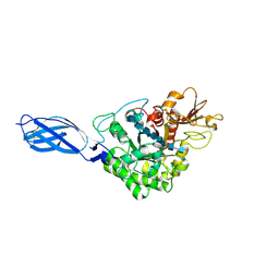
CRYSTAL STRUCTURE OF CHITINASE A FROM S. MARCESCENS AT 1.55 ANGSTROMS
Descriptor: CHITINASE A
Authors:Papanikolau, Y, Petratos, K.
Deposit date:2000-01-28
Release date:2000-02-18
Last modified:2024-10-16
Method:X-RAY DIFFRACTION (1.55 Å)
Cite:De novo purification scheme and crystallization conditions yield high-resolution structures of chitinase A and its complex with the inhibitor allosamidin.
Acta Crystallogr.,Sect.D, 59, 2003
2FSH
DownloadVisualize
BU of 2fsh by Molmil
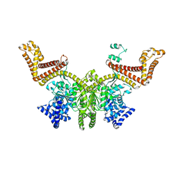
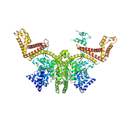
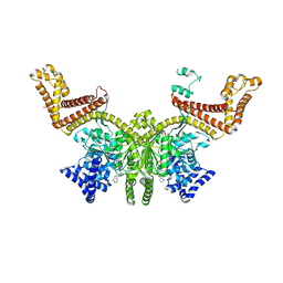
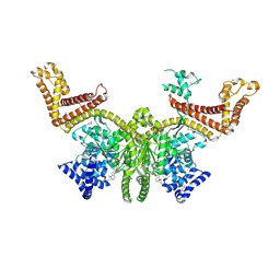
Complex SecA:AMP-PNP from Escherichia coli
Descriptor: PHOSPHOAMINOPHOSPHONIC ACID-ADENYLATE ESTER, Preprotein translocase secA subunit
Authors:Papanikolau, Y, Petratos, K, Economou, A.
Deposit date:2006-01-23
Release date:2007-01-16
Last modified:2023-10-25
Method:X-RAY DIFFRACTION (2 Å)
Cite:Structure of dimeric SecA, the Escherichia coli preprotein translocase motor.
J.Mol.Biol., 366, 2007


12>

227933

數據於2024-11-27公開中

PDB statisticsPDBj update infoContact PDBjnumon