Loading
PDBj
MenuPDBj@FacebookPDBj@TwitterPDBj@YouTubewwPDB FoundationwwPDB
RCSB PDBPDBeBMRBAdv. SearchSearch help
PDB: 34 results

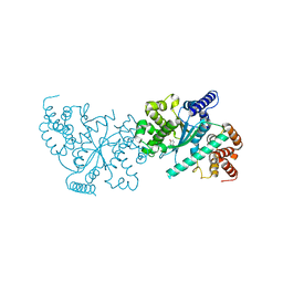
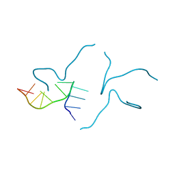
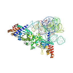
2RKJ
DownloadVisualize
BU of 2rkj by Molmil
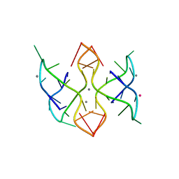
Cocrystal structure of a tyrosyl-tRNA synthetase splicing factor with a group I intron RNA
Descriptor: RNA (238-MER), RNA (5'-R(P*GP*CP*UP*U)-3'), Tyrosyl-tRNA synthetase
Authors:Paukstelis, P.J, Chen, J.-H, Chase, E, Lambowitz, A.M, Golden, B.L.
Deposit date:2007-10-16
Release date:2008-01-08
Last modified:2023-08-30
Method:X-RAY DIFFRACTION (4.5 Å)
Cite:Structure of a tyrosyl-tRNA synthetase splicing factor bound to a group I intron RNA.
Nature, 451, 2008
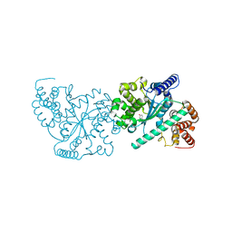
1Y42
DownloadVisualize
BU of 1y42 by Molmil
Crystal structure of a C-terminally truncated CYT-18 protein
Descriptor: TYROSINE, Tyrosyl-tRNA synthetase, mitochondrial
Authors:Paukstelis, P.J, Coon, R, Madabusi, L, Nowakowski, J, Monzingo, A, Robertus, J, Lambowitz, A.M.
Deposit date:2004-11-29
Release date:2005-02-15
Last modified:2023-08-23
Method:X-RAY DIFFRACTION (1.95 Å)
Cite:A tyrosyl-tRNA synthetase adapted to function in group I intron splicing by acquiring a new RNA binding surface.
Mol.Cell, 17, 2005
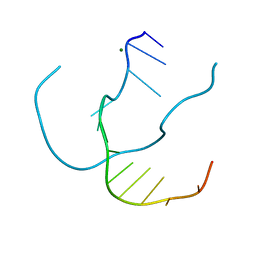
4OJM
DownloadVisualize
BU of 4ojm by Molmil
Crystal structure of a C-terminally truncated CYT-18 protein including N-terminal residues
Descriptor: TYROSINE, Tyrosine--tRNA ligase, mitochondrial
Authors:Paukstelis, P.J, Geng, C.
Deposit date:2014-01-21
Release date:2014-04-16
Last modified:2023-09-20
Method:X-RAY DIFFRACTION (1.95 Å)
Cite:An in Vitro Peptide Complementation Assay for CYT-18-Dependent Group I Intron Splicing Reveals a New Role for the N-Terminus.
Biochemistry, 53, 2014
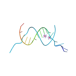
1P1Y
DownloadVisualize
BU of 1p1y by Molmil
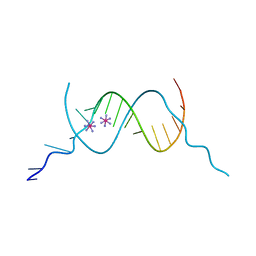
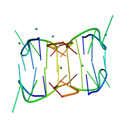
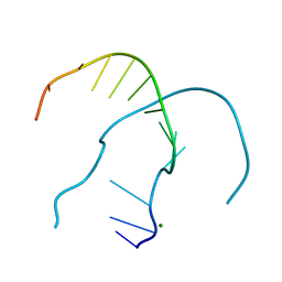
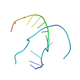
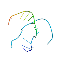
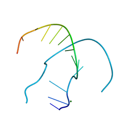
Crystal structure of a continuous three-dimensional DNA lattice from d(GGACAGATGGGAG)
Descriptor: 5'-D(*GP*GP*AP*(CBR)P*AP*GP*AP*(BRU)P*GP*GP*GP*AP*G)-3', MAGNESIUM ION
Authors:Paukstelis, P.J, Nowakowski, J, Birktoft, J.J, Seeman, N.C.
Deposit date:2003-04-14
Release date:2004-06-01
Last modified:2024-02-14
Method:X-RAY DIFFRACTION (2.1 Å)
Cite:Crystal structure of a continuous three-dimensional DNA lattice.
Chem.Biol., 11, 2004
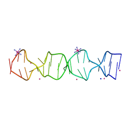
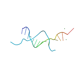
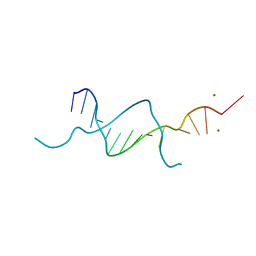
4F8G
DownloadVisualize
BU of 4f8g by Molmil
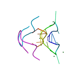
Three-dimensional DNA lattices with non-canonical base pairs
Descriptor: COBALT HEXAMMINE(III), DNA (5'-D(*G*CP*GP*AP*AP*AP*GP*GP*GP*CP*AP*CP*GP*TP*GP*CP*CP*CP*T)-3')
Authors:Paukstelis, P.J, Muser, S.E.
Deposit date:2012-05-17
Release date:2012-07-25
Last modified:2024-02-28
Method:X-RAY DIFFRACTION (1.93 Å)
Cite:Three-Dimensional DNA Crystals with pH-Responsive Noncanonical Junctions.
J.Am.Chem.Soc., 134, 2012
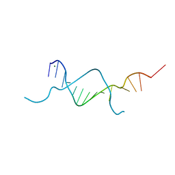
4F8I
DownloadVisualize
BU of 4f8i by Molmil
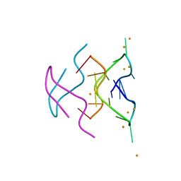
Three-dimensional DNA lattices with non-canonical base pairs
Descriptor: COBALT HEXAMMINE(III), DNA (5'-D(*G*CP*GP*AP*AP*AP*GP*GP*GP*CP*AP*CP*GP*TP*GP*CP*CP*CP*T)-3')
Authors:Paukstelis, P.J, Muser, S.E.
Deposit date:2012-05-17
Release date:2012-07-25
Last modified:2024-02-28
Method:X-RAY DIFFRACTION (2 Å)
Cite:Three-Dimensional DNA Crystals with pH-Responsive Noncanonical Junctions.
J.Am.Chem.Soc., 134, 2012
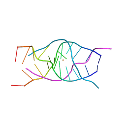
4U92
DownloadVisualize
BU of 4u92 by Molmil
Crystal structure of a DNA/Ba2+ G-quadruplex containing a water-mediated C-tetrad
Descriptor: BARIUM ION, DNA (5'-D(*CP*CP*AP*KP*GP*CP*GP*TP*GP*G)-3'), MAGNESIUM ION
Authors:Paukstelis, P.J, Zhang, D, Huang, T, Lukeman, P.
Deposit date:2014-08-05
Release date:2014-11-26
Last modified:2023-12-27
Method:X-RAY DIFFRACTION (1.5 Å)
Cite:Crystal structure of a DNA/Ba2+ G-quadruplex containing a water-mediated C-tetrad.
Nucleic Acids Res., 42, 2014
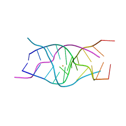
7T6Y
DownloadVisualize
BU of 7t6y by Molmil
d((CGA)5TGA) parallel-stranded homo-duplex
Descriptor: BARIUM ION, DNA (5'-D(*CP*GP*AP*CP*GP*AP*CP*GP*AP*CP*GP*AP*CP*GP*AP*TP*GP*A)-3')
Authors:Luteran, E.M, Paukstelis, P.J.
Deposit date:2021-12-14
Release date:2021-12-22
Last modified:2023-10-18
Method:X-RAY DIFFRACTION (2.3 Å)
Cite:The parallel-stranded d(CGA) duplex is a highly predictable structural motif with two conformationally distinct strands.
Acta Crystallogr D Struct Biol, 78, 2022
5BZY
DownloadVisualize
BU of 5bzy by Molmil
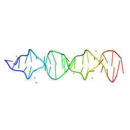
X-ray crystal structure of a continuously hydrogen bonded 14mer DNA lattice.
Descriptor: DNA (5'-D(*GP*GP*AP*AP*AP*TP*AP*TP*TP*GP*GP*AP*GP*A)-3'), MAGNESIUM ION
Authors:Saoji, M, Paukstelis, P.J.
Deposit date:2015-06-11
Release date:2015-11-11
Last modified:2023-09-27
Method:X-RAY DIFFRACTION (2.4 Å)
Cite:Sequence-dependent structural changes in a self-assembling DNA oligonucleotide.
Acta Crystallogr.,Sect.D, 71, 2015
7SB8
DownloadVisualize
BU of 7sb8 by Molmil
d(GA(CGA)5) parallel-stranded homo-duplex
Descriptor: COBALT HEXAMMINE(III), GA(CGA)5, SODIUM ION, ...
Authors:Luteran, E.M, Paukstelis, P.J.
Deposit date:2021-09-24
Release date:2021-10-06
Last modified:2023-10-18
Method:X-RAY DIFFRACTION (1.317 Å)
Cite:The parallel-stranded d(CGA) duplex is a highly predictable structural motif with two conformationally distinct strands.
Acta Crystallogr D Struct Biol, 78, 2022
6TZS
DownloadVisualize
BU of 6tzs by Molmil
A DNA i-motif/duplex hybrid
Descriptor: DNA (5'-D(*CP*CP*AP*GP*GP*CP*TP*GP*(CBR)P*AP*A)-3')
Authors:Chu, B, Paukstelis, P.J.
Deposit date:2019-08-13
Release date:2019-10-16
Last modified:2024-03-13
Method:X-RAY DIFFRACTION (2.6 Å)
Cite:A DNA G-quadruplex/i-motif hybrid.
Nucleic Acids Res., 47, 2019
6TZQ
DownloadVisualize
BU of 6tzq by Molmil
A DNA G-quadruplex/i-motif hybrid
Descriptor: BARIUM ION, DNA (5'-D(*CP*CP*AP*GP*GP*CP*TP*GP*CP*AP*A)-3')
Authors:Chu, B, Paukstelis, P.J.
Deposit date:2019-08-13
Release date:2019-10-16
Last modified:2023-10-11
Method:X-RAY DIFFRACTION (2.29 Å)
Cite:A DNA G-quadruplex/i-motif hybrid.
Nucleic Acids Res., 47, 2019
6TZR
DownloadVisualize
BU of 6tzr by Molmil
A DNA G-quadruplex/i-motif hybrid
Descriptor: BARIUM ION, DNA (5'-D(*CP*CP*AP*GP*GP*CP*(BRU)P*GP*CP*AP*A)-3')
Authors:Chu, B, Paukstelis, P.J.
Deposit date:2019-08-13
Release date:2019-10-16
Last modified:2024-03-13
Method:X-RAY DIFFRACTION (2.4 Å)
Cite:A DNA G-quadruplex/i-motif hybrid.
Nucleic Acids Res., 47, 2019
6MC4
DownloadVisualize
BU of 6mc4 by Molmil
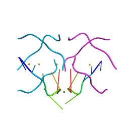
Crystal structure of a tetrameric DNA fold-back quadruplex
Descriptor: CALCIUM ION, DNA (5'-D(*CP*GP*TP*CP*AP*GP*GP*CP*G)-3'), POTASSIUM ION
Authors:Chu, B, Paukstelis, P.J.
Deposit date:2018-08-30
Release date:2018-11-14
Last modified:2024-03-13
Method:X-RAY DIFFRACTION (2.25 Å)
Cite:Crystal Structure of a Tetrameric DNA Fold-Back Quadruplex.
J. Am. Chem. Soc., 140, 2018
6MC3
DownloadVisualize
BU of 6mc3 by Molmil
Crystal structure of a tetrameric DNA fold-back quadruplex
Descriptor: DNA (5'-D(*CP*GP*TP*GP*AP*GP*GP*CP*G)-3'), MAGNESIUM ION
Authors:Chu, B, Paukstelis, P.J.
Deposit date:2018-08-30
Release date:2018-11-14
Last modified:2024-03-13
Method:X-RAY DIFFRACTION (1.57 Å)
Cite:Crystal Structure of a Tetrameric DNA Fold-Back Quadruplex.
J. Am. Chem. Soc., 140, 2018
6MC2
DownloadVisualize
BU of 6mc2 by Molmil
Crystal structure of a tetrameric DNA fold-back quadruplex
Descriptor: DNA (5'-D(*CP*GP*TP*AP*AP*GP*GP*CP*G)-3'), MAGNESIUM ION
Authors:Chu, B, Paukstelis, P.J.
Deposit date:2018-08-30
Release date:2018-11-14
Last modified:2024-03-13
Method:X-RAY DIFFRACTION (1.05 Å)
Cite:Crystal Structure of a Tetrameric DNA Fold-Back Quadruplex.
J. Am. Chem. Soc., 140, 2018
6N4G
DownloadVisualize
BU of 6n4g by Molmil
Crystal structure of a tetrameric DNA fold-back quadruplex
Descriptor: BARIUM ION, DNA (5'-D(*CP*GP*TP*TP*AP*GP*GP*CP*G)-3')
Authors:Chu, B, Paukstelis, P.J.
Deposit date:2018-11-19
Release date:2018-12-05
Last modified:2024-03-13
Method:X-RAY DIFFRACTION (1.4 Å)
Cite:Crystal Structure of a Tetrameric DNA Fold-Back Quadruplex.
J. Am. Chem. Soc., 140, 2018
5BZ7
DownloadVisualize
BU of 5bz7 by Molmil
X-ray crystal structure of a continuously hydrogen bonded 14mer DNA lattice.
Descriptor: DNA (5'-D(*GP*GP*AP*AP*AP*AP*TP*TP*TP*GP*GP*AP*GP*A)-3'), MAGNESIUM ION
Authors:Saoji, M, Paukstelis, P.J.
Deposit date:2015-06-11
Release date:2015-11-11
Last modified:2023-09-27
Method:X-RAY DIFFRACTION (2.03 Å)
Cite:Sequence-dependent structural changes in a self-assembling DNA oligonucleotide.
Acta Crystallogr.,Sect.D, 71, 2015
5BXW
DownloadVisualize
BU of 5bxw by Molmil
X-ray crystal structure of a continuously hydrogen bonded 14mer DNA lattice.
Descriptor: DNA (5'-D(*GP*GP*AP*AP*AP*GP*CP*TP*TP*GP*GP*AP*GP*A)-3'), MAGNESIUM ION
Authors:Saoji, M, Paukstelis, P.J.
Deposit date:2015-06-09
Release date:2015-11-11
Last modified:2024-03-06
Method:X-RAY DIFFRACTION (2.15 Å)
Cite:Sequence-dependent structural changes in a self-assembling DNA oligonucleotide.
Acta Crystallogr.,Sect.D, 71, 2015
5BZ9
DownloadVisualize
BU of 5bz9 by Molmil
X-ray crystal structure of a continuously hydrogen bonded 14mer DNA lattice.
Descriptor: DNA (5'-D(GGAAACGTTGGAGA), MAGNESIUM ION
Authors:Saoji, M, Paukstelis, P.J.
Deposit date:2015-06-11
Release date:2015-11-11
Last modified:2023-09-27
Method:X-RAY DIFFRACTION (2.1 Å)
Cite:Sequence-dependent structural changes in a self-assembling DNA oligonucleotide.
Acta Crystallogr.,Sect.D, 71, 2015
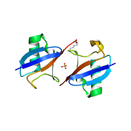
5EMZ
DownloadVisualize
BU of 5emz by Molmil
Crystal structure of K48-linked diubiquitin with F45W mutation in the proximal unit
Descriptor: Polyubiquitin-B, SULFATE ION
Authors:Nakasone, M.A, Paukstelis, P.J, Fushman, D.
Deposit date:2015-11-07
Release date:2016-11-16
Last modified:2023-09-27
Method:X-RAY DIFFRACTION (1.66 Å)
Cite:Structural characterization and practical fluorescence applications of the F45W ubiquitin mutant
To Be Published
4RP2
DownloadVisualize
BU of 4rp2 by Molmil
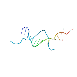
Sequence and structure of a self-assembled 3-D DNA crystal: D(GGATAGCTAGGAG)
Descriptor: D(GGATAGCTAGGAG), MAGNESIUM ION
Authors:Saoji, M.M, Paukstelis, P.J.
Deposit date:2014-10-29
Release date:2015-06-17
Last modified:2023-09-20
Method:X-RAY DIFFRACTION (2.32 Å)
Cite:Probing the role of sequence in the assembly of three-dimensional DNA crystals.
Biopolymers, 103, 2015
4ROY
DownloadVisualize
BU of 4roy by Molmil
Sequence and structure of a self-assembled 3-D DNA crystal: D(GGACGATCGGGAG)
Descriptor: D(GGACGATCGGGAG), MAGNESIUM ION
Authors:Saoji, M.M, Paukstelis, P.J.
Deposit date:2014-10-29
Release date:2015-06-17
Last modified:2023-09-20
Method:X-RAY DIFFRACTION (2.09 Å)
Cite:Probing the role of sequence in the assembly of three-dimensional DNA crystals.
Biopolymers, 103, 2015
4ROG
DownloadVisualize
BU of 4rog by Molmil
Sequence and structure of a self-assembled 3-D DNA crystal: D(GGACACGTGGGAG)
Descriptor: D(GGACACGTGGGAG), MAGNESIUM ION
Authors:Saoji, M.M, Paukstelis, P.J.
Deposit date:2014-10-28
Release date:2015-06-17
Last modified:2023-09-20
Method:X-RAY DIFFRACTION (2.08 Å)
Cite:Probing the role of sequence in the assembly of three-dimensional DNA crystals.
Biopolymers, 103, 2015
4ROK
DownloadVisualize
BU of 4rok by Molmil
Sequence and structure of a self-assembled 3-D DNA crystal: D(GGACAGCTGGGAG)
Descriptor: D(GGACAGCTGGGAG), MAGNESIUM ION
Authors:Saoji, M.M, Paukstelis, P.J.
Deposit date:2014-10-28
Release date:2015-06-17
Last modified:2023-09-20
Method:X-RAY DIFFRACTION (2.16 Å)
Cite:Probing the role of sequence in the assembly of three-dimensional DNA crystals.
Biopolymers, 103, 2015


12>

227111

数据于2024-11-06公开中

PDB statisticsPDBj update infoContact PDBjnumon