8G55
 
 | |
7FT5
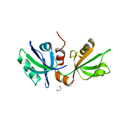
 | | SDCBP PanDDA analysis group deposition -- The PDZ domans of SDCBP in complex with POB0093 | | 分子名称: | 1,2-ETHANEDIOL, 4-(4-methoxyphenyl)oxane-4-carboxylic acid, ALANINE, ... | | 著者 | Bradshaw, W.J, Katis, V.L, Bountra, C, von Delft, F, Brennan, P.E. | | 登録日 | 2023-01-24 | | 公開日 | 2023-02-15 | | 最終更新日 | 2024-05-22 | | 実験手法 | X-RAY DIFFRACTION (1.77 Å) | | 主引用文献 | SDCBP PanDDA analysis group deposition
To Be Published
|
|
7FSO
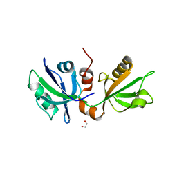
 | | SDCBP PanDDA analysis group deposition -- The PDZ domans of SDCBP in complex with Z198195770 | | 分子名称: | 1,2-ETHANEDIOL, D-GLUTAMIC ACID, GLYCINE, ... | | 著者 | Bradshaw, W.J, Katis, V.L, Bountra, C, von Delft, F, Brennan, P.E. | | 登録日 | 2023-01-24 | | 公開日 | 2023-02-15 | | 最終更新日 | 2024-05-22 | | 実験手法 | X-RAY DIFFRACTION (1.9 Å) | | 主引用文献 | SDCBP PanDDA analysis group deposition
To Be Published
|
|
7FSQ
 | | SDCBP PanDDA analysis group deposition -- The PDZ domans of SDCBP in complex with Z1429867185 | | 分子名称: | 1,2-ETHANEDIOL, 4-methoxy-1H-indole, ALANINE, ... | | 著者 | Bradshaw, W.J, Katis, V.L, Bountra, C, von Delft, F, Brennan, P.E. | | 登録日 | 2023-01-24 | | 公開日 | 2023-02-15 | | 最終更新日 | 2024-05-22 | | 実験手法 | X-RAY DIFFRACTION (2.07 Å) | | 主引用文献 | SDCBP PanDDA analysis group deposition
To Be Published
|
|
7FSS
 | | SDCBP PanDDA analysis group deposition -- The PDZ domans of SDCBP in complex with Z839706072 | | 分子名称: | 1,2-ETHANEDIOL, 2-(4-bromanylpyrazol-1-yl)-~{N}-cyclopropyl-~{N}-methyl-ethanamide, ALANINE, ... | | 著者 | Bradshaw, W.J, Katis, V.L, Bountra, C, von Delft, F, Brennan, P.E. | | 登録日 | 2023-01-24 | | 公開日 | 2023-02-15 | | 最終更新日 | 2024-05-22 | | 実験手法 | X-RAY DIFFRACTION (2.11 Å) | | 主引用文献 | SDCBP PanDDA analysis group deposition
To Be Published
|
|
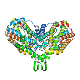
5AZJ
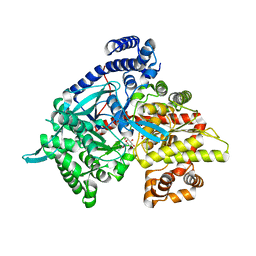
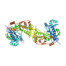 | | Crystal structure of glycerol kinase from Trypanosoma brucei gambiense complexed with 4NP (with disulfide bridge) | | 分子名称: | 4-NITROPHENYL PHOSPHATE, GLYCEROL, Glycerol kinase | | 著者 | Balogun, E.O, Inaoka, D.K, Shiba, T, Tokuoka, S.M, Tokumasu, F, Sakamoto, K, Michels, P.A.M, Harada, S, Kita, K. | | 登録日 | 2015-10-08 | | 公開日 | 2016-10-19 | | 最終更新日 | 2023-11-08 | | 実験手法 | X-RAY DIFFRACTION (2.61 Å) | | 主引用文献 | Glycerol kinase of African trypanosomes possesses an intrinsic phosphatase activity.
Biochim Biophys Acta Gen Subj, 1861, 2017
|
|
7FSZ
 | | SDCBP PanDDA analysis group deposition -- The PDZ domans of SDCBP in complex with Z3006151474 | | 分子名称: | (5S)-5-(difluoromethoxy)pyridin-2(5H)-one, 1,2-ETHANEDIOL, D-GLUTAMIC ACID, ... | | 著者 | Bradshaw, W.J, Katis, V.L, Bountra, C, von Delft, F, Brennan, P.E. | | 登録日 | 2023-01-24 | | 公開日 | 2023-02-15 | | 最終更新日 | 2024-05-22 | | 実験手法 | X-RAY DIFFRACTION (2.05 Å) | | 主引用文献 | SDCBP PanDDA analysis group deposition
To Be Published
|
|
8G54
 | |
7FT4
 | | SDCBP PanDDA analysis group deposition -- The PDZ domans of SDCBP in complex with Z48847594 | | 分子名称: | 1,2-ETHANEDIOL, ALANINE, D-GLUTAMIC ACID, ... | | 著者 | Bradshaw, W.J, Katis, V.L, Bountra, C, von Delft, F, Brennan, P.E. | | 登録日 | 2023-01-24 | | 公開日 | 2023-02-15 | | 最終更新日 | 2024-05-22 | | 実験手法 | X-RAY DIFFRACTION (2.17 Å) | | 主引用文献 | SDCBP PanDDA analysis group deposition
To Be Published
|
|
3ZLG
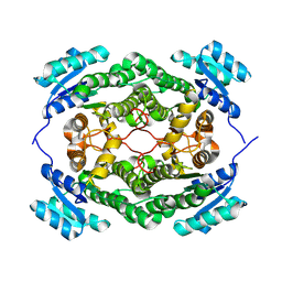
 | | Structure of group A Streptococcal enolase K362A mutant | | 分子名称: | ENOLASE, PHOSPHATE ION | | 著者 | Cork, A.J, Ericsson, D.J, Law, R.H.P, Casey, L.W, Valkov, E, Bertozzi, C, Stamp, A, Aquilina, J.A, Whisstock, J.C, Walker, M.J, Kobe, B. | | 登録日 | 2013-01-31 | | 公開日 | 2014-02-05 | | 最終更新日 | 2023-12-20 | | 実験手法 | X-RAY DIFFRACTION (2.1 Å) | | 主引用文献 | Stability of the Octameric Structure Affects Plasminogen-Binding Capacity of Streptococcal Enolase.
Plos One, 10, 2015
|
|
7FT3
 | | SDCBP PanDDA analysis group deposition -- The PDZ domans of SDCBP in complex with Z291279160 | | 分子名称: | 1,2-ETHANEDIOL, 1-[2-(trifluoromethyloxy)phenyl]thiourea, ALANINE, ... | | 著者 | Bradshaw, W.J, Katis, V.L, Bountra, C, von Delft, F, Brennan, P.E. | | 登録日 | 2023-01-24 | | 公開日 | 2023-02-15 | | 最終更新日 | 2024-05-22 | | 実験手法 | X-RAY DIFFRACTION (2.05 Å) | | 主引用文献 | SDCBP PanDDA analysis group deposition
To Be Published
|
|
1TLB
 | | Yeast coproporphyrinogen oxidase | | 分子名称: | Coproporphyrinogen III oxidase, SULFATE ION | | 著者 | Phillip, J.D, Whitby, F.G, Warby, C.A, Labbe, P, Yang, C, Pflugrath, J.W, Ferrara, J.D, Robinson, H, Kushner, J.P, Hill, C.P. | | 登録日 | 2004-06-09 | | 公開日 | 2004-07-20 | | 最終更新日 | 2024-02-14 | | 実験手法 | X-RAY DIFFRACTION (2.4 Å) | | 主引用文献 | Crystal structure of the oxygen-dependent coproporphyrinogen oxidase (Hem13p) of Saccharomyces cerevisiae
J.Biol.Chem., 279, 2004
|
|
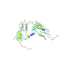
1XF2
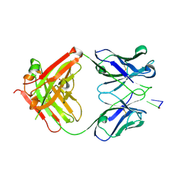 | | Structure of Fab DNA-1 complexed with dT3 | | 分子名称: | 5'-D(*TP*TP*T)-3', SULFATE ION, antibody heavy chain Fab, ... | | 著者 | Schuermann, J.P, Prewitt, S.P, Deutscher, S.L, Tanner, J.J. | | 登録日 | 2004-09-13 | | 公開日 | 2005-04-12 | | 最終更新日 | 2023-08-23 | | 実験手法 | X-RAY DIFFRACTION (2.3 Å) | | 主引用文献 | Evidence for Structural Plasticity of Heavy Chain Complementarity-determining Region 3 in Antibody-ssDNA Recognition
J.Mol.Biol., 347, 2005
|
|
1JB7
 | |
4IRI
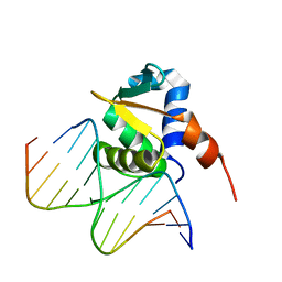 | | Auto-inhibited ERG Ets Domain-DNA Complex | | 分子名称: | DNA (5'-D(*CP*CP*AP*CP*TP*TP*CP*CP*GP*GP*TP*C)-3'), DNA (5'-D(*GP*AP*CP*CP*GP*GP*AP*AP*GP*TP*GP*G)-3'), Transcriptional regulator ERG | | 著者 | Regan, M.C, Horanyi, P.S, Pryor, E.E, Sarver, J.L, Cafiso, D.S, Bushweller, J.H. | | 登録日 | 2013-01-14 | | 公開日 | 2013-07-31 | | 最終更新日 | 2024-02-28 | | 実験手法 | X-RAY DIFFRACTION (2.77 Å) | | 主引用文献 | Structural and dynamic studies of the transcription factor ERG reveal DNA binding is allosterically autoinhibited.
Proc.Natl.Acad.Sci.USA, 110, 2013
|
|
3ZSZ
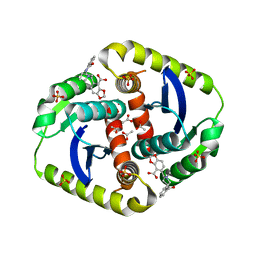 | | Small molecule inhibitors of the LEDGF site of HIV type 1 integrase identified by fragment screening and structure based drug design | | 分子名称: | (R)-[2-[[(2S)-BUTAN-2-YL]CARBAMOYL]PHENYL]METHYL-[(4-CARBOXY-1,3-BENZODIOXOL-5-YL)METHYL]-METHYL-AZANIUM, ACETATE ION, CHLORIDE ION, ... | | 著者 | Peat, T.S, Newman, J, Rhodes, D.I, Vandergraaff, N, Le, G, Jones, E.D, Smith, J.A, Coates, J.A.V, Thienthong, N, Dolezal, O, Ryan, J.H, Savage, G.P, Francis, C.L, Deadman, J.J. | | 登録日 | 2011-07-01 | | 公開日 | 2012-07-11 | | 最終更新日 | 2023-12-20 | | 実験手法 | X-RAY DIFFRACTION (2 Å) | | 主引用文献 | Small Molecule Inhibitors of the Ledgf Site of Human Immunodeficiency Virus Integrase Identified by Fragment Screening and Structure Based Design.
Plos One, 7, 2012
|
|
4L0U
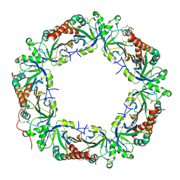 | | Crystal structure of Plasmodium vivax Prx1a | | 分子名称: | 2-Cys peroxiredoxin, putative, ACETATE ION | | 著者 | Gretes, M.C, Karplus, P.A. | | 登録日 | 2013-06-01 | | 公開日 | 2016-11-09 | | 最終更新日 | 2023-09-20 | | 実験手法 | X-RAY DIFFRACTION (2.5 Å) | | 主引用文献 | Observed octameric assembly of a Plasmodium yoelii peroxiredoxin can be explained by the replacement of native "ball-and-socket" interacting residues by an affinity tag.
Protein Sci., 22, 2013
|
|
6R3A
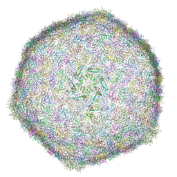 | | BACTERIOPHAGE SPP1 MATURE CAPSID PROTEIN | | 分子名称: | Major capsid protein | | 著者 | Ignatiou, A, El Sadek Fadel, M, Buerger, J, Mielke, T, Topf, M, Tavares, P. | | 登録日 | 2019-03-19 | | 公開日 | 2019-10-23 | | 最終更新日 | 2024-05-15 | | 実験手法 | ELECTRON MICROSCOPY (4 Å) | | 主引用文献 | Structural transitions during the scaffolding-driven assembly of a viral capsid.
Nat Commun, 10, 2019
|
|
1MRR
 | |
7OA6
 | | Pseudo-atomic model for Hsp26 residues 63 to 214. Please be advised that the target map is not of sufficient resolution to unambiguously position backbone or side chain atoms. This model represents a likely fit. | | 分子名称: | Heat shock protein 26 | | 著者 | Muehlhofer, M, Peters, C, Kriehuber, T, Kreuzeder, M, Kazman, P, Rodina, N, Reif, B, Haslbeck, M, Weinkauf, S, Buchner, J. | | 登録日 | 2021-04-19 | | 公開日 | 2021-11-24 | | 最終更新日 | 2024-07-10 | | 実験手法 | ELECTRON MICROSCOPY (7.8 Å) | | 主引用文献 | Phosphorylation activates the yeast small heat shock protein Hsp26 by weakening domain contacts in the oligomer ensemble.
Nat Commun, 12, 2021
|
|
8GJY
 | | Structure of a cGAS-like receptor Sp-cGLR1 from S. pistillata | | 分子名称: | cGAS-like receptor 1 | | 著者 | Li, Y, Toyoda, H, Slavik, K.M, Morehouse, B.R, Kranzusch, P.J. | | 登録日 | 2023-03-16 | | 公開日 | 2023-07-05 | | 最終更新日 | 2024-05-22 | | 実験手法 | X-RAY DIFFRACTION (1.5 Å) | | 主引用文献 | cGLRs are a diverse family of pattern recognition receptors in innate immunity.
Cell, 186, 2023
|
|
4L53
 | | Crystal Structure of (1R,4R)-4-{4-[7-amino-2-(1,2,3-benzothiadiazol-7-yl)-3-chlorofuro[2,3-c]pyridin-4-yl]-1H-pyrazol-1-yl}cyclohexan-1-ol bound to TAK1-TAB1 | | 分子名称: | 1,2-ETHANEDIOL, Mitogen-activated protein kinase kinase kinase 7, TGF-beta-activated kinase 1 and MAP3K7-binding protein 1 chimera, ... | | 著者 | Wang, J, Hornberger, K.R, Crew, A.P, Jestel, A, Maskos, K, Moertl, M. | | 登録日 | 2013-06-10 | | 公開日 | 2013-07-03 | | 最終更新日 | 2024-02-28 | | 実験手法 | X-RAY DIFFRACTION (2.55 Å) | | 主引用文献 | Discovery of 7-aminofuro[2,3-c]pyridine inhibitors of TAK1: Optimization of kinase selectivity and pharmacokinetics.
Bioorg.Med.Chem.Lett., 23, 2013
|
|
7O6P
 | |
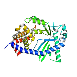
3ZSN
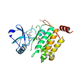
 | | Structure of the mixed-function P450 MycG F286A mutant in complex with mycinamicin IV | | 分子名称: | BENZAMIDINE, GLYCEROL, MYCINAMICIN IV, ... | | 著者 | Li, S, Kells, P.M, Rutaganira, F.U, Anzai, Y, Kato, F, Sherman, D.H, Podust, L.M. | | 登録日 | 2011-06-29 | | 公開日 | 2012-05-09 | | 最終更新日 | 2023-12-20 | | 実験手法 | X-RAY DIFFRACTION (1.9 Å) | | 主引用文献 | Substrate Recognition by the Multifunctional Cytochrome P450 Mycg in Mycinamicin Hydroxylation and Epoxidation Reactions.
J.Biol.Chem., 287, 2012
|
|
1XL0
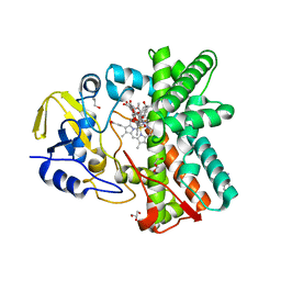
 | | Kinetic and crystallographic studies on 2-(beta-D-glucopyranosyl)-5-methyl-1,3,4-oxadiazole,-benzothiazole, and-benzimidazole, inhibitors of muscle glycogen phosphorylase b. Evidence for a new binding site. | | 分子名称: | (1R)-1,5-anhydro-1-(5-methyl-1,3,4-oxadiazol-2-yl)-D-glucitol, Glycogen phosphorylase, muscle form, ... | | 著者 | Chrysina, E.D, Kosmopoulou, M.N, Tiraidis, C, Kardakaris, R, Bischler, N, Leonidas, D.D, Hadady, Z, Somsak, L, Docsa, T, Gergely, P, Oikonomakos, N.G. | | 登録日 | 2004-09-30 | | 公開日 | 2005-03-15 | | 最終更新日 | 2020-07-29 | | 実験手法 | X-RAY DIFFRACTION (1.92 Å) | | 主引用文献 | Kinetic and crystallographic studies on 2-(beta-D-glucopyranosyl)-5-methyl-1, 3, 4-oxadiazole, -benzothiazole, and -benzimidazole, inhibitors of muscle glycogen phosphorylase b. Evidence for a new binding site
Protein Sci., 14, 2005
|
|


