Loading
PDBj
メニューPDBj@FacebookPDBj@TwitterPDBj@YouTubewwPDB FoundationwwPDB
RCSB PDBPDBeBMRBAdv. SearchSearch help
PDB: 45955 件

7A7Z
DownloadVisualize
BU of 7a7z by Molmil
rsGreen0.7-K206A-E222G in the green-on state
分子名称: Green fluorescent protein
著者De Zitter, E, Dedecker, P, Van Meervelt, L.
登録日2020-08-30
公開日2021-02-17
最終更新日2023-11-15
実験手法X-RAY DIFFRACTION (1.1 Å)
主引用文献Structure-Function Dataset Reveals Environment Effects within a Fluorescent Protein Model System*.
Angew.Chem.Int.Ed.Engl., 60, 2021
7A8L
DownloadVisualize
BU of 7a8l by Molmil
rsGreen0.7-K206A-N205C in the green-on state
分子名称: Green fluorescent protein
著者De Zitter, E, Dedecker, P, Van Meervelt, L.
登録日2020-08-30
公開日2021-02-17
最終更新日2021-05-05
実験手法X-RAY DIFFRACTION (1.75 Å)
主引用文献Structure-Function Dataset Reveals Environment Effects within a Fluorescent Protein Model System*.
Angew.Chem.Int.Ed.Engl., 60, 2021
2CTB
DownloadVisualize
BU of 2ctb by Molmil
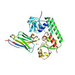
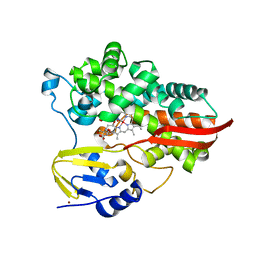
THE HIGH RESOLUTION CRYSTAL STRUCTURE OF THE COMPLEX BETWEEN CARBOXYPEPTIDASE A AND L-PHENYL LACTATE
分子名称: CARBOXYPEPTIDASE A, ZINC ION
著者Teplyakov, A, Wilson, K.S, Orioli, P, Mangani, S.
登録日1993-04-02
公開日1994-01-31
最終更新日2017-11-29
実験手法X-RAY DIFFRACTION (1.5 Å)
主引用文献High-resolution structure of the complex between carboxypeptidase A and L-phenyl lactate.
Acta Crystallogr.,Sect.D, 49, 1993
7UEK
DownloadVisualize
BU of 7uek by Molmil
Crystal structure of a de novo designed ovoid TIM barrel OT3
分子名称: OT3
著者Chu, A.E, Fernandez, D, Huang, P.-S.
登録日2022-03-21
公開日2023-01-18
最終更新日2024-04-03
実験手法X-RAY DIFFRACTION (2.1 Å)
主引用文献De Novo Design of a Highly Stable Ovoid TIM Barrel: Unlocking Pocket Shape towards Functional Design.
Biodes Res, 2022, 2022
1D7M
DownloadVisualize
BU of 1d7m by Molmil
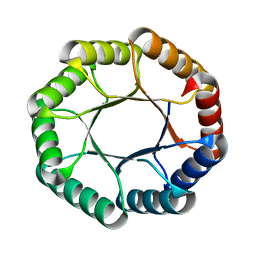
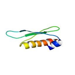
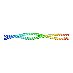
COILED-COIL DIMERIZATION DOMAIN FROM CORTEXILLIN I
分子名称: CORTEXILLIN I
著者Burkhard, P, Kammerer, R.A, Steinmetz, M.O, Bourenkov, G.P, Aebi, U.
登録日1999-10-19
公開日2000-03-27
最終更新日2024-02-07
実験手法X-RAY DIFFRACTION (2.7 Å)
主引用文献The coiled-coil trigger site of the rod domain of cortexillin I unveils a distinct network of interhelical and intrahelical salt bridges.
Structure Fold.Des., 8, 2000
7A8N
DownloadVisualize
BU of 7a8n by Molmil
rsGreen0.7-K206A-N205L in the green-on state
分子名称: Green fluorescent protein
著者De Zitter, E, Dedecker, P, Van Meervelt, L.
登録日2020-08-30
公開日2021-02-17
最終更新日2021-05-05
実験手法X-RAY DIFFRACTION (2.1 Å)
主引用文献Structure-Function Dataset Reveals Environment Effects within a Fluorescent Protein Model System*.
Angew.Chem.Int.Ed.Engl., 60, 2021
7A85
DownloadVisualize
BU of 7a85 by Molmil
rsGreen0.7-K206A-F145L in the green-on state
分子名称: DI(HYDROXYETHYL)ETHER, Green fluorescent protein, TRIETHYLENE GLYCOL
著者De Zitter, E, Dedecker, P, Van Meervelt, L.
登録日2020-08-30
公開日2021-02-17
最終更新日2021-05-05
実験手法X-RAY DIFFRACTION (1.52 Å)
主引用文献Structure-Function Dataset Reveals Environment Effects within a Fluorescent Protein Model System*.
Angew.Chem.Int.Ed.Engl., 60, 2021
7A8E
DownloadVisualize
BU of 7a8e by Molmil
rsGreen0.7-K206A-F165W partially in the green-off state
分子名称: Green fluorescent protein
著者De Zitter, E, Dedecker, P, Van Meervelt, L.
登録日2020-08-30
公開日2021-02-17
最終更新日2021-05-05
実験手法X-RAY DIFFRACTION (2.2 Å)
主引用文献Structure-Function Dataset Reveals Environment Effects within a Fluorescent Protein Model System*.
Angew.Chem.Int.Ed.Engl., 60, 2021
7A8K
DownloadVisualize
BU of 7a8k by Molmil
rsGreen0.7-K206A-H148V partially in the green-off state
分子名称: DI(HYDROXYETHYL)ETHER, Green fluorescent protein
著者De Zitter, E, Dedecker, P, Van Meervelt, L.
登録日2020-08-30
公開日2021-02-17
最終更新日2024-01-31
実験手法X-RAY DIFFRACTION (2.25 Å)
主引用文献Structure-Function Dataset Reveals Environment Effects within a Fluorescent Protein Model System*.
Angew.Chem.Int.Ed.Engl., 60, 2021
7A7O
DownloadVisualize
BU of 7a7o by Molmil
rsGreen0.7-K206A in the green-on state
分子名称: Green fluorescent protein
著者De Zitter, E, Dedecker, P, Van Meervelt, L.
登録日2020-08-30
公開日2021-02-17
最終更新日2021-05-05
実験手法X-RAY DIFFRACTION (1.8 Å)
主引用文献Structure-Function Dataset Reveals Environment Effects within a Fluorescent Protein Model System*.
Angew.Chem.Int.Ed.Engl., 60, 2021
2LRS
DownloadVisualize
BU of 2lrs by Molmil
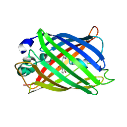
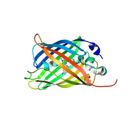
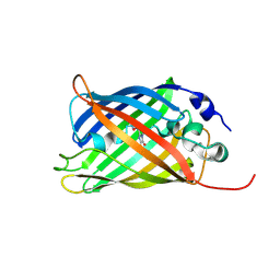
The second dsRBD domain from A. thaliana DICER-LIKE 1
分子名称: Endoribonuclease Dicer homolog 1
著者Burdisso, P, Suarez, I, Bersch, B, Bologna, N, Palatnik, J, Boisbouvier, J, Rasia, R.
登録日2012-04-12
公開日2013-01-23
最終更新日2024-05-01
実験手法SOLUTION NMR
主引用文献Second Double-Stranded RNA Binding Domain of Dicer-like Ribonuclease 1: Structural and Biochemical Characterization.
Biochemistry, 51, 2012
7A82
DownloadVisualize
BU of 7a82 by Molmil
rsGreen0.7-K206A-F145A partially in the green-off state
分子名称: Green fluorescent protein
著者De Zitter, E, Dedecker, P, Van Meervelt, L.
登録日2020-08-30
公開日2021-02-17
最終更新日2021-05-05
実験手法X-RAY DIFFRACTION (1.9 Å)
主引用文献Structure-Function Dataset Reveals Environment Effects within a Fluorescent Protein Model System*.
Angew.Chem.Int.Ed.Engl., 60, 2021
7A87
DownloadVisualize
BU of 7a87 by Molmil
rsGreen0.7-K206A-F145M in the green-on state
分子名称: DI(HYDROXYETHYL)ETHER, Green fluorescent protein
著者De Zitter, E, Dedecker, P, Van Meervelt, L.
登録日2020-08-30
公開日2021-02-17
最終更新日2021-05-05
実験手法X-RAY DIFFRACTION (1.75 Å)
主引用文献Structure-Function Dataset Reveals Environment Effects within a Fluorescent Protein Model System*.
Angew.Chem.Int.Ed.Engl., 60, 2021
7A8B
DownloadVisualize
BU of 7a8b by Molmil
rsGreen0.7-K206A-F145S partially in the green-on state
分子名称: Green fluorescent protein
著者De Zitter, E, Dedecker, P, Van Meervelt, L.
登録日2020-08-30
公開日2021-02-17
最終更新日2021-05-05
実験手法X-RAY DIFFRACTION (1.55 Å)
主引用文献Structure-Function Dataset Reveals Environment Effects within a Fluorescent Protein Model System*.
Angew.Chem.Int.Ed.Engl., 60, 2021
1MRN
DownloadVisualize
BU of 1mrn by Molmil
CRYSTAL STRUCTURE OF MYCOBACTERIUM TUBERCULOSIS THYMIDYLATE KINASE COMPLEXED WITH BISUBSTRATE INHIBITOR (TP5A)
分子名称: MAGNESIUM ION, P1-(5'-ADENOSYL)P5-(5'-THYMIDYL)PENTAPHOSPHATE, SULFATE ION, ...
著者Haouz, A, Vanheusden, V, Munier-Lehmann, H, Froeyen, M, Herdewijn, P, Van Calenbergh, S, Delarue, M.
登録日2002-09-18
公開日2003-01-07
最終更新日2024-02-14
実験手法X-RAY DIFFRACTION (2.45 Å)
主引用文献Enzymatic and structural analysis of inhibitors designed against Mycobacterium tuberculosis thymidylate kinase. New insights into the phosphoryl transfer mechanism.
J.Biol.Chem., 278, 2003
1MZV
DownloadVisualize
BU of 1mzv by Molmil
Crystal Structure of Adenine Phosphoribosyltransferase (APRT) From Leishmania tarentolae
分子名称: ADENOSINE MONOPHOSPHATE, Adenine Phosphoribosyltransferase, PHOSPHATE ION
著者Thiemann, O.H, Silva, M, Oliva, G, Silva, C.H.T.P, Iulek, J.
登録日2002-10-10
公開日2003-10-28
最終更新日2024-02-14
実験手法X-RAY DIFFRACTION (2.2 Å)
主引用文献Crystal structure of adenine phosphoribosyltransferase from Leishmania tarentolae: potential implications for APRT catalytic mechanism.
Biochim.Biophys.Acta, 1696, 2004
7A83
DownloadVisualize
BU of 7a83 by Molmil
rsGreen0.7-K206A-F145H in the green-on-state
分子名称: Green fluorescent protein, TRIETHYLENE GLYCOL
著者De Zitter, E, Dedecker, P, Van Meervelt, L.
登録日2020-08-30
公開日2021-02-17
最終更新日2021-05-05
実験手法X-RAY DIFFRACTION (1.73 Å)
主引用文献Structure-Function Dataset Reveals Environment Effects within a Fluorescent Protein Model System*.
Angew.Chem.Int.Ed.Engl., 60, 2021
5OVW
DownloadVisualize
BU of 5ovw by Molmil
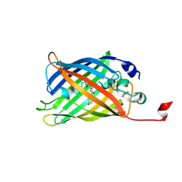
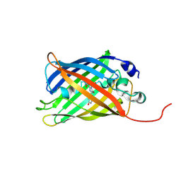
Nanobody-bound BtuF, the vitamin B12 binding protein in Escherichia coli
分子名称: GLYCEROL, Nanobody, Vitamin B12-binding protein
著者Mireku, S.A, Sauer, M.M, Glockshuber, R, Locher, K.P.
登録日2017-08-30
公開日2017-11-08
最終更新日2019-10-16
実験手法X-RAY DIFFRACTION (2.653 Å)
主引用文献Structural basis of nanobody-mediated blocking of BtuF, the cognate substrate-binding protein of the Escherichia coli vitamin B12 transporter BtuCD.
Sci Rep, 7, 2017
2CYF
DownloadVisualize
BU of 2cyf by Molmil
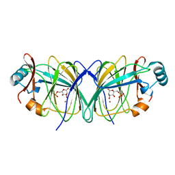
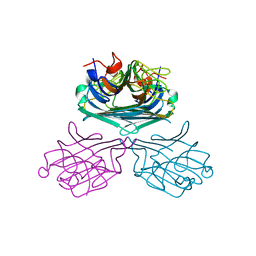
The Crystal Structure of Canavalia Maritima Lectin (ConM) in Complex with Trehalose and Maltose
分子名称: CALCIUM ION, Lectin, MANGANESE (II) ION, ...
著者Delatorre, P, Rocha, B.A.M, Sousa, E.P, Gadelha, C.A.A, Azevedo Jr, W.F, Cavada, B.S.
登録日2005-07-06
公開日2006-06-13
最終更新日2024-03-13
実験手法X-RAY DIFFRACTION (1.8 Å)
主引用文献Crystal structure of a lectin from Canavalia maritima (ConM) in complex with trehalose and maltose reveals relevant mutation in ConA-like lectins
J.Struct.Biol., 154, 2006
7A8D
DownloadVisualize
BU of 7a8d by Molmil
rsGreen0.7-K206A-F165W partially in the green-on state
分子名称: Green fluorescent protein
著者De Zitter, E, Dedecker, P, Van Meervelt, L.
登録日2020-08-30
公開日2021-02-17
最終更新日2021-05-05
実験手法X-RAY DIFFRACTION (1.65 Å)
主引用文献Structure-Function Dataset Reveals Environment Effects within a Fluorescent Protein Model System*.
Angew.Chem.Int.Ed.Engl., 60, 2021
4CQ8
DownloadVisualize
BU of 4cq8 by Molmil
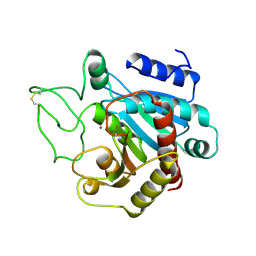
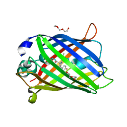
Plasmodium falciparum dihydroorotate dehydrogenase (DHODH) in complex with Genz-669178
分子名称: 5-(4-cyano-2-methyl-1H-benzimidazol-1-yl)-N-cyclopropylthiophene-2-carboxamide, DIHYDROOROTATE DEHYDROGENASE, FLAVIN MONONUCLEOTIDE, ...
著者Rowland, P.
登録日2014-02-12
公開日2014-04-30
最終更新日2024-05-08
実験手法X-RAY DIFFRACTION (1.98 Å)
主引用文献In Vitro Resistance Selections for Plasmodium Falciparum Dihydroorotate Dehydrogenase Inhibitors Give Mutants with Multiple Point Mutations in the Drug-Binding Site and Altered Growth.
J.Biol.Chem., 289, 2014
7A8A
DownloadVisualize
BU of 7a8a by Molmil
rsGreen0.7-K206A-F145Q partially in the green-off state
分子名称: Green fluorescent protein
著者De Zitter, E, Dedecker, P, Van Meervelt, L.
登録日2020-08-30
公開日2021-02-17
最終更新日2021-05-05
実験手法X-RAY DIFFRACTION (1.93 Å)
主引用文献Structure-Function Dataset Reveals Environment Effects within a Fluorescent Protein Model System*.
Angew.Chem.Int.Ed.Engl., 60, 2021
7UOR
DownloadVisualize
BU of 7uor by Molmil
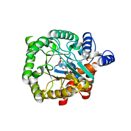
Crystal structure of cytochrome P450 enzyme CYP119 in complex with methyliridium(III) mesoporphyrin.
分子名称: 2-[BIS-(2-HYDROXY-ETHYL)-AMINO]-2-HYDROXYMETHYL-PROPANE-1,3-DIOL, Cytochrome, NICKEL (II) ION, ...
著者Pereira, J.H, Bloomer, B.J, Hartwig, J.F, Adams, P.D.
登録日2022-04-13
公開日2023-02-22
最終更新日2023-10-25
実験手法X-RAY DIFFRACTION (3.16 Å)
主引用文献Mechanistic and structural characterization of an iridium-containing cytochrome reveals kinetically relevant cofactor dynamics
Nat Catal, 6, 2023
7A8M
DownloadVisualize
BU of 7a8m by Molmil
rsGreen0.7-K206A-N205G in the green-on state
分子名称: Green fluorescent protein
著者De Zitter, E, Dedecker, P, Van Meervelt, L.
登録日2020-08-30
公開日2021-02-17
最終更新日2021-05-05
実験手法X-RAY DIFFRACTION (1.6 Å)
主引用文献Structure-Function Dataset Reveals Environment Effects within a Fluorescent Protein Model System*.
Angew.Chem.Int.Ed.Engl., 60, 2021
2GC3
DownloadVisualize
BU of 2gc3 by Molmil
The crystal structure of phosphoglucose isomerase from Pyrococcus furiosus in complex with mannose 6-phosphate and zinc
分子名称: 6-O-phosphono-alpha-D-mannopyranose, Glucose-6-phosphate isomerase, ZINC ION
著者Berrisford, J.M, Rice, D.W, Baker, P.J.
登録日2006-03-13
公開日2006-04-11
最終更新日2023-08-30
実験手法X-RAY DIFFRACTION (2.1 Å)
主引用文献Evidence Supporting a cis-enediol-based Mechanism for Pyrococcus furiosus Phosphoglucose Isomerase
J.Mol.Biol., 358, 2006

224004

件を2024-08-21に公開中

PDB statisticsPDBj update infoContact PDBjnumon