2J1R
 
 | |
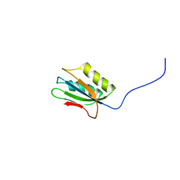
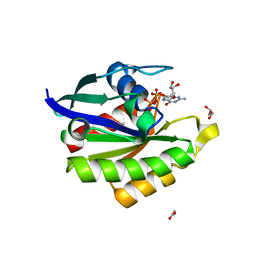
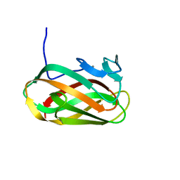
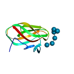
1OF3
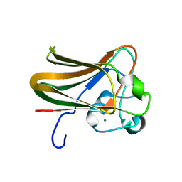
 | | Structural and thermodynamic dissection of specific mannan recognition by a carbohydrate-binding module, TmCBM27 | | 分子名称: | BETA-MANNOSIDASE, CALCIUM ION | | 著者 | Boraston, A.B, Revett, T.J, Boraston, C.M, Nurizzo, D, Davies, G.J. | | 登録日 | 2003-04-07 | | 公開日 | 2003-04-17 | | 最終更新日 | 2011-07-13 | | 実験手法 | X-RAY DIFFRACTION (2 Å) | | 主引用文献 | Structural and Thermodynamic Dissection of Specific Mannan Recognition by a Carbohydrate Binding Module, Tmcbm27
Structure, 11, 2003
|
|
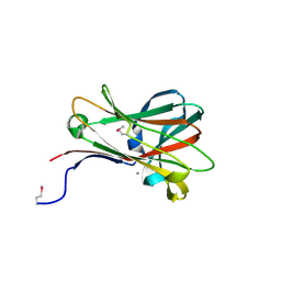
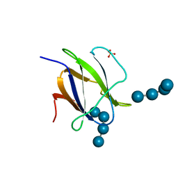
1OH4
 | | Structural and thermodynamic dissection of specific mannan recognition by a carbohydrate-binding module | | 分子名称: | BETA-MANNOSIDASE, CALCIUM ION, GLYCEROL, ... | | 著者 | Boraston, A.B, Revett, T.J, Boraston, C.M, Nurizzo, D, Davies, G.J. | | 登録日 | 2003-05-21 | | 公開日 | 2004-03-16 | | 最終更新日 | 2023-12-13 | | 実験手法 | X-RAY DIFFRACTION (1.35 Å) | | 主引用文献 | Structural and Thermodynamic Dissection of Specific Mannan Recognition by a Carbohydrate Binding Module, Tmcbm27
Structure, 11, 2003
|
|
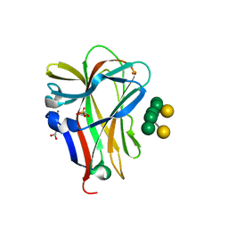
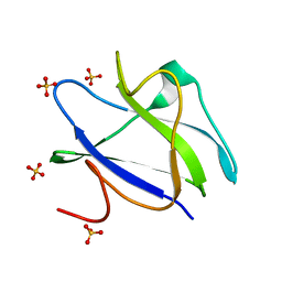
1OF4
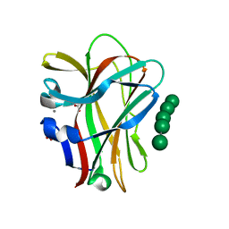 | | Structural and thermodynamic dissection of specific mannan recognition by a carbohydrate-binding module, TmCBM27 | | 分子名称: | BETA-MANNOSIDASE, CALCIUM ION, GLYCEROL, ... | | 著者 | Boraston, A.B, Revett, T.J, Boraston, C.M, Nurizzo, D, Davies, G.J. | | 登録日 | 2003-04-07 | | 公開日 | 2003-04-17 | | 最終更新日 | 2024-05-08 | | 実験手法 | X-RAY DIFFRACTION (1.6 Å) | | 主引用文献 | Structural and Thermodynamic Dissection of Specific Mannan Recognition by a Carbohydrate Binding Module, Tmcbm27
Structure, 11, 2003
|
|
1Y9O
 | | 1H NMR Structure of Acylphosphatase from the hyperthermophile Sulfolobus Solfataricus | | 分子名称: | Acylphosphatase | | 著者 | Corazza, A, Rosano, C, Pagano, K, Alverdi, V, Esposito, G, Capanni, C, Bemporad, F, Plakoutsi, G, Stefani, M, Chiti, F, Zuccotti, S, Bolognesi, M, Viglino, P. | | 登録日 | 2004-12-16 | | 公開日 | 2005-11-29 | | 最終更新日 | 2024-05-29 | | 実験手法 | SOLUTION NMR | | 主引用文献 | Structure, conformational stability, and enzymatic properties of acylphosphatase from the hyperthermophile Sulfolobus solfataricus
Proteins, 62, 2006
|
|
8DMY
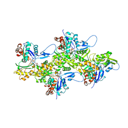 | | Cryo-EM structure of cardiac muscle alpha-actin | | 分子名称: | ADENOSINE-5'-DIPHOSPHATE, Actin, alpha cardiac muscle 1, ... | | 著者 | Arora, A.S, Huang, H.L, Heissler, S.M, Chinthalapudi, K. | | 登録日 | 2022-07-09 | | 公開日 | 2023-04-12 | | 実験手法 | ELECTRON MICROSCOPY (3.07 Å) | | 主引用文献 | Structural insights into actin isoforms.
Elife, 12, 2023
|
|
8DNF
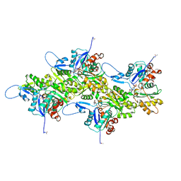 | | Cryo-EM structure of nonmuscle gamma-actin | | 分子名称: | ADENOSINE-5'-DIPHOSPHATE, Actin, cytoplasmic 2, ... | | 著者 | Arora, A.S, Huang, H.L, Heissler, S.M, Chinthalapudi, K. | | 登録日 | 2022-07-11 | | 公開日 | 2023-04-12 | | 実験手法 | ELECTRON MICROSCOPY (3.38 Å) | | 主引用文献 | Structural insights into actin isoforms.
Elife, 12, 2023
|
|
8DMX
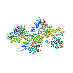 | | Cryo-EM structure of skeletal muscle alpha-actin | | 分子名称: | ADENOSINE-5'-DIPHOSPHATE, Actin, alpha skeletal muscle, ... | | 著者 | Arora, A.S, Huang, H.L, Heissler, S.M, Chinthalapudi, K. | | 登録日 | 2022-07-08 | | 公開日 | 2023-04-12 | | 実験手法 | ELECTRON MICROSCOPY (3.37 Å) | | 主引用文献 | Structural insights into actin isoforms.
Elife, 12, 2023
|
|
8DNH
 | | Cryo-EM structure of nonmuscle beta-actin | | 分子名称: | ADENOSINE-5'-DIPHOSPHATE, Actin, cytoplasmic 1, ... | | 著者 | Arora, A.S, Huang, H.L, Heissler, S.M, Chinthalapudi, K. | | 登録日 | 2022-07-11 | | 公開日 | 2023-04-12 | | 実験手法 | ELECTRON MICROSCOPY (2.99 Å) | | 主引用文献 | Structural insights into actin isoforms.
Elife, 12, 2023
|
|
8U36
 | |
6M8N
 | | Endo-fucoidan hydrolase P5AFcnA from glycoside hydrolase family 107 | | 分子名称: | CALCIUM ION, MALONATE ION, P5AFcnA | | 著者 | Boraston, A.B, Vickers, C.J, Abe, K, Salama-Alber, O. | | 登録日 | 2018-08-22 | | 公開日 | 2018-10-03 | | 最終更新日 | 2024-03-13 | | 実験手法 | X-RAY DIFFRACTION (1.55 Å) | | 主引用文献 | Endo-fucoidan hydrolases from glycoside hydrolase family 107 (GH107) display structural and mechanistic similarities to alpha-l-fucosidases from GH29.
J. Biol. Chem., 293, 2018
|
|
6ALK
 | | NMR solution structure of the major beech pollen allergen Fag s 1 | | 分子名称: | Fag s 1 pollen allergen | | 著者 | Moraes, A.H, Asam, A, Almeida, F.C.L, Wallner, M, Ferreira, F, Valente, A.P. | | 登録日 | 2017-08-08 | | 公開日 | 2018-08-08 | | 最終更新日 | 2024-05-15 | | 実験手法 | SOLUTION NMR | | 主引用文献 | Structural basis for cross-reactivity and conformation fluctuation of the major beech pollen allergen Fag s 1.
Sci Rep, 8, 2018
|
|
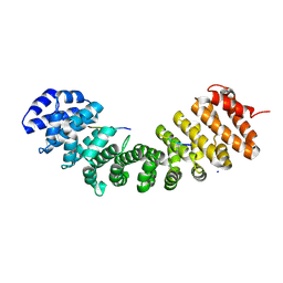
6L6O
 | | Crystal structure of stabilized Rab5a GTPase domain from Leishmania donovani | | 分子名称: | ACETATE ION, GLYCEROL, GUANOSINE-5'-DIPHOSPHATE, ... | | 著者 | Arora, A, Zohib, M, Biswal, B.K, Maheshwari, D, Pal, R.K. | | 登録日 | 2019-10-29 | | 公開日 | 2020-11-11 | | 最終更新日 | 2023-11-22 | | 実験手法 | X-RAY DIFFRACTION (1.8 Å) | | 主引用文献 | Crystal structure of the GDP-bound GTPase domain of Rab5a from Leishmania donovani.
Acta Crystallogr.,Sect.F, 76, 2020
|
|
8SV0
 | |
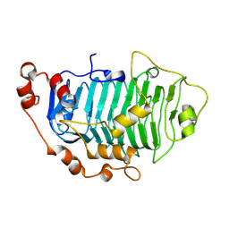
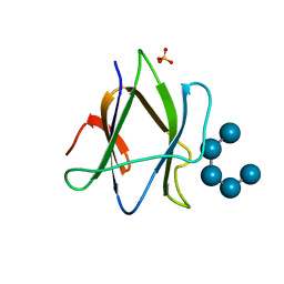
1NAE
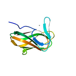 | | Structure of CsCBM6-3 from Clostridium stercorarium in complex with xylotriose | | 分子名称: | CALCIUM ION, beta-D-xylopyranose-(1-4)-beta-D-xylopyranose-(1-4)-beta-D-xylopyranose, putative xylanase | | 著者 | Boraston, A.B, Notenboom, V, Warren, R.A.J, Kilburn, D.G, Rose, D.R, Davies, G. | | 登録日 | 2002-11-27 | | 公開日 | 2003-03-18 | | 最終更新日 | 2023-08-16 | | 実験手法 | X-RAY DIFFRACTION (2.05 Å) | | 主引用文献 | Structure and ligand binding of carbohydrate-binding module CsCBM6-3 reveals similarities with fucose-specific lectins and "galactose-binding" domains
J.Mol.Biol., 327, 2003
|
|
1OD3
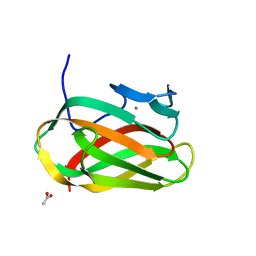 | | Structure of CSCBM6-3 From Clostridium stercorarium in complex with laminaribiose | | 分子名称: | ACETIC ACID, CALCIUM ION, PUTATIVE XYLANASE, ... | | 著者 | Boraston, A.B, Notenboom, V, Warren, R.A.J, Kilburn, D.G, Rose, D.R, Davies, G.J. | | 登録日 | 2003-02-12 | | 公開日 | 2003-03-13 | | 最終更新日 | 2023-12-13 | | 実験手法 | X-RAY DIFFRACTION (1 Å) | | 主引用文献 | Structure and Ligand Binding of Carbohydrate-Binding Module Cscbm6-3 Reveals Similarities with Fucose-Specific Lectins and Galactose-Binding Domains
J.Mol.Biol., 327, 2003
|
|
1O8S
 | | Structure of CsCBM6-3 from Clostridium stercorarium in complex with cellobiose | | 分子名称: | CALCIUM ION, PUTATIVE ENDO-XYLANASE, beta-D-glucopyranose-(1-4)-beta-D-glucopyranose | | 著者 | Boraston, A.B, Notenboom, V, Warren, R.A.J, Kilbrun, D.G, Rose, D.R, Davies, G.J. | | 登録日 | 2002-11-28 | | 公開日 | 2003-03-13 | | 最終更新日 | 2023-12-13 | | 実験手法 | X-RAY DIFFRACTION (1.15 Å) | | 主引用文献 | Structure and Ligand Binding of Carbohydrate-Binding Module Cscbm6-3 Reveals Similarities with Fucose-Specific Lectins and Galactose-Binding Domains
J.Mol.Biol., 327, 2003
|
|
9BUJ
 | |
1W9S
 | |
1W9T
 | | Structure of a beta-1,3-glucan binding CBM6 from Bacillus halodurans in complex with xylobiose | | 分子名称: | BH0236 PROTEIN, SODIUM ION, alpha-D-xylopyranose, ... | | 著者 | Boraston, A.B, van Bueren, A.L. | | 登録日 | 2004-10-18 | | 公開日 | 2004-11-03 | | 最終更新日 | 2023-12-13 | | 実験手法 | X-RAY DIFFRACTION (1.62 Å) | | 主引用文献 | Family 6 Carbohydrate Binding Modules Recognize the Non-Reducing End of Beta-1,3-Linked Glucans by Presenting a Unique Ligand Binding Surface
J.Biol.Chem., 280, 2005
|
|
1W9W
 | |
2C3W
 | | Structure of CBM25 from Bacillus halodurans amylase in complex with maltotetraose | | 分子名称: | ALPHA-AMYLASE G-6, SULFATE ION, alpha-D-glucopyranose-(1-4)-alpha-D-glucopyranose-(1-4)-alpha-D-glucopyranose, ... | | 著者 | Boraston, A.B, Healey, M, Klassen, J, Ficko-Blean, E, Lammerts van Bueren, A, Law, V. | | 登録日 | 2005-10-12 | | 公開日 | 2005-10-17 | | 最終更新日 | 2024-05-08 | | 実験手法 | X-RAY DIFFRACTION (1.81 Å) | | 主引用文献 | A Structural and Functional Analysis of Alpha-Glucan Recognition by Family 25 and 26 Carbohydrate-Binding Modules Reveals a Conserved Mode of Starch Recognition
J.Biol.Chem., 281, 2006
|
|
2C3H
 | | Structure of CBM26 from Bacillus halodurans amylase in complex with maltose | | 分子名称: | ALPHA-AMYLASE G-6, SULFATE ION, alpha-D-glucopyranose, ... | | 著者 | Boraston, A.B, Healey, M, Klassen, J, Ficko-Blean, E, Lammerts van Bueren, A, Law, V. | | 登録日 | 2005-10-07 | | 公開日 | 2005-10-17 | | 最終更新日 | 2024-05-08 | | 実験手法 | X-RAY DIFFRACTION (2.24 Å) | | 主引用文献 | A Structural and Functional Analysis of Alpha-Glucan Recognition by Family 25 and 26 Carbohydrate-Binding Modules Reveals a Conserved Mode of Starch Recognition
J.Biol.Chem., 281, 2006
|
|
2C3X
 | | Structure of iodinated CBM25 from Bacillus halodurans amylase in complex with maltotetraose | | 分子名称: | ALPHA-AMYLASE G-6, SULFATE ION, alpha-D-glucopyranose-(1-4)-alpha-D-glucopyranose-(1-4)-alpha-D-glucopyranose-(1-4)-alpha-D-glucopyranose-(1-4)-alpha-D-glucopyranose | | 著者 | Boraston, A.B, Healey, M, Klassen, J, Ficko-Blean, E, Lammerts van Bueren, A, Law, V. | | 登録日 | 2005-10-12 | | 公開日 | 2005-10-17 | | 最終更新日 | 2024-05-08 | | 実験手法 | X-RAY DIFFRACTION (2.7 Å) | | 主引用文献 | A Structural and Functional Analysis of Alpha-Glucan Recognition by Family 25 and 26 Carbohydrate-Binding Modules Reveals a Conserved Mode of Starch Recognition
J.Biol.Chem., 281, 2006
|
|
2C3V
 | | Structure of iodinated CBM25 from Bacillus halodurans amylase | | 分子名称: | ALPHA-AMYLASE G-6, IODIDE ION | | 著者 | Boraston, A.B, Healey, M, Klassen, J, Ficko-Blean, E, Lammerts van Bueren, A, Law, V. | | 登録日 | 2005-10-12 | | 公開日 | 2005-10-17 | | 最終更新日 | 2011-07-13 | | 実験手法 | X-RAY DIFFRACTION (1.39 Å) | | 主引用文献 | A Structural and Functional Analysis of Alpha-Glucan Recognition by Family 25 and 26 Carbohydrate-Binding Modules Reveals a Conserved Mode of Starch Recognition
J.Biol.Chem., 281, 2006
|
|


