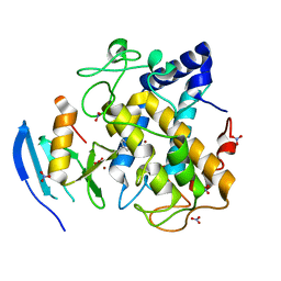
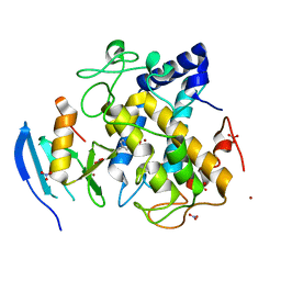
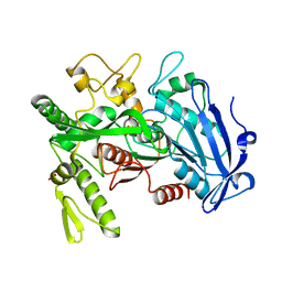
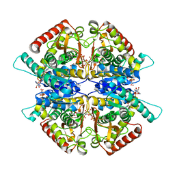
6K4D
 
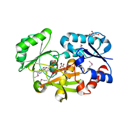
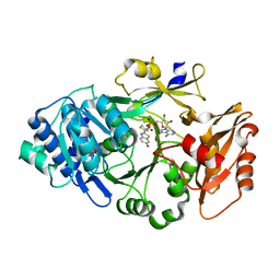 | | Ancestral luciferase AncLamp in complex with ATP and D-luciferin | | 分子名称: | (4S)-2-(6-hydroxy-1,3-benzothiazol-2-yl)-4,5-dihydro-1,3-thiazole-4-carboxylic acid, Ancestral luciferase AncLamp, [[(2R,3S,4R,5R)-5-(6-aminopurin-9-yl)-3,4-bis(oxidanyl)oxolan-2-yl]methoxy-oxidanyl-phosphoryl] (4S)-2-(6-oxidanyl-1,3-benzothiazol-2-yl)-4,5-dihydro-1,3-thiazole-4-carboxylate | | 著者 | Oba, Y, Konishi, K, Yano, D, Kato, D, Shirai, T. | | 登録日 | 2019-05-23 | | 公開日 | 2020-05-27 | | 最終更新日 | 2023-11-22 | | 実験手法 | X-RAY DIFFRACTION (1.7 Å) | | 主引用文献 | Resurrecting the ancient glow of the fireflies.
Sci Adv, 6, 2020
|
|
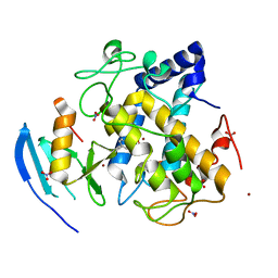
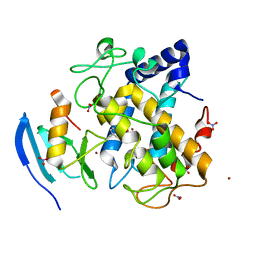
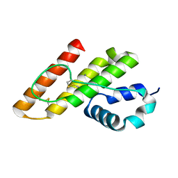
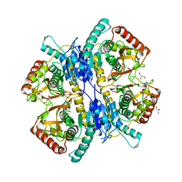
6K4C
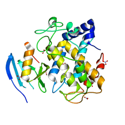
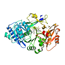 | | Ancestral luciferase AncLamp in complex with DLSA | | 分子名称: | 5'-O-[N-(DEHYDROLUCIFERYL)-SULFAMOYL] ADENOSINE, Ancestral luciferase AncLamp, MAGNESIUM ION | | 著者 | Oba, Y, Konishi, K, Yano, D, Kato, D, Shirai, T. | | 登録日 | 2019-05-23 | | 公開日 | 2020-05-27 | | 最終更新日 | 2023-11-22 | | 実験手法 | X-RAY DIFFRACTION (2.1 Å) | | 主引用文献 | Resurrecting the ancient glow of the fireflies.
Sci Adv, 6, 2020
|
|
2EYY
 | |
5B1H
 | |
5B1I
 | |
6JIL
 | | Crystal structure of D-cycloserine synthetase DcsG | | 分子名称: | ADENOSINE-5'-DIPHOSPHATE, Cycloserine biosynthesis protein DcsG, L(+)-TARTARIC ACID, ... | | 著者 | Matoba, Y, Sugiyama, M. | | 登録日 | 2019-02-22 | | 公開日 | 2019-12-18 | | 最終更新日 | 2020-07-22 | | 実験手法 | X-RAY DIFFRACTION (2.32 Å) | | 主引用文献 | Cyclization mechanism catalyzed by an ATP-grasp enzyme essential for d-cycloserine biosynthesis.
Febs J., 287, 2020
|
|
5Z0E
 | |
5Z0F
 | |
5Z0L
 | |
5Z0M
 | |
5Z0K
 | |
5Z0D
 | |
5Z0J
 | |
5Z0G
 | |
5Z0I
 | |
5Z0H
 | |
2RSE
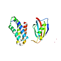 | | NMR structure of FKBP12-mTOR FRB domain-rapamycin complex structure determined based on PCS | | 分子名称: | Peptidyl-prolyl cis-trans isomerase FKBP1A, Serine/threonine-protein kinase mTOR, TERBIUM(III) ION | | 著者 | Kobashigawa, Y, Ushio, M, Saio, T, Inagaki, F. | | 登録日 | 2012-01-25 | | 公開日 | 2012-05-30 | | 最終更新日 | 2024-05-15 | | 実験手法 | SOLUTION NMR | | 主引用文献 | Convenient method for resolving degeneracies due to symmetry of the magnetic susceptibility tensor and its application to pseudo contact shift-based protein-protein complex structure determination.
J.Biomol.Nmr, 53, 2012
|
|
3WA6
 | |
1KP4
 | |
3X44
 | | Crystal structure of O-ureido-L-serine-bound K43A mutant of O-ureido-L-serine synthase | | 分子名称: | (E)-O-(carbamoylamino)-N-({3-hydroxy-2-methyl-5-[(phosphonooxy)methyl]pyridin-4-yl}methylidene)-L-serine, O-ureido-L-serine synthase | | 著者 | Matoba, Y, Uda, N, Oda, K, Sugiyama, M. | | 登録日 | 2015-03-13 | | 公開日 | 2015-07-29 | | 最終更新日 | 2024-05-29 | | 実験手法 | X-RAY DIFFRACTION (1.9 Å) | | 主引用文献 | The structural and mutational analyses of O-ureido-L-serine synthase necessary for D-cycloserine biosynthesis.
Febs J., 282, 2015
|
|
1LWB
 | |
3VGO
 | |
3WSW
 | |
3WSV
 | |
3WA7
 | | Crystal structure of selenomethionine-labeled tannase from Lactobacillus plantarum in the orthorhombic crystal | | 分子名称: | ACETATE ION, GLYCEROL, SULFATE ION, ... | | 著者 | Matoba, Y, Tanaka, N, Sugiyama, M. | | 登録日 | 2013-04-27 | | 公開日 | 2013-07-24 | | 最終更新日 | 2013-11-06 | | 実験手法 | X-RAY DIFFRACTION (1.7 Å) | | 主引用文献 | Crystallographic and mutational analyses of tannase from Lactobacillus plantarum.
Proteins, 81, 2013
|
|


