Loading
PDBj
MenuPDBj@FacebookPDBj@TwitterPDBj@YouTubewwPDB FoundationwwPDB
RCSB PDBPDBeBMRBAdv. SearchSearch help
PDB: 51 results

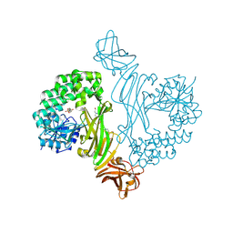
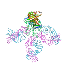
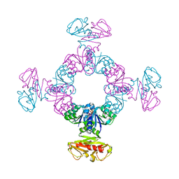
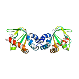
6S5O
DownloadVisualize
BU of 6s5o by Molmil
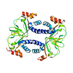
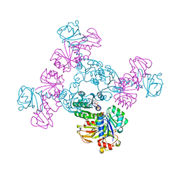
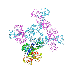
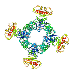
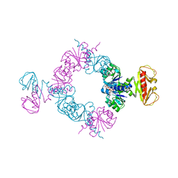
Non-square conformations of KtrA E125Q mutant rings with bound ADP
Descriptor: ADENOSINE-5'-DIPHOSPHATE, Ktr system potassium uptake protein A
Authors:Teixeira-Duarte, C.M, Fonseca, F, Morais-Cabral, J.H.
Deposit date:2019-07-02
Release date:2020-01-08
Last modified:2024-01-24
Method:X-RAY DIFFRACTION (3.983 Å)
Cite:Activation of a nucleotide-dependent RCK domain requires binding of a cation cofactor to a conserved site.
Elife, 8, 2019
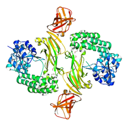
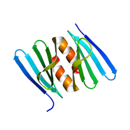
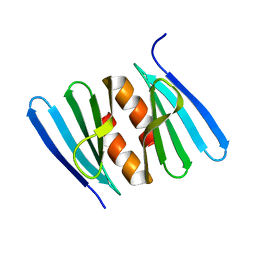
3CLP
DownloadVisualize
BU of 3clp by Molmil
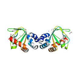
M. loti cyclic-nucleotide binding domain mutant 2
Descriptor: ADENOSINE-3',5'-CYCLIC-MONOPHOSPHATE, Mll3241 protein
Authors:Clayton, G.M, Altieri, S.L, Thomas, L.R, Morais-Cabral, J.H.
Deposit date:2008-03-19
Release date:2008-08-05
Last modified:2024-02-21
Method:X-RAY DIFFRACTION (2 Å)
Cite:Structural and Energetic Analysis of Activation by a Cyclic Nucleotide Binding Domain.
J.Mol.Biol., 381, 2008
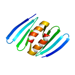
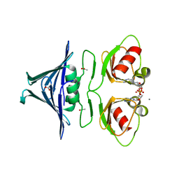
3CO2
DownloadVisualize
BU of 3co2 by Molmil
Mlotik1 ion channel cyclic-nucleotide binding domain mutant
Descriptor: Mlotik1 ion channel protein
Authors:Clayton, G.M, Alteiri, S.L, Thomas, L.R, Morais-Cabral, J.H.
Deposit date:2008-03-27
Release date:2008-08-05
Last modified:2024-02-21
Method:X-RAY DIFFRACTION (2.9 Å)
Cite:Structural and Energetic Analysis of Activation by a Cyclic Nucleotide Binding Domain.
J.Mol.Biol., 381, 2008
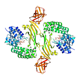
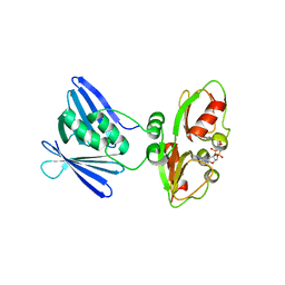
3CL1
DownloadVisualize
BU of 3cl1 by Molmil
M. loti cyclic-nucleotide binding domain, cyclic-GMP bound
Descriptor: CHLORIDE ION, CYCLIC GUANOSINE MONOPHOSPHATE, Mll3241 protein, ...
Authors:Clayton, G.M, Alteiri, S.L, Thomas, L.R, Morais-Cabral, J.H.
Deposit date:2008-03-18
Release date:2008-08-05
Last modified:2024-02-21
Method:X-RAY DIFFRACTION (2.4 Å)
Cite:Structural and Energetic Analysis of Activation by a Cyclic Nucleotide Binding Domain.
J.Mol.Biol., 381, 2008
8BZ6
DownloadVisualize
BU of 8bz6 by Molmil
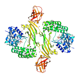
Crystal structure of the L. monocytogenes RmlT in complex with UDP-glucose
Descriptor: ACETATE ION, CHLORIDE ION, Glycosyltransferase, ...
Authors:Cereija, T.B, Morais-Cabral, J.H.
Deposit date:2022-12-14
Release date:2023-12-27
Method:X-RAY DIFFRACTION (2.4 Å)
Cite:Crystal structure of the L. monocytogenes RmlT in complex with UDP-glucose
To Be Published
8BZ8
DownloadVisualize
BU of 8bz8 by Molmil
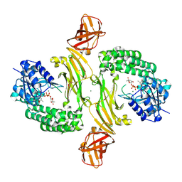
Crystal structure of the L. monocytogenes RmlT D198A variant in complex with TDP-rhamnose
Descriptor: 2'-DEOXY-THYMIDINE-BETA-L-RHAMNOSE, CHLORIDE ION, D(-)-TARTARIC ACID, ...
Authors:Cereija, T.B, Morais-Cabral, J.H.
Deposit date:2022-12-14
Release date:2023-12-27
Method:X-RAY DIFFRACTION (2.52 Å)
Cite:Crystal structure of the L. monocytogenes RmlT D198A variant in complex with TDP-rhamnose
To Be Published
8BZ5
DownloadVisualize
BU of 8bz5 by Molmil
Crystal structure of the L. monocytogenes RmlT in complex with HEPES
Descriptor: 4-(2-HYDROXYETHYL)-1-PIPERAZINE ETHANESULFONIC ACID, Glycosyltransferase
Authors:Cereija, T.B, Morais-Cabral, J.H.
Deposit date:2022-12-14
Release date:2023-12-27
Method:X-RAY DIFFRACTION (2.3 Å)
Cite:Crystal structure of the L. monocytogenes RmlT in complex with HEPES
To Be Published
8BZ4
DownloadVisualize
BU of 8bz4 by Molmil
Crystal structure of the apo form of the L. monocytogenes RmlT
Descriptor: CHLORIDE ION, Glycosyltransferase
Authors:Cereija, T.B, Morais-Cabral, J.H.
Deposit date:2022-12-14
Release date:2023-12-27
Last modified:2024-11-06
Method:X-RAY DIFFRACTION (2.52 Å)
Cite:Crystal structure of the apo form of the L. monocytogenes RmlT
To Be Published
8BZ7
DownloadVisualize
BU of 8bz7 by Molmil
Crystal structure of the L. monocytogenes RmlT in complex with TDP-rhamnose
Descriptor: 2'-DEOXY-THYMIDINE-BETA-L-RHAMNOSE, CHLORIDE ION, D(-)-TARTARIC ACID, ...
Authors:Cereija, T.B, Morais-Cabral, J.H.
Deposit date:2022-12-14
Release date:2023-12-27
Method:X-RAY DIFFRACTION (2.2 Å)
Cite:Crystal structure of the L. monocytogenes RmlT in complex with TDP-rhamnose
To Be Published
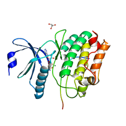
5H9B
DownloadVisualize
BU of 5h9b by Molmil
Drosophila CaMKII-wt in complex with a fragment of the Eag potassium channel and Mg2+/AMPPN
Descriptor: AMP PHOSPHORAMIDATE, Calcium/calmodulin-dependent protein kinase II, isoform C, ...
Authors:Castro-Rodrigues, A.F, Morais-Cabral, J.H.
Deposit date:2015-12-27
Release date:2017-01-11
Last modified:2024-05-08
Method:X-RAY DIFFRACTION (2.25 Å)
Cite:The Interaction between the Drosophila EAG Potassium Channel and the Protein Kinase CaMKII Involves an Extensive Interface at the Active Site of the Kinase.
J.Mol.Biol., 430, 2018
5HIT
DownloadVisualize
BU of 5hit by Molmil
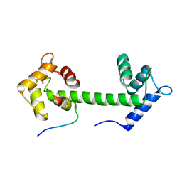
Crystal Structure Analysis of Ca2+-calmodulin and a C-terminal EAG1 channel fragment
Descriptor: CALCIUM ION, Calmodulin, Potassium voltage-gated channel subfamily H member 1
Authors:Marques-Carvalho, M.J, Morais-Cabral, J.H.
Deposit date:2016-01-12
Release date:2016-09-14
Last modified:2024-01-10
Method:X-RAY DIFFRACTION (2.85 Å)
Cite:Molecular Insights into the Mechanism of Calmodulin Inhibition of the EAG1 Potassium Channel.
Structure, 24, 2016
5HU3
DownloadVisualize
BU of 5hu3 by Molmil
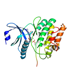
Drosophila CaMKII-D136N in complex with a phosphorylated fragment of the Eag potassium channel and Mg2+/ADP
Descriptor: ADENOSINE-5'-DIPHOSPHATE, Calcium/calmodulin-dependent protein kinase type II alpha chain, MAGNESIUM ION, ...
Authors:Castro-Rodrigues, A.F, Morais-Cabral, J.H.
Deposit date:2016-01-27
Release date:2017-02-01
Last modified:2024-11-06
Method:X-RAY DIFFRACTION (1.885 Å)
Cite:The Interaction between the Drosophila EAG Potassium Channel and the Protein Kinase CaMKII Involves an Extensive Interface at the Active Site of the Kinase.
J.Mol.Biol., 430, 2018
1BEB
DownloadVisualize
BU of 1beb by Molmil
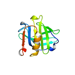
BOVINE BETA-LACTOGLOBULIN, LATTICE X
Descriptor: BETA-LACTOGLOBULIN, SULFATE ION
Authors:Brownlow, S, Morais-Cabral, J.H, Sawyer, L.
Deposit date:1996-12-20
Release date:1997-05-15
Last modified:2024-10-09
Method:X-RAY DIFFRACTION (1.8 Å)
Cite:Bovine beta-lactoglobulin at 1.8 A resolution--still an enigmatic lipocalin.
Structure, 5, 1997
1K4C
DownloadVisualize
BU of 1k4c by Molmil
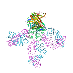
Potassium Channel KcsA-Fab complex in high concentration of K+
Descriptor: DIACYL GLYCEROL, NONAN-1-OL, POTASSIUM ION, ...
Authors:Zhou, Y, Morais-Cabral, J.H, Kaufman, A, MacKinnon, R.
Deposit date:2001-10-07
Release date:2001-11-14
Last modified:2024-10-30
Method:X-RAY DIFFRACTION (2 Å)
Cite:Chemistry of ion coordination and hydration revealed by a K+ channel-Fab complex at 2.0 A resolution.
Nature, 414, 2001
1K4D
DownloadVisualize
BU of 1k4d by Molmil
Potassium Channel KcsA-Fab complex in low concentration of K+
Descriptor: DIACYL GLYCEROL, NONAN-1-OL, POTASSIUM ION, ...
Authors:Zhou, Y, Morais-Cabral, J.H, Kaufman, A, MacKinnon, R.
Deposit date:2001-10-07
Release date:2001-11-14
Last modified:2021-10-27
Method:X-RAY DIFFRACTION (2.3 Å)
Cite:Chemistry of ion coordination and hydration revealed by a K+ channel-Fab complex at 2.0 A resolution.
Nature, 414, 2001
7AGY
DownloadVisualize
BU of 7agy by Molmil
Structure of the N-domain of the K+/H+ antiporter subunit KhtT at pH 8.5
Descriptor: K(+)/H(+) antiporter subunit KhtT
Authors:Cereija, T.B, Morais-Cabral, J.H.
Deposit date:2020-09-23
Release date:2021-03-31
Last modified:2024-01-31
Method:X-RAY DIFFRACTION (1.54 Å)
Cite:c-di-AMP, a likely master regulator of bacterial K + homeostasis machinery, activates a K + exporter.
Proc.Natl.Acad.Sci.USA, 118, 2021
7AHT
DownloadVisualize
BU of 7aht by Molmil
Structure of the N-domain of the K+/H+ antiporter subunit KhtT at pH 7.5
Descriptor: K(+)/H(+) antiporter subunit KhtT
Authors:Cereija, T.B, Morais-Cabral, J.H.
Deposit date:2020-09-25
Release date:2021-03-31
Last modified:2024-01-31
Method:X-RAY DIFFRACTION (2.16 Å)
Cite:c-di-AMP, a likely master regulator of bacterial K + homeostasis machinery, activates a K + exporter.
Proc.Natl.Acad.Sci.USA, 118, 2021
7AHM
DownloadVisualize
BU of 7ahm by Molmil
Low-resolution structure of the K+/H+ antiporter subunit KhtT in complex with c-di-AMP
Descriptor: (2R,3R,3aS,5R,7aR,9R,10R,10aS,12R,14aR)-2,9-bis(6-amino-9H-purin-9-yl)octahydro-2H,7H-difuro[3,2-d:3',2'-j][1,3,7,9,2,8 ]tetraoxadiphosphacyclododecine-3,5,10,12-tetrol 5,12-dioxide, K(+)/H(+) antiporter subunit KhtT
Authors:Cereija, T.B, Guerra, J.P, Morais-Cabral, J.H.
Deposit date:2020-09-24
Release date:2021-03-31
Last modified:2024-01-31
Method:X-RAY DIFFRACTION (3.14 Å)
Cite:c-di-AMP, a likely master regulator of bacterial K + homeostasis machinery, activates a K + exporter.
Proc.Natl.Acad.Sci.USA, 118, 2021
7AGV
DownloadVisualize
BU of 7agv by Molmil
High-resolution structure of the K+/H+ antiporter subunit KhtT in complex with c-di-AMP
Descriptor: (2R,3R,3aS,5R,7aR,9R,10R,10aS,12R,14aR)-2,9-bis(6-amino-9H-purin-9-yl)octahydro-2H,7H-difuro[3,2-d:3',2'-j][1,3,7,9,2,8 ]tetraoxadiphosphacyclododecine-3,5,10,12-tetrol 5,12-dioxide, ACETATE ION, CALCIUM ION, ...
Authors:Cereija, T.B, Guerra, J.P, Morais-Cabral, J.H.
Deposit date:2020-09-23
Release date:2021-03-31
Last modified:2024-05-01
Method:X-RAY DIFFRACTION (1.85 Å)
Cite:c-di-AMP, a likely master regulator of bacterial K + homeostasis machinery, activates a K + exporter.
Proc.Natl.Acad.Sci.USA, 118, 2021
7AGW
DownloadVisualize
BU of 7agw by Molmil
Structure of the N-domain of the K+/H+ antiporter subunit KhtT at pH 6.5
Descriptor: K(+)/H(+) antiporter subunit KhtT
Authors:Cereija, T.B, Morais-Cabral, J.H.
Deposit date:2020-09-23
Release date:2021-03-31
Last modified:2024-01-31
Method:X-RAY DIFFRACTION (1.511 Å)
Cite:c-di-AMP, a likely master regulator of bacterial K + homeostasis machinery, activates a K + exporter.
Proc.Natl.Acad.Sci.USA, 118, 2021
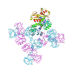
6S5D
DownloadVisualize
BU of 6s5d by Molmil
Square conformation of KtrA R16A mutant ring with bound ATP
Descriptor: ADENOSINE-5'-TRIPHOSPHATE, Ktr system potassium uptake protein A, MAGNESIUM ION
Authors:Teixeira-Duarte, C.M, Fonseca, F, Morais-Cabral, J.H.
Deposit date:2019-07-01
Release date:2020-01-08
Last modified:2024-01-24
Method:X-RAY DIFFRACTION (3.393 Å)
Cite:Activation of a nucleotide-dependent RCK domain requires binding of a cation cofactor to a conserved site.
Elife, 8, 2019
6S5G
DownloadVisualize
BU of 6s5g by Molmil
Non-square conformation of KtrA A80P mutant ring with bound ADP
Descriptor: ADENOSINE-5'-DIPHOSPHATE, Ktr system potassium uptake protein A
Authors:Teixeira-Duarte, C.M, Fonseca, F, Morais-Cabral, J.H.
Deposit date:2019-07-01
Release date:2020-01-08
Last modified:2024-01-24
Method:X-RAY DIFFRACTION (4.33 Å)
Cite:Activation of a nucleotide-dependent RCK domain requires binding of a cation cofactor to a conserved site.
Elife, 8, 2019
6S5E
DownloadVisualize
BU of 6s5e by Molmil
Non-square conformation of KtrA A80P mutant ring with bound ATP
Descriptor: ADENOSINE-5'-TRIPHOSPHATE, Ktr system potassium uptake protein A
Authors:Teixeira-Duarte, C.M, Fonseca, F, Morais-Cabral, J.H.
Deposit date:2019-07-01
Release date:2020-01-08
Last modified:2024-01-24
Method:X-RAY DIFFRACTION (3.893 Å)
Cite:Activation of a nucleotide-dependent RCK domain requires binding of a cation cofactor to a conserved site.
Elife, 8, 2019
6S5B
DownloadVisualize
BU of 6s5b by Molmil
Non-square conformation of KtrA R16K mutant ring with bound ADP
Descriptor: ADENOSINE-5'-DIPHOSPHATE, Ktr system potassium uptake protein A
Authors:Teixeira-Duarte, C.M, Fonseca, F, Morais-Cabral, J.H.
Deposit date:2019-07-01
Release date:2020-01-08
Last modified:2024-01-24
Method:X-RAY DIFFRACTION (3.052 Å)
Cite:Activation of a nucleotide-dependent RCK domain requires binding of a cation cofactor to a conserved site.
Elife, 8, 2019
6S7R
DownloadVisualize
BU of 6s7r by Molmil
Non-square conformations of KtrA R16A mutant rings with bound ADP
Descriptor: ADENOSINE-5'-DIPHOSPHATE, Ktr system potassium uptake protein A
Authors:Teixeira-Duarte, C.M, Fonseca, F, Morais-Cabral, J.H.
Deposit date:2019-07-05
Release date:2020-01-08
Last modified:2024-01-24
Method:X-RAY DIFFRACTION (3.73 Å)
Cite:Activation of a nucleotide-dependent RCK domain requires binding of a cation cofactor to a conserved site.
Elife, 8, 2019

227111

PDB entries from 2024-11-06

PDB statisticsPDBj update infoContact PDBjnumon