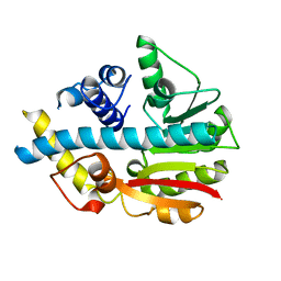
2WAM
 
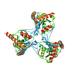 | | Crystal structure of Mycobacterium tuberculosis unknown function protein Rv2714 | | 分子名称: | CONSERVED HYPOTHETICAL ALANINE AND LEUCINE RICH PROTEIN, GLYCEROL | | 著者 | Bellinzoni, M, Grana, M, Buschiazzo, A, Miras, I, Haouz, A, Alzari, P.M. | | 登録日 | 2009-02-09 | | 公開日 | 2009-10-06 | | 最終更新日 | 2024-05-01 | | 実験手法 | X-RAY DIFFRACTION (2.6 Å) | | 主引用文献 | Structure of Mycobacterium Tuberculosis Rv2714, a Representative of a Duplicated Gene Family in Actinobacteria.
Acta Crystallogr.,Sect.F, 65, 2009
|
|
4ADL
 | | Crystal structures of Rv1098c in complex with malate | | 分子名称: | (2S)-2-hydroxybutanedioic acid, FUMARATE HYDRATASE CLASS II | | 著者 | Mechaly, A.E, Haouz, A, Miras, I, Weber, P, Shepard, W, Cole, S, Alzari, P.M, Bellinzoni, M. | | 登録日 | 2011-12-26 | | 公開日 | 2012-04-25 | | 最終更新日 | 2023-12-20 | | 実験手法 | X-RAY DIFFRACTION (2.2 Å) | | 主引用文献 | Conformational Changes Upon Ligand Binding in the Essential Class II Fumarase Rv1098C from Mycobacterium Tuberculosis.
FEBS Lett., 586, 2012
|
|
4ADM
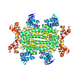 | | Crystal structure of Rv1098c in complex with meso-tartrate | | 分子名称: | FUMARATE HYDRATASE CLASS II, GLYCEROL, S,R MESO-TARTARIC ACID | | 著者 | Mechaly, A.E, Haouz, A, Miras, I, Weber, P, Shepard, W, Cole, S, Alzari, P.M, Bellinzoni, M. | | 登録日 | 2011-12-27 | | 公開日 | 2012-04-25 | | 最終更新日 | 2023-12-20 | | 実験手法 | X-RAY DIFFRACTION (1.65 Å) | | 主引用文献 | Conformational Changes Upon Ligand Binding in the Essential Class II Fumarase Rv1098C from Mycobacterium Tuberculosis.
FEBS Lett., 586, 2012
|
|
2V7S
 | | Crystal structure of the putative lipoprotein LppA from Mycobacterium tuberculosis | | 分子名称: | GLYCEROL, PROBABLE CONSERVED LIPOPROTEIN LPPA | | 著者 | Grana, M, Miras, I, Haouz, A, Winter, N, Buschiazzo, A, Bellinzoni, M, Alzari, P.M. | | 登録日 | 2007-08-01 | | 公開日 | 2008-08-26 | | 最終更新日 | 2011-09-28 | | 実験手法 | X-RAY DIFFRACTION (1.96 Å) | | 主引用文献 | Crystal Structure of Mycobacterium Tuberculosis Lppa, a Lipoprotein Confined to Pathogenic Mycobacteria.
Proteins, 78, 2010
|
|
2UYQ
 | | Crystal structure of ML2640c from Mycobacterium leprae in complex with S-adenosylmethionine | | 分子名称: | HYPOTHETICAL PROTEIN ML2640, S-ADENOSYLMETHIONINE | | 著者 | Grana, M, Buschiazzo, A, Wehenkel, A, Haouz, A, Miras, I, Shepard, W, Alzari, P.M. | | 登録日 | 2007-04-11 | | 公開日 | 2007-08-07 | | 最終更新日 | 2023-12-13 | | 実験手法 | X-RAY DIFFRACTION (1.8 Å) | | 主引用文献 | The Crystal Structure of M. Leprae Ml2640C Defines a Large Family of Putative S-Adenosylmethionine- Dependent Methyltransferases in Mycobacteria.
Protein Sci., 16, 2007
|
|
2UYO
 | | Crystal structure of ML2640c from Mycobacterium leprae in an hexagonal crystal form | | 分子名称: | HYPOTHETICAL PROTEIN ML2640 | | 著者 | Grana, M, Buschiazzo, A, Wehenkel, A, Haouz, A, Miras, I, Shepard, W, Alzari, P.M. | | 登録日 | 2007-04-11 | | 公開日 | 2007-08-07 | | 最終更新日 | 2023-12-13 | | 実験手法 | X-RAY DIFFRACTION (1.7 Å) | | 主引用文献 | The Crystal Structure of M. Leprae Ml2640C Defines a Large Family of Putative S-Adenosylmethionine- Dependent Methyltransferases in Mycobacteria.
Protein Sci., 16, 2007
|
|
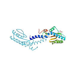
3OSG
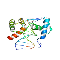 | | The structure of protozoan parasite Trichomonas vaginalis Myb2 in complex with MRE-1-12 DNA | | 分子名称: | 5'-D(*AP*AP*AP*TP*AP*TP*CP*GP*TP*TP*AP*T)-3', 5'-D(*AP*TP*AP*AP*CP*GP*AP*TP*AP*TP*TP*T)-3', MYB21 | | 著者 | Jiang, I, Tsai, C.K, Chen, S.C, Wang, S.H, Amiraslanov, I, Chang, C.F, Wu, W.J, Tai, J.H, Liaw, Y.C, Huang, T.H. | | 登録日 | 2010-09-09 | | 公開日 | 2011-08-03 | | 最終更新日 | 2024-03-20 | | 実験手法 | X-RAY DIFFRACTION (1.997 Å) | | 主引用文献 | Molecular basis of the recognition of the ap65-1 gene transcription promoter elements by a Myb protein from the protozoan parasite Trichomonas vaginalis.
Nucleic Acids Res., 39, 2011
|
|
3OSF
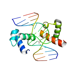 | | The structure of protozoan parasite Trichomonas vaginalis Myb2 in complex with MRE-2f-13 DNA | | 分子名称: | 5'-D(*CP*AP*AP*GP*AP*CP*GP*AP*TP*AP*CP*AP*G)-3', 5'-D(*CP*TP*GP*TP*AP*TP*CP*GP*TP*CP*TP*TP*G)-3', ISOPROPYL ALCOHOL, ... | | 著者 | Jiang, I, Tsai, C.K, Chen, S.C, Wang, S.H, Amiraslanov, I, Chang, C.F, Wu, W.J, Tai, J.H, Liaw, Y.C, Huang, T.H. | | 登録日 | 2010-09-09 | | 公開日 | 2011-08-03 | | 最終更新日 | 2024-03-20 | | 実験手法 | X-RAY DIFFRACTION (2.032 Å) | | 主引用文献 | Molecular basis of the recognition of the ap65-1 gene transcription promoter elements by a Myb protein from the protozoan parasite Trichomonas vaginalis.
Nucleic Acids Res., 39, 2011
|
|
1M07
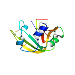 | | RESIDUES INVOLVED IN THE CATALYSIS AND BASE SPECIFICITY OF CYTOTOXIC RIBONUCLEASE FROM BULLFROG (RANA CATESBEIANA) | | 分子名称: | 5'-D(*AP*CP*GP*A)-3', Ribonuclease | | 著者 | Leu, Y.-J, Chern, S.-S, Wang, S.-C, Hsiao, Y.-Y, Amiraslanov, I, Liaw, Y.-C, Liao, Y.-D. | | 登録日 | 2002-06-12 | | 公開日 | 2003-01-21 | | 最終更新日 | 2019-12-25 | | 実験手法 | X-RAY DIFFRACTION (1.8 Å) | | 主引用文献 | Residues involved in the catalysis, base specificity, and cytotoxicity of ribonuclease from Rana catesbeiana based upon mutagenesis and X-ray crystallography
J.Biol.Chem., 278, 2003
|
|
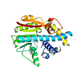
1KM9
 | | The Structure of a Cytotoxic Ribonuclease From the Oocyte of Rana Catesbeiana (Bullfrog) | | 分子名称: | PHOSPHATE ION, RIBONUCLEASE, OOCYTES | | 著者 | Chern, S.-S, Musayev, F.N, Amiraslanov, I.R, Liao, Y.-D, Liaw, Y.-C. | | 登録日 | 2001-12-14 | | 公開日 | 2003-09-09 | | 最終更新日 | 2023-08-16 | | 実験手法 | X-RAY DIFFRACTION (1.96 Å) | | 主引用文献 | The Structure of a Cytotoxic Ribonuclease From the Oocyte of Rana Catesbeiana (Bullfrog)
To be Published
|
|
1KM8
 | | The Structure of a Cytotoxic Ribonuclease From the Oocyte of Rana Catesbeiana (Bullfrog) | | 分子名称: | PHOSPHATE ION, RIBONUCLEASE, OOCYTES | | 著者 | Chern, S.-S, Musayev, F.N, Amiraslanov, I.R, Liao, Y.-D, Liaw, Y.-C. | | 登録日 | 2001-12-14 | | 公開日 | 2003-09-09 | | 最終更新日 | 2023-08-16 | | 実験手法 | X-RAY DIFFRACTION (1.9 Å) | | 主引用文献 | The Structure of a Cytotoxic Ribonuclease From the Oocyte of Rana Catesbeiana (Bullfrog)
To be Published
|
|
2QLK
 | | Adenovirus AD35 fibre head | | 分子名称: | Fiber, GLYCEROL | | 著者 | Liaw, Y.-C, Amiraslanov, I, Wang, H, Lieber, A. | | 登録日 | 2007-07-13 | | 公開日 | 2008-02-19 | | 最終更新日 | 2023-08-30 | | 実験手法 | X-RAY DIFFRACTION (2.02 Å) | | 主引用文献 | Identification of CD46 binding sites within the adenovirus serotype 35 fiber knob
J.Virol., 81, 2007
|
|


