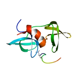
3Q68
 
 | |
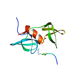
3Q66
 | |
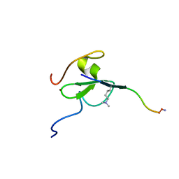
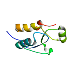
2K2W
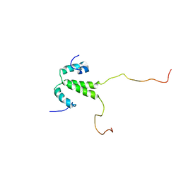
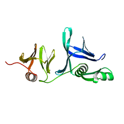
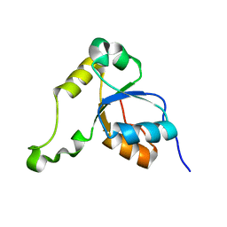 | | Second BRCT domain of NBS1 | | 分子名称: | Recombination and DNA repair protein | | 著者 | Xu, C, Cui, G, Botuyan, M, Mer, G. | | 登録日 | 2008-04-14 | | 公開日 | 2008-06-17 | | 最終更新日 | 2024-05-29 | | 実験手法 | SOLUTION NMR | | 主引用文献 | Structure of a second BRCT domain identified in the nijmegen breakage syndrome protein Nbs1 and its function in an MDC1-dependent localization of Nbs1 to DNA damage sites.
J.Mol.Biol., 381, 2008
|
|
2KTF
 | |
2L0F
 | |
2L0G
 | |
2LH9
 | |
2LDM
 | |
2LH0
 | |
2LVM
 | |
2LY1
 | |
2LY2
 | |
2MWO
 | |
2MWP
 | |
5TBN
 | |
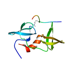
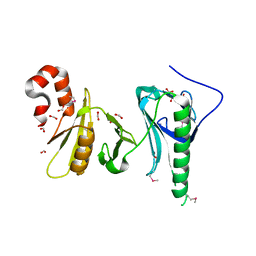
5VEY
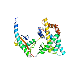 | | Solution NMR structure of histone H2A-H2B mono-ubiquitylated at H2A Lys15 in complex with RNF169 (653-708) | | 分子名称: | E3 ubiquitin-protein ligase RNF169, Histone H2B type 1-J,Histone H2A type 1-B/E, Polyubiquitin-B | | 著者 | Hu, Q, Botuyan, M.V, Cui, G, Mer, G. | | 登録日 | 2017-04-06 | | 公開日 | 2017-05-17 | | 最終更新日 | 2022-02-23 | | 実験手法 | SOLUTION NMR | | 主引用文献 | Mechanisms of Ubiquitin-Nucleosome Recognition and Regulation of 53BP1 Chromatin Recruitment by RNF168/169 and RAD18.
Mol. Cell, 66, 2017
|
|
5VX7
 | |
5UMV
 | |
2KDK
 | |
2KRE
 | |
5UMS
 | |
5UMU
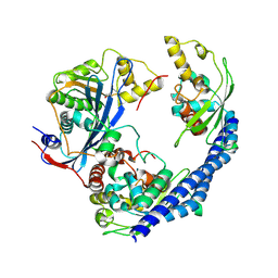
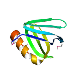
 | | Crystal structure of the middle double PH domain of human FACT complex subunit SPT16 | | 分子名称: | ACETATE ION, FACT complex subunit SPT16, FORMIC ACID | | 著者 | Hu, Q, Thompson, J.R, Heroux, A, Su, D, Botuyan, M.V, Mer, G. | | 登録日 | 2017-01-29 | | 公開日 | 2018-01-31 | | 最終更新日 | 2019-12-04 | | 実験手法 | X-RAY DIFFRACTION (1.903 Å) | | 主引用文献 | Crystal structure of the middle double PH domain of human FACT complex subunit SPT16
To Be Published
|
|
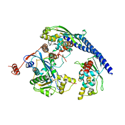
5UMR
 | | Crystal structure of N-terminal domain of human FACT complex subunit SSRP1 | | 分子名称: | FACT complex subunit SSRP1 | | 著者 | Su, D, Hu, Q, Thompson, J.R, Heroux, A, Botuyan, M.V, Mer, G. | | 登録日 | 2017-01-29 | | 公開日 | 2018-01-31 | | 最終更新日 | 2019-12-04 | | 実験手法 | X-RAY DIFFRACTION (1.501 Å) | | 主引用文献 | Crystal structure of N-terminal domain of human FACT complex subunit SSRP1
To Be Published
|
|
5UMT
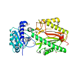 | | Crystal structure of N-terminal domain of human FACT complex subunit SPT16 | | 分子名称: | 1,2-ETHANEDIOL, FACT complex subunit SPT16, GLYCEROL | | 著者 | Su, D, Hu, Q, Thompson, J.R, Botuyan, M.V, Mer, G. | | 登録日 | 2017-01-29 | | 公開日 | 2018-01-31 | | 最終更新日 | 2023-10-04 | | 実験手法 | X-RAY DIFFRACTION (2.092 Å) | | 主引用文献 | Crystal structure of N-terminal domain of human FACT complex subunit SPT16
To Be Published
|
|
5VWE
 | |


