Loading
PDBj
MenuPDBj@FacebookPDBj@X(formerly Twitter)PDBj@BlueSkyPDBj@YouTubewwPDB FoundationwwPDB
RCSB PDBPDBeBMRBAdv. SearchSearch help
PDB: 99 results

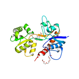
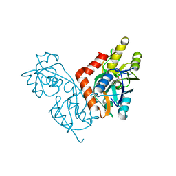
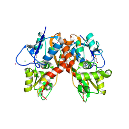
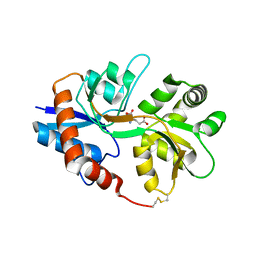
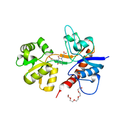
2F36
DownloadVisualize
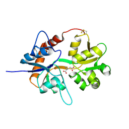
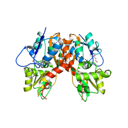
BU of 2f36 by Molmil
Crystal Structure of the GluR5 Ligand Binding Core Dimer with Glutamate At 2.1 Angstroms Resolution
Descriptor: GLUTAMATE RECEPTOR, IONOTROPIC KAINATE 1, GLUTAMIC ACID, ...
Authors:Mayer, M.L.
Deposit date:2005-11-18
Release date:2006-04-04
Last modified:2024-11-06
Method:X-RAY DIFFRACTION (2.11 Å)
Cite:Crystal structures of the kainate receptor GluR5 ligand binding core dimer with novel GluR5-selective antagonists.
J.Neurosci., 26, 2006
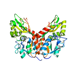
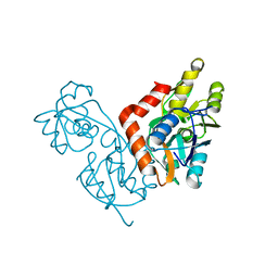
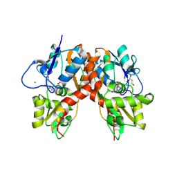
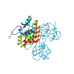
2F34
DownloadVisualize
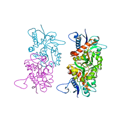
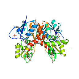
BU of 2f34 by Molmil
Crystal Structure of the GluR5 Ligand Binding Core Dimer with UBP310 At 1.74 Angstroms Resolution
Descriptor: (S)-1-(2-AMINO-2-CARBOXYETHYL)-3(2-CARBOXYTHIOPHENE-3-YL-METHYL)-5-METHYLPYRIMIDINE-2,4-DIONE, CHLORIDE ION, GLUTAMATE RECEPTOR, ...
Authors:Mayer, M.L.
Deposit date:2005-11-18
Release date:2006-04-04
Last modified:2023-08-23
Method:X-RAY DIFFRACTION (1.74 Å)
Cite:Crystal structures of the kainate receptor GluR5 ligand binding core dimer with novel GluR5-selective antagonists.
J.Neurosci., 26, 2006
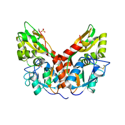
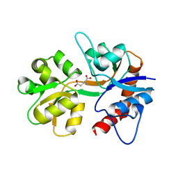
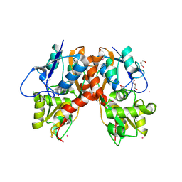
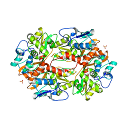
2F35
DownloadVisualize
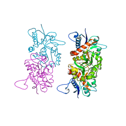
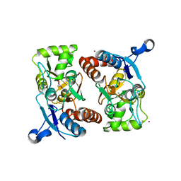
BU of 2f35 by Molmil
Crystal Structure of the GluR5 Ligand Binding Core with UBP302 At 1.87 Angstroms Resolution
Descriptor: (S)-1-(2-AMINO-2-CARBOXYETHYL)-3-(2-CARBOXYBENZYL)PYRIMIDINE-2,4-DIONE, CHLORIDE ION, GLUTAMATE RECEPTOR, ...
Authors:Mayer, M.L.
Deposit date:2005-11-18
Release date:2006-04-04
Last modified:2024-04-03
Method:X-RAY DIFFRACTION (1.87 Å)
Cite:Crystal structures of the kainate receptor GluR5 ligand binding core dimer with novel GluR5-selective antagonists.
J.Neurosci., 26, 2006
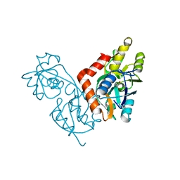
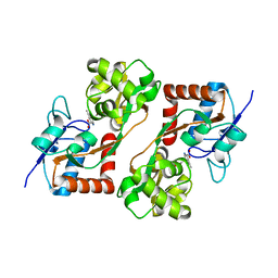
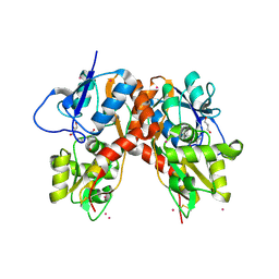
3C33
DownloadVisualize
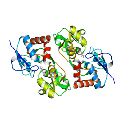
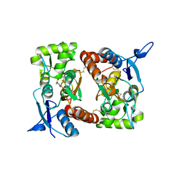
BU of 3c33 by Molmil
Crystal structure of GluR5 ligand-binding core in complex with potassium at 1.78 Angstrom resolution
Descriptor: 3-(CARBOXYMETHYL)-4-ISOPROPENYLPROLINE, CHLORIDE ION, GLUTAMATE RECEPTOR, ...
Authors:Mayer, M.L.
Deposit date:2008-01-27
Release date:2008-06-17
Last modified:2024-11-13
Method:X-RAY DIFFRACTION (1.72 Å)
Cite:Molecular basis of kainate receptor modulation by sodium.
Neuron, 58, 2008
3C36
DownloadVisualize
BU of 3c36 by Molmil
Crystal structure of GluR5 ligand-binding core in complex with ammonium ions at 1.68 Angstrom resolution
Descriptor: 3-(CARBOXYMETHYL)-4-ISOPROPENYLPROLINE, AMMONIUM ION, CHLORIDE ION, ...
Authors:Mayer, M.L.
Deposit date:2008-01-27
Release date:2008-06-17
Last modified:2024-11-13
Method:X-RAY DIFFRACTION (1.68 Å)
Cite:Molecular basis of kainate receptor modulation by sodium.
Neuron, 58, 2008
3C34
DownloadVisualize
BU of 3c34 by Molmil
Crystal structure of GluR5 ligand-binding core in complex with rubidium at 1.82 Angstrom resolution
Descriptor: 3-(CARBOXYMETHYL)-4-ISOPROPENYLPROLINE, CHLORIDE ION, GLUTAMATE RECEPTOR, ...
Authors:Mayer, M.L.
Deposit date:2008-01-27
Release date:2008-06-17
Last modified:2024-10-30
Method:X-RAY DIFFRACTION (1.82 Å)
Cite:Molecular basis of kainate receptor modulation by sodium.
Neuron, 58, 2008
5CMB
DownloadVisualize
BU of 5cmb by Molmil
Mnemiopsis leidyi ML032222a iGluR LBD R703K mutant glycine complex
Descriptor: GLYCINE, MAGNESIUM ION, ML032222a iGluR, ...
Authors:Mayer, M.L, Thomas, A.
Deposit date:2015-07-16
Release date:2016-07-20
Last modified:2024-10-30
Method:X-RAY DIFFRACTION (1.34 Å)
Cite:Molecular lock regulates binding of glycine to a primitive NMDA receptor.
Proc.Natl.Acad.Sci.USA, 113, 2016
5CMC
DownloadVisualize
BU of 5cmc by Molmil
Mnemiopsis leidyi ML032222a iGluR LBD E423S mutant glycine complex
Descriptor: GLYCINE, MAGNESIUM ION, ML032222a iGluR, ...
Authors:Mayer, M.L, Thomas, A.
Deposit date:2015-07-16
Release date:2016-07-20
Last modified:2024-10-23
Method:X-RAY DIFFRACTION (1.28 Å)
Cite:Molecular lock regulates binding of glycine to a primitive NMDA receptor.
Proc.Natl.Acad.Sci.USA, 113, 2016
2OJT
DownloadVisualize
BU of 2ojt by Molmil
Structure and mechanism of kainate receptor modulation by anions
Descriptor: (S)-1-(2-AMINO-2-CARBOXYETHYL)-3(2-CARBOXYTHIOPHENE-3-YL-METHYL)-5-METHYLPYRIMIDINE-2,4-DIONE, BROMIDE ION, Glutamate receptor, ...
Authors:Mayer, M.L.
Deposit date:2007-01-14
Release date:2007-04-03
Last modified:2023-08-30
Method:X-RAY DIFFRACTION (1.95 Å)
Cite:Structure and mechanism of kainate receptor modulation by anions.
Neuron, 53, 2007
1IIT
DownloadVisualize
BU of 1iit by Molmil
GLUR0 LIGAND BINDING CORE COMPLEX WITH L-SERINE
Descriptor: SERINE, Slr1257 protein
Authors:Mayer, M.L, Olson, R, Gouaux, E.
Deposit date:2001-04-24
Release date:2001-09-19
Last modified:2024-02-07
Method:X-RAY DIFFRACTION (1.9 Å)
Cite:Mechanisms for ligand binding to GluR0 ion channels: crystal structures of the glutamate and serine complexes and a closed apo state.
J.Mol.Biol., 311, 2001
1II5
DownloadVisualize
BU of 1ii5 by Molmil
CRYSTAL STRUCTURE OF THE GLUR0 LIGAND BINDING CORE COMPLEX WITH L-GLUTAMATE
Descriptor: GLUTAMIC ACID, Slr1257 protein
Authors:Mayer, M.L, Olson, R, Gouaux, E.
Deposit date:2001-04-20
Release date:2001-09-19
Last modified:2024-02-07
Method:X-RAY DIFFRACTION (1.6 Å)
Cite:Mechanisms for ligand binding to GluR0 ion channels: crystal structures of the glutamate and serine complexes and a closed apo state.
J.Mol.Biol., 311, 2001
1IIW
DownloadVisualize
BU of 1iiw by Molmil
GLUR0 LIGAND BINDING CORE: CLOSED-CLEFT LIGAND-FREE STRUCTURE
Descriptor: Slr1257 protein
Authors:Mayer, M.L, Olson, R, Gouaux, E.
Deposit date:2001-04-24
Release date:2001-09-19
Last modified:2024-02-07
Method:X-RAY DIFFRACTION (1.9 Å)
Cite:Mechanisms for ligand binding to GluR0 ion channels: crystal structures of the glutamate and serine complexes and a closed apo state.
J.Mol.Biol., 311, 2001
1SD3
DownloadVisualize
BU of 1sd3 by Molmil
Crystal structure of the GLUR6 ligand binding core in complex with 2S,4R-4-methylglutamate at 1.8 Angstrom resolution
Descriptor: 2S,4R-4-METHYLGLUTAMATE, Glutamate receptor, ionotropic kainate 2
Authors:Mayer, M.L.
Deposit date:2004-02-12
Release date:2005-02-15
Last modified:2023-10-25
Method:X-RAY DIFFRACTION (1.8 Å)
Cite:Crystal structures of the GluR5 and GluR6 ligand binding cores: molecular mechanisms underlying kainate receptor selectivity.
Neuron, 45, 2005
1S50
DownloadVisualize
BU of 1s50 by Molmil
X-ray structure of the GluR6 ligand binding core (S1S2A) in complex with glutamate at 1.65 A resolution
Descriptor: GLUTAMIC ACID, Glutamate Receptor 6
Authors:Mayer, M.L.
Deposit date:2004-01-19
Release date:2005-02-08
Last modified:2024-11-06
Method:X-RAY DIFFRACTION (1.65 Å)
Cite:Crystal structures of the GluR5 and GluR6 ligand binding cores: Molecular mechanisms underlying kainate receptor selectivity
Neuron, 45, 2005
1TXF
DownloadVisualize
BU of 1txf by Molmil
CRYSTAL STRUCTURE OF THE GLUR5 LIGAND BINDING CORE IN COMPLEX WITH GLUTAMATE AT 2.1 ANGSTROM RESOLUTION
Descriptor: GLUTAMIC ACID, Glutamate receptor, ionotropic kainate 1
Authors:Mayer, M.L.
Deposit date:2004-07-04
Release date:2005-02-08
Last modified:2023-08-23
Method:X-RAY DIFFRACTION (2.1 Å)
Cite:Crystal structures of the GluR5 and GluR6 ligand binding cores: Molecular mechanisms underlying kainate receptor selectivity
Neuron, 45, 2005
1TT1
DownloadVisualize
BU of 1tt1 by Molmil
CRYSTAL STRUCTURE OF THE GLUR6 LIGAND BINDING CORE IN COMPLEX WITH KAINATE 1.93 A RESOLUTION
Descriptor: 3-(CARBOXYMETHYL)-4-ISOPROPENYLPROLINE, Glutamate receptor, ionotropic kainate 2
Authors:Mayer, M.L.
Deposit date:2004-06-21
Release date:2005-02-08
Last modified:2023-08-23
Method:X-RAY DIFFRACTION (1.93 Å)
Cite:Crystal structures of the GluR5 and GluR6 ligand binding cores: Molecular mechanisms underlying kainate receptor selectivity
Neuron, 45, 2005
1S9T
DownloadVisualize
BU of 1s9t by Molmil
Crystal structure of the GLUR6 ligand binding core in complex with quisqualate at 1.8A resolution
Descriptor: (S)-2-AMINO-3-(3,5-DIOXO-[1,2,4]OXADIAZOLIDIN-2-YL)-PROPIONIC ACID, CHLORIDE ION, Glutamate receptor, ...
Authors:Mayer, M.L.
Deposit date:2004-02-05
Release date:2005-02-08
Last modified:2023-10-25
Method:X-RAY DIFFRACTION (1.8 Å)
Cite:Crystal structures of the GluR5 and GluR6 ligand binding cores: molecular mechanisms underlying kainate receptor selectivity.
Neuron, 45, 2005
1S7Y
DownloadVisualize
BU of 1s7y by Molmil
Crystal structure of the GluR6 ligand binding core in complex with glutamate at 1.75 A resolution orthorhombic form
Descriptor: GLUTAMIC ACID, Glutamate receptor, ionotropic kainate 2 precursor
Authors:Mayer, M.L.
Deposit date:2004-01-30
Release date:2005-02-08
Last modified:2024-02-14
Method:X-RAY DIFFRACTION (1.75 Å)
Cite:Crystal structures of the GluR5 and GluR6 ligand binding cores: Molecular mechanisms underlying kainate receptor selectivity
Neuron, 45, 2005
3C35
DownloadVisualize
BU of 3c35 by Molmil
Crystal structure of GluR5 ligand-binding core in complex with cesium at 1.97 Angstrom resolution
Descriptor: 3-(CARBOXYMETHYL)-4-ISOPROPENYLPROLINE, CESIUM ION, CHLORIDE ION, ...
Authors:Mayer, M.L.
Deposit date:2008-01-27
Release date:2008-06-17
Last modified:2024-11-20
Method:X-RAY DIFFRACTION (1.97 Å)
Cite:Molecular basis of kainate receptor modulation by sodium.
Neuron, 58, 2008
3C32
DownloadVisualize
BU of 3c32 by Molmil
Crystal structure of GluR5 ligand-binding core in complex with sodium at 1.72 Angstrom resolution
Descriptor: 3-(CARBOXYMETHYL)-4-ISOPROPENYLPROLINE, CHLORIDE ION, GLUTAMATE RECEPTOR, ...
Authors:Mayer, M.L.
Deposit date:2008-01-27
Release date:2008-06-17
Last modified:2024-10-16
Method:X-RAY DIFFRACTION (1.72 Å)
Cite:Molecular basis of kainate receptor modulation by sodium.
Neuron, 58, 2008
3C31
DownloadVisualize
BU of 3c31 by Molmil
Crystal structure of GluR5 ligand-binding core in complex with lithium at 1.49 Angstrom resolution
Descriptor: 3-(CARBOXYMETHYL)-4-ISOPROPENYLPROLINE, CHLORIDE ION, GLUTAMATE RECEPTOR, ...
Authors:Mayer, M.L.
Deposit date:2008-01-27
Release date:2008-06-17
Last modified:2024-11-13
Method:X-RAY DIFFRACTION (1.49 Å)
Cite:Molecular basis of kainate receptor modulation by sodium.
Neuron, 58, 2008
2I0C
DownloadVisualize
BU of 2i0c by Molmil
Crystal structure of the GluR6 ligand binding core dimer crosslinked by disulfide bonds between Y490C and L752C at 2.25 Angstroms Resolution
Descriptor: GLUTAMIC ACID, Glutamate receptor, ionotropic kainate 2
Authors:Mayer, M.L.
Deposit date:2006-08-10
Release date:2006-11-21
Last modified:2024-11-20
Method:X-RAY DIFFRACTION (2.25 Å)
Cite:Conformational restriction blocks glutamate receptor desensitization.
Nat.Struct.Mol.Biol., 13, 2006
2I0B
DownloadVisualize
BU of 2i0b by Molmil
Crystal structure of the GluR6 ligand binding core ELKQ mutant dimer at 1.96 Angstroms Resolution
Descriptor: GLUTAMIC ACID, Glutamate receptor, ionotropic kainate 2, ...
Authors:Mayer, M.L.
Deposit date:2006-08-10
Release date:2006-11-21
Last modified:2024-11-20
Method:X-RAY DIFFRACTION (1.96 Å)
Cite:Conformational restriction blocks glutamate receptor desensitization.
Nat.Struct.Mol.Biol., 13, 2006
2GFE
DownloadVisualize
BU of 2gfe by Molmil
Crystal structure of the GluR2 A476E S673D Ligand Binding Core Mutant at 1.54 Angstroms Resolution
Descriptor: GLUTAMIC ACID, Glutamate receptor 2, ZINC ION
Authors:Mayer, M.L.
Deposit date:2006-03-21
Release date:2006-08-22
Last modified:2024-11-20
Method:X-RAY DIFFRACTION (1.54 Å)
Cite:Interdomain interactions in AMPA and kainate receptors regulate affinity for glutamate.
J.Neurosci., 26, 2006
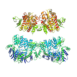
4UQJ
DownloadVisualize
BU of 4uqj by Molmil
Cryo-EM density map of GluA2em in complex with ZK200775
Descriptor: GLUTAMATE RECEPTOR 2, {[7-morpholin-4-yl-2,3-dioxo-6-(trifluoromethyl)-3,4-dihydroquinoxalin-1(2H)-yl]methyl}phosphonic acid
Authors:Meyerson, J.R, Kumar, J, Chittori, S, Rao, P, Pierson, J, Bartesaghi, A, Mayer, M.L, Subramaniam, S.
Deposit date:2014-06-24
Release date:2014-08-13
Last modified:2024-11-13
Method:ELECTRON MICROSCOPY (10.4 Å)
Cite:Structural Mechanism of Glutamate Receptor Activation and Desensitization
Nature, 514, 2014


1234>

227561

数据于2024-11-20公开中

PDB statisticsPDBj update infoContact PDBjnumon