4N73
 
 | |
3NY2
 | | Structure of the ubr-box of UBR2 ubiquitin ligase | | 分子名称: | E3 ubiquitin-protein ligase UBR2, ZINC ION | | 著者 | Matta-Camacho, E, Kozlov, G, Li, F, Gehring, K. | | 登録日 | 2010-07-14 | | 公開日 | 2010-08-11 | | 最終更新日 | 2024-02-21 | | 実験手法 | X-RAY DIFFRACTION (2.61 Å) | | 主引用文献 | Structural basis of substrate recognition and specificity in the N-end rule pathway.
Nat.Struct.Mol.Biol., 17, 2010
|
|
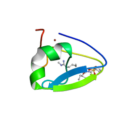
3NY3
 | | Structure of the ubr-box of UBR2 in complex with N-degron | | 分子名称: | E3 ubiquitin-protein ligase UBR2, N-degron, ZINC ION | | 著者 | Matta-Camacho, E, Kozlov, G, Li, F, Gehring, K. | | 登録日 | 2010-07-14 | | 公開日 | 2010-08-11 | | 最終更新日 | 2024-02-21 | | 実験手法 | X-RAY DIFFRACTION (1.6 Å) | | 主引用文献 | Structural basis of substrate recognition and specificity in the N-end rule pathway.
Nat.Struct.Mol.Biol., 17, 2010
|
|
3NY1
 | | Structure of the ubr-box of the UBR1 ubiquitin ligase | | 分子名称: | E3 ubiquitin-protein ligase UBR1, ZINC ION | | 著者 | Matta-Camacho, E, Kozlov, G, Li, F, Gehring, K. | | 登録日 | 2010-07-14 | | 公開日 | 2010-08-11 | | 最終更新日 | 2024-02-21 | | 実験手法 | X-RAY DIFFRACTION (2.085 Å) | | 主引用文献 | Structural basis of substrate recognition and specificity in the N-end rule pathway.
Nat.Struct.Mol.Biol., 17, 2010
|
|
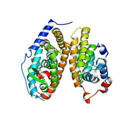
3PT3
 | | Crystal structure of the C-terminal lobe of the human UBR5 HECT domain | | 分子名称: | E3 ubiquitin-protein ligase UBR5 | | 著者 | Matta-Camacho, E, Kozlov, G, Menade, M, Gehring, K. | | 登録日 | 2010-12-02 | | 公開日 | 2012-01-25 | | 最終更新日 | 2023-09-06 | | 実験手法 | X-RAY DIFFRACTION (1.97 Å) | | 主引用文献 | Structure of the HECT C-lobe of the UBR5 E3 ubiquitin ligase.
Acta Crystallogr.,Sect.F, 68, 2012
|
|
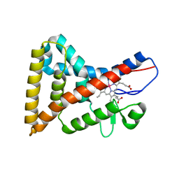
4ZO1
 | | Crystal Structure of the T3-bound TR-beta Ligand-binding Domain in complex with RXR-alpha | | 分子名称: | 3,5,3'TRIIODOTHYRONINE, Nuclear receptor coactivator 2, Retinoic acid receptor RXR-alpha, ... | | 著者 | Bruning, J.B, Kojetin, D.J, Matta-Camacho, E, Hughes, T.S, Srinivasan, S, Nwachukwu, J.C, Cavett, V, Nowak, J, Chalmers, M.J, Marciano, D.P, Kamenecka, T.M, Rance, M, Shulman, A.I, Mangelsdorf, D.J, Griffin, P.R, Nettles, K.W. | | 登録日 | 2015-05-05 | | 公開日 | 2015-09-02 | | 最終更新日 | 2023-11-15 | | 実験手法 | X-RAY DIFFRACTION (3.221 Å) | | 主引用文献 | Structural mechanism for signal transduction in RXR nuclear receptor heterodimers.
Nat Commun, 6, 2015
|
|
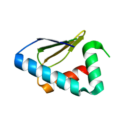
2KBW
 | | Solution Structure of human Mcl-1 complexed with human Bid_BH3 peptide | | 分子名称: | BH3-interacting domain death agonist, Induced myeloid leukemia cell differentiation protein Mcl-1 | | 著者 | Liu, Q, Moldoveanu, T, Sprules, T, Matta-Camacho, E, Mansur-Azzam, N, Gehring, K. | | 登録日 | 2008-12-09 | | 公開日 | 2009-12-15 | | 最終更新日 | 2024-05-22 | | 実験手法 | SOLUTION NMR | | 主引用文献 | Apoptotic regulation by MCL-1 through heterodimerization.
J.Biol.Chem., 285, 2010
|
|
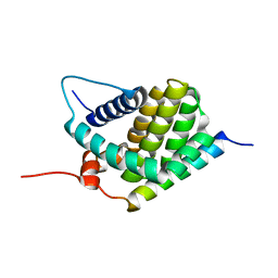
5TDC
 | | Crystal structure of the human UBR-box domain from UBR1 in complex with monomethylated arginine peptide. | | 分子名称: | E3 ubiquitin-protein ligase UBR1, NMM-ILE-PHE-SER peptide, SULFATE ION, ... | | 著者 | Kozlov, G, Munoz-Escobar, J, Matta-Camacho, E, Gehring, K. | | 登録日 | 2016-09-19 | | 公開日 | 2017-03-22 | | 最終更新日 | 2023-10-04 | | 実験手法 | X-RAY DIFFRACTION (1.607 Å) | | 主引用文献 | Bound Waters Mediate Binding of Diverse Substrates to a Ubiquitin Ligase.
Structure, 25, 2017
|
|


