7FDS
 
 | | High resolution crystal structure of LpqH from Mycobacterium tuberculosis | | 分子名称: | Lipoprotein LpqH | | 著者 | Kundapura, S.V, Chatterjee, S, Samanta, D, Ramagopal, U.A. | | 登録日 | 2021-07-17 | | 公開日 | 2021-12-15 | | 最終更新日 | 2023-11-29 | | 実験手法 | X-RAY DIFFRACTION (1.258 Å) | | 主引用文献 | High-resolution crystal structure of LpqH, an immunomodulatory surface lipoprotein of Mycobacterium tuberculosis reveals a distinct fold and a conserved cleft on its surface.
Int.J.Biol.Macromol., 210, 2022
|
|
2YKN
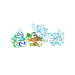
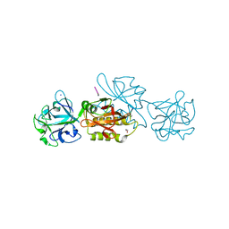
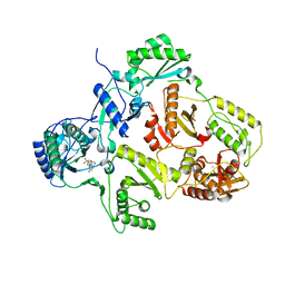 | | Crystal structure of HIV-1 Reverse Transcriptase (RT) in complex with a Difluoromethylbenzoxazole (DFMB) Pyrimidine Thioether derivative, a non-nucleoside RT inhibitor (NNRTI) | | 分子名称: | 2-[DIFLUORO-[(4-METHYL-PYRIMIDINYL)-THIO]METHYL]-BENZOXAZOLE, CALCIUM ION, REVERSE TRANSCRIPTASE/RIBONUCLEASE H | | 著者 | Boyer, J, Arnoult, E, Medebielle, M, Guillemont, J, Unge, T, Unge, J, Jochmans, D. | | 登録日 | 2011-05-28 | | 公開日 | 2011-08-17 | | 最終更新日 | 2024-05-01 | | 実験手法 | X-RAY DIFFRACTION (2.12 Å) | | 主引用文献 | Difluoromethylbenzoxazole Pyrimidine Thioether Derivatives: A Novel Class of Potent Non-Nucleoside HIV-1 Reverse Transcriptase Inhibitors.
J.Med.Chem., 54, 2011
|
|
1Z93
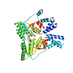
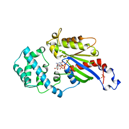
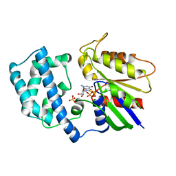
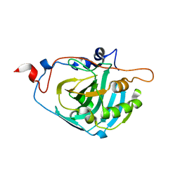 | | Human Carbonic Anhydrase III:Structural and Kinetic study of Catalysis and Proton Transfer. | | 分子名称: | Carbonic anhydrase III, ZINC ION | | 著者 | Duda, D.M, Tu, C, Fisher, S.Z, An, H, Yoshioka, C, Govindasamy, L, Laipis, P.J, Agbandje-McKenna, M, Silverman, D.N, McKenna, R. | | 登録日 | 2005-03-31 | | 公開日 | 2005-08-09 | | 最終更新日 | 2023-08-23 | | 実験手法 | X-RAY DIFFRACTION (2.1 Å) | | 主引用文献 | Human Carbonic Anhydrase III: Structural and Kinetic Study of Catalysis and Proton Transfer
Biochemistry, 44, 2005
|
|
1ZLJ
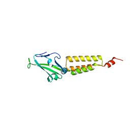
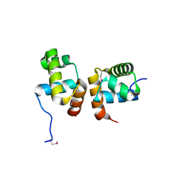 | | Crystal Structure of the Mycobacterium tuberculosis Hypoxic Response Regulator DosR C-terminal Domain | | 分子名称: | Dormancy Survival Regulator | | 著者 | Wisedchaisri, G, Wu, M, Rice, A.E, Roberts, D.M, Sherman, D.R, Hol, W.G.J. | | 登録日 | 2005-05-06 | | 公開日 | 2006-01-31 | | 最終更新日 | 2011-07-13 | | 実験手法 | X-RAY DIFFRACTION (2 Å) | | 主引用文献 | Structures of Mycobacterium tuberculosis DosR and DosR-DNA complex involved in gene activation during adaptation to hypoxic latency.
J.Mol.Biol., 354, 2005
|
|
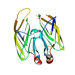
2O0I
 | | crystal structure of the R185A mutant of the N-terminal domain of the Group B Streptococcus Alpha C protein | | 分子名称: | C protein alpha-antigen | | 著者 | Hogle, J.M, Filman, D.J, Baron, M.J, Madoff, L.C, Iglesias, A. | | 登録日 | 2006-11-27 | | 公開日 | 2007-02-06 | | 最終更新日 | 2023-08-30 | | 実験手法 | X-RAY DIFFRACTION (3.1 Å) | | 主引用文献 | Identification of a glycosaminoglycan binding region of the alpha C protein that mediates entry of group B streptococci into host cells.
J.Biol.Chem., 282, 2007
|
|
2CNW
 | | GDPALF4 complex of the SRP GTPases Ffh and FtsY | | 分子名称: | CELL DIVISION PROTEIN FTSY, GUANOSINE-5'-DIPHOSPHATE, GUANOSINE-5'-MONOPHOSPHATE, ... | | 著者 | Focia, P.J, Gawronski-Salerno, J, Coon V, J.S, Freymann, D.M. | | 登録日 | 2006-05-24 | | 公開日 | 2006-10-11 | | 最終更新日 | 2023-12-13 | | 実験手法 | X-RAY DIFFRACTION (2.39 Å) | | 主引用文献 | Structure of a Gdp:Alf(4) Complex of the Srp Gtpases Ffh and Ftsy, and Identification of a Peripheral Nucleotide Interaction Site.
J.Mol.Biol., 360, 2006
|
|
6TSQ
 | | Marasmius oreades agglutinin (MOA) activated by manganese (II) | | 分子名称: | 1,2-ETHANEDIOL, Agglutinin, CALCIUM ION, ... | | 著者 | Cordara, G, Manna, D, Krengel, U. | | 登録日 | 2019-12-21 | | 公開日 | 2020-07-29 | | 最終更新日 | 2024-01-24 | | 実験手法 | X-RAY DIFFRACTION (1.85 Å) | | 主引用文献 | Crystal structure of MOA in complex with a peptide fragment: A protease caught in flagranti .
Curr Res Struct Biol, 2, 2020
|
|
1PIV
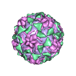 | | BINDING OF THE ANTIVIRAL DRUG WIN51711 TO THE SABIN STRAIN OF TYPE 3 POLIOVIRUS: STRUCTURAL COMPARISON WITH DRUG BINDING IN RHINOVIRUS 14 | | 分子名称: | 5-(7-(4-(4,5-DIHYDRO-2-OXAZOLYL)PHENOXY)HEPTYL)-3-METHYL ISOXAZOLE, MYRISTIC ACID, POLIOVIRUS TYPE 3 (SUBUNIT VP1), ... | | 著者 | Hiremath, C.N, Grant, R.A, Filman, D.J, Hogle, J.M. | | 登録日 | 1995-02-02 | | 公開日 | 1995-06-03 | | 最終更新日 | 2024-06-05 | | 実験手法 | X-RAY DIFFRACTION (2.9 Å) | | 主引用文献 | Binding of the antiviral drug WIN51711 to the sabin strain of type 3 poliovirus: structural comparison with drug binding in rhinovirus 14.
Acta Crystallogr.,Sect.D, 51, 1995
|
|
1PVC
 | |
6TSL
 | | Marasmius oreades agglutinin (MOA) in complex with the truncated PVPRAHS synthetic substrate | | 分子名称: | 1,2-ETHANEDIOL, Agglutinin, CALCIUM ION, ... | | 著者 | Cordara, G, Manna, D, Krengel, U. | | 登録日 | 2019-12-20 | | 公開日 | 2020-07-29 | | 最終更新日 | 2024-01-24 | | 実験手法 | X-RAY DIFFRACTION (1.4 Å) | | 主引用文献 | Crystal structure of MOA in complex with a peptide fragment: A protease caught in flagranti .
Curr Res Struct Biol, 2, 2020
|
|
1AS3
 | | GDP BOUND G42V GIA1 | | 分子名称: | GIA1, GUANOSINE-5'-DIPHOSPHATE, SULFATE ION | | 著者 | Raw, A.S, Coleman, D.E, Gilman, A.G, Sprang, S.R. | | 登録日 | 1997-08-11 | | 公開日 | 1997-11-12 | | 最終更新日 | 2024-05-22 | | 実験手法 | X-RAY DIFFRACTION (2.4 Å) | | 主引用文献 | Structural and biochemical characterization of the GTPgammaS-, GDP.Pi-, and GDP-bound forms of a GTPase-deficient Gly42 --> Val mutant of Gialpha1.
Biochemistry, 36, 1997
|
|
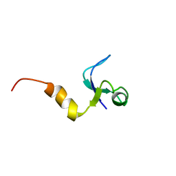
1YTR
 | | NMR structure of plantaricin a in dpc micelles, 20 structures | | 分子名称: | Bacteriocin plantaricin A | | 著者 | Kristiansen, P.E, Fimland, G, Mantzilas, D, Nissen-Meyer, J. | | 登録日 | 2005-02-11 | | 公開日 | 2005-05-17 | | 最終更新日 | 2024-05-29 | | 実験手法 | SOLUTION NMR | | 主引用文献 | Structure and mode of action of the membrane-permeabilizing antimicrobial peptide pheromone plantaricin A
J.Biol.Chem., 280, 2005
|
|
1AS0
 | | GTP-GAMMA-S BOUND G42V GIA1 | | 分子名称: | 5'-GUANOSINE-DIPHOSPHATE-MONOTHIOPHOSPHATE, GIA1, MAGNESIUM ION, ... | | 著者 | Raw, A.S, Coleman, D.E, Gilman, A.G, Sprang, S.R. | | 登録日 | 1997-08-11 | | 公開日 | 1997-11-12 | | 最終更新日 | 2024-05-22 | | 実験手法 | X-RAY DIFFRACTION (2 Å) | | 主引用文献 | Structural and biochemical characterization of the GTPgammaS-, GDP.Pi-, and GDP-bound forms of a GTPase-deficient Gly42 --> Val mutant of Gialpha1.
Biochemistry, 36, 1997
|
|
6TSM
 | | Marasmius oreades agglutinin (MOA) in complex with the truncated PVVRAHS synthetic substrate | | 分子名称: | 1,2-ETHANEDIOL, Agglutinin, CALCIUM ION, ... | | 著者 | Cordara, G, Manna, D, Krengel, U. | | 登録日 | 2019-12-20 | | 公開日 | 2020-07-29 | | 最終更新日 | 2024-01-24 | | 実験手法 | X-RAY DIFFRACTION (1.4 Å) | | 主引用文献 | Crystal structure of MOA in complex with a peptide fragment: A protease caught in flagranti .
Curr Res Struct Biol, 2, 2020
|
|
1AS2
 | | GDP+PI BOUND G42V GIA1 | | 分子名称: | GIA1, GUANOSINE-5'-DIPHOSPHATE, PHOSPHATE ION | | 著者 | Raw, A.S, Coleman, D.E, Gilman, A.G, Sprang, S.R. | | 登録日 | 1997-08-11 | | 公開日 | 1997-11-12 | | 最終更新日 | 2024-05-22 | | 実験手法 | X-RAY DIFFRACTION (2.8 Å) | | 主引用文献 | Structural and biochemical characterization of the GTPgammaS-, GDP.Pi-, and GDP-bound forms of a GTPase-deficient Gly42 --> Val mutant of Gialpha1.
Biochemistry, 36, 1997
|
|
5LRX
 | | Structure of A20 OTU domain bound to ubiquitin | | 分子名称: | Polyubiquitin-B, Tumor necrosis factor alpha-induced protein 3 | | 著者 | Mevissen, T.E.T, Kulathu, Y, Mulder, M.P.C, Geurink, P.P, Maslen, S.L, Gersch, M, Elliott, P.R, Burke, J.E, van Tol, B.D.M, Akutsu, M, El Oualid, F, Kawasaki, M, Freund, S.M.V, Ovaa, H, Komander, D. | | 登録日 | 2016-08-22 | | 公開日 | 2016-10-19 | | 最終更新日 | 2017-09-13 | | 実験手法 | X-RAY DIFFRACTION (2.85 Å) | | 主引用文献 | Molecular basis of Lys11-polyubiquitin specificity in the deubiquitinase Cezanne.
Nature, 538, 2016
|
|
6TSR
 | | Marasmius oreades agglutinin (MOA) activated by manganese (II) and calcium | | 分子名称: | 1,2-ETHANEDIOL, Agglutinin, CALCIUM ION, ... | | 著者 | Cordara, G, Manna, D, Krengel, U. | | 登録日 | 2019-12-21 | | 公開日 | 2020-07-29 | | 最終更新日 | 2024-01-24 | | 実験手法 | X-RAY DIFFRACTION (1.85 Å) | | 主引用文献 | Crystal structure of MOA in complex with a peptide fragment: A protease caught in flagranti .
Curr Res Struct Biol, 2, 2020
|
|
6TSO
 | | Marasmius oreades agglutinin (MOA) inhibited by cadmium | | 分子名称: | 1,2-ETHANEDIOL, Agglutinin, CADMIUM ION, ... | | 著者 | Cordara, G, Manna, D, Krengel, U. | | 登録日 | 2019-12-20 | | 公開日 | 2020-07-29 | | 最終更新日 | 2024-01-24 | | 実験手法 | X-RAY DIFFRACTION (2.1 Å) | | 主引用文献 | Crystal structure of MOA in complex with a peptide fragment: A protease caught in flagranti .
Curr Res Struct Biol, 2, 2020
|
|
8DI5
 | | Cryo-EM structure of SARS-CoV-2 Beta (B.1.351) spike protein in complex with VH domain F6 (focused refinement of RBD and VH F6) | | 分子名称: | 2-acetamido-2-deoxy-beta-D-glucopyranose, Spike glycoprotein, VH F6 | | 著者 | Zhu, X, Saville, J.W, Mannar, D, Berezuk, A.M, Subramaniam, S. | | 登録日 | 2022-06-28 | | 公開日 | 2022-08-24 | | 実験手法 | ELECTRON MICROSCOPY (3.04 Å) | | 主引用文献 | Potent and broad neutralization of SARS-CoV-2 variants of concern (VOCs) including omicron sub-lineages BA.1 and BA.2 by biparatopic human VH domains.
Iscience, 25, 2022
|
|
2ADP
 | | Nitrated Human Manganese Superoxide Dismutase | | 分子名称: | MANGANESE (II) ION, POTASSIUM ION, Superoxide dismutase [Mn] | | 著者 | Quint, P, Reutzel, R, Mikulski, R, McKenna, R, Silverman, D.N. | | 登録日 | 2005-07-20 | | 公開日 | 2006-07-04 | | 最終更新日 | 2024-04-03 | | 実験手法 | X-RAY DIFFRACTION (2.4 Å) | | 主引用文献 | Crystal structure of nitrated human manganese superoxide dismutase: mechanism of inactivation.
FREE RADIC.BIOL.MED., 40, 2006
|
|
1M3A
 | | Solution structure of a circular form of the truncated N-terminal SH3 domain from oncogene protein c-Crk. | | 分子名称: | Proto-oncogene C-crk | | 著者 | Schumann, F.H, Varadan, R, Tayakuniyil, P.P, Hall, J.B, Camarero, J.A, Fushman, D. | | 登録日 | 2002-06-27 | | 公開日 | 2003-08-05 | | 最終更新日 | 2021-10-27 | | 実験手法 | SOLUTION NMR | | 主引用文献 | Changing protein backbone topology: Structural and dynamic consequences of the backbone cyclization in SH3 domain
To be Published
|
|
2ADQ
 | | Human Manganese Superoxide Dismutase | | 分子名称: | MANGANESE (II) ION, POTASSIUM ION, Superoxide dismutase [Mn] | | 著者 | Quint, P, Reutzel, R, Mikulski, R, McKenna, R, Silverman, D.N. | | 登録日 | 2005-07-20 | | 公開日 | 2006-07-04 | | 最終更新日 | 2024-04-03 | | 実験手法 | X-RAY DIFFRACTION (2.4 Å) | | 主引用文献 | Crystal structure of nitrated human manganese superoxide dismutase: mechanism of inactivation.
FREE RADIC.BIOL.MED., 40, 2006
|
|
1CQU
 | |
5MU9
 | | MOA-E-64 complex | | 分子名称: | 1,2-ETHANEDIOL, ACETATE ION, Agglutinin, ... | | 著者 | Cordara, G, Manna, D, Krengel, U. | | 登録日 | 2017-01-12 | | 公開日 | 2017-07-12 | | 最終更新日 | 2024-01-17 | | 実験手法 | X-RAY DIFFRACTION (1.3 Å) | | 主引用文献 | Family of Papain-Like Fungal Chimerolectins with Distinct Ca(2+)-Dependent Activation Mechanism.
Biochemistry, 56, 2017
|
|
6RV3
 | | Crystal structure of the human two pore domain potassium ion channel TASK-1 (K2P3.1) in a closed conformation with a bound inhibitor BAY 1000493 | | 分子名称: | 1,2-DIACYL-SN-GLYCERO-3-PHOSPHOCHOLINE, CHOLESTEROL HEMISUCCINATE, DECYL-BETA-D-MALTOPYRANOSIDE, ... | | 著者 | Rodstrom, K.E.J, Pike, A.C.W, Zhang, W, Quigley, A, Speedman, D, Mukhopadhyay, S.M.M, Shrestha, L, Chalk, R, Venkaya, S, Bushell, S.R, Tessitore, A, Burgess-Brown, N, Arrowsmith, C.H, Edwards, A.M, Bountra, C, Carpenter, E.P, Structural Genomics Consortium (SGC) | | 登録日 | 2019-05-30 | | 公開日 | 2019-08-07 | | 最終更新日 | 2024-01-24 | | 実験手法 | X-RAY DIFFRACTION (2.9 Å) | | 主引用文献 | A lower X-gate in TASK channels traps inhibitors within the vestibule.
Nature, 582, 2020
|
|


