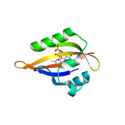
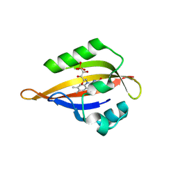
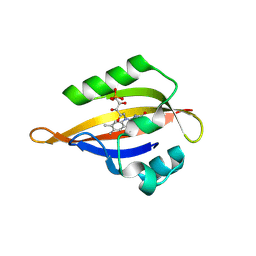
8QIT
 
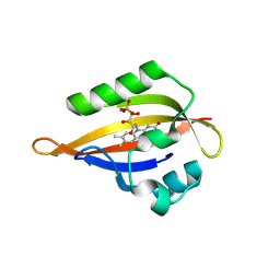 | | CrPhotLOV1 light state structure 77.5 ms (75-80 ms) after illumination determined by time-resolved serial synchrotron crystallography at room temperature | | 分子名称: | FLAVIN MONONUCLEOTIDE, Phototropin | | 著者 | Gotthard, G, Mous, S, Weinert, T, Maia, R.N.A, James, D, Dworkowski, F, Gashi, D, Antonia, F, Wang, M, Panepucci, E, Ozerov, D, Schertler, G.F.X, Heberle, J, Standfuss, J, Nogly, P. | | 登録日 | 2023-09-12 | | 公開日 | 2024-07-24 | | 最終更新日 | 2024-09-11 | | 実験手法 | X-RAY DIFFRACTION (2.9 Å) | | 主引用文献 | Capturing the blue-light activated state of the Phot-LOV1 domain from Chlamydomonas reinhardtii using time-resolved serial synchrotron crystallography.
Iucrj, 11, 2024
|
|
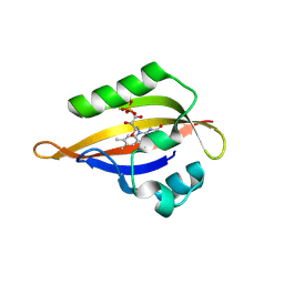
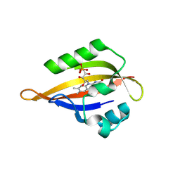
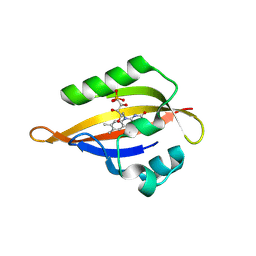
8QIA
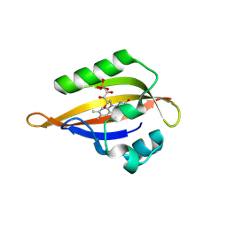 | | CrPhotLOV1 light state structure 2.5 ms (0-5 ms) after illumination determined by time-resolved serial synchrotron crystallography at room temperature | | 分子名称: | FLAVIN MONONUCLEOTIDE, Phototropin | | 著者 | Gotthard, G, Mous, S, Weinert, T, Maia, R.N.A, James, D, Dworkowski, F, Gashi, D, Antonia, F, Wang, M, Panepucci, E, Ozerov, D, Schertler, G.F.X, Heberle, J, Standfuss, J, Nogly, P. | | 登録日 | 2023-09-11 | | 公開日 | 2024-07-24 | | 最終更新日 | 2024-09-11 | | 実験手法 | X-RAY DIFFRACTION (2.5 Å) | | 主引用文献 | Capturing the blue-light activated state of the Phot-LOV1 domain from Chlamydomonas reinhardtii using time-resolved serial synchrotron crystallography.
Iucrj, 11, 2024
|
|
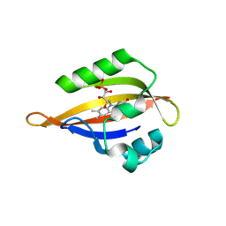
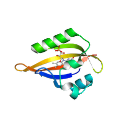
8QIU
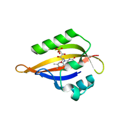 | | CrPhotLOV1 light state structure 82.5 ms (80-85 ms) after illumination determined by time-resolved serial synchrotron crystallography at room temperature | | 分子名称: | FLAVIN MONONUCLEOTIDE, Phototropin | | 著者 | Gotthard, G, Mous, S, Weinert, T, Maia, R.N.A, James, D, Dworkowski, F, Gashi, D, Antonia, F, Wang, M, Panepucci, E, Ozerov, D, Schertler, G.F.X, Heberle, J, Standfuss, J, Nogly, P. | | 登録日 | 2023-09-12 | | 公開日 | 2024-07-24 | | 最終更新日 | 2024-09-11 | | 実験手法 | X-RAY DIFFRACTION (3 Å) | | 主引用文献 | Capturing the blue-light activated state of the Phot-LOV1 domain from Chlamydomonas reinhardtii using time-resolved serial synchrotron crystallography.
Iucrj, 11, 2024
|
|
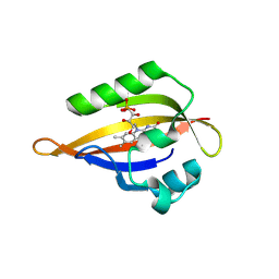
8QII
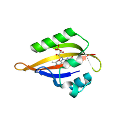 | | CrPhotLOV1 light state structure 27.5 ms (25-30 ms) after illumination determined by time-resolved serial synchrotron crystallography at room temperature | | 分子名称: | FLAVIN MONONUCLEOTIDE, Phototropin | | 著者 | Gotthard, G, Mous, S, Weinert, T, Maia, R.N.A, James, D, Dworkowski, F, Gashi, D, Antonia, F, Wang, M, Panepucci, E, Ozerov, D, Schertler, G.F.X, Heberle, J, Standfuss, J, Nogly, P. | | 登録日 | 2023-09-12 | | 公開日 | 2024-07-24 | | 最終更新日 | 2024-09-11 | | 実験手法 | X-RAY DIFFRACTION (2.5 Å) | | 主引用文献 | Capturing the blue-light activated state of the Phot-LOV1 domain from Chlamydomonas reinhardtii using time-resolved serial synchrotron crystallography.
Iucrj, 11, 2024
|
|
8QIW
 | | CrPhotLOV1 light state structure 92.5 ms (90-95 ms) after illumination determined by time-resolved serial synchrotron crystallography at room temperature | | 分子名称: | FLAVIN MONONUCLEOTIDE, Phototropin | | 著者 | Gotthard, G, Mous, S, Weinert, T, Maia, R.N.A, James, D, Dworkowski, F, Gashi, D, Antonia, F, Wang, M, Panepucci, E, Ozerov, D, Schertler, G.F.X, Heberle, J, Standfuss, J, Nogly, P. | | 登録日 | 2023-09-12 | | 公開日 | 2024-07-24 | | 最終更新日 | 2024-09-11 | | 実験手法 | X-RAY DIFFRACTION (3.05 Å) | | 主引用文献 | Capturing the blue-light activated state of the Phot-LOV1 domain from Chlamydomonas reinhardtii using time-resolved serial synchrotron crystallography.
Iucrj, 11, 2024
|
|
8QIQ
 | | CrPhotLOV1 light state structure 62.5 ms (60-65 ms) after illumination determined by time-resolved serial synchrotron crystallography at room temperature | | 分子名称: | FLAVIN MONONUCLEOTIDE, Phototropin | | 著者 | Gotthard, G, Mous, S, Weinert, T, Maia, R.N.A, James, D, Dworkowski, F, Gashi, D, Antonia, F, Wang, M, Panepucci, E, Ozerov, D, Schertler, G.F.X, Heberle, J, Standfuss, J, Nogly, P. | | 登録日 | 2023-09-12 | | 公開日 | 2024-07-24 | | 最終更新日 | 2024-09-11 | | 実験手法 | X-RAY DIFFRACTION (2.9 Å) | | 主引用文献 | Capturing the blue-light activated state of the Phot-LOV1 domain from Chlamydomonas reinhardtii using time-resolved serial synchrotron crystallography.
Iucrj, 11, 2024
|
|
8QIB
 | | CrPhotLOV1 light state structure 7.5 ms (5-10 ms) after illumination determined by time-resolved serial synchrotron crystallography at room temperature | | 分子名称: | FLAVIN MONONUCLEOTIDE, Phototropin | | 著者 | Gotthard, G, Mous, S, Weinert, T, Maia, R.N.A, James, D, Dworkowski, F, Gashi, D, Antonia, F, Wang, M, Panepucci, E, Ozerov, D, Schertler, G.F.X, Heberle, J, Standfuss, J, Nogly, P. | | 登録日 | 2023-09-11 | | 公開日 | 2024-07-24 | | 最終更新日 | 2024-09-11 | | 実験手法 | X-RAY DIFFRACTION (2.45 Å) | | 主引用文献 | Capturing the blue-light activated state of the Phot-LOV1 domain from Chlamydomonas reinhardtii using time-resolved serial synchrotron crystallography.
Iucrj, 11, 2024
|
|
8QIS
 | | CrPhotLOV1 light state structure 72.5 ms (70-75 ms) after illumination determined by time-resolved serial synchrotron crystallography at room temperature | | 分子名称: | FLAVIN MONONUCLEOTIDE, Phototropin | | 著者 | Gotthard, G, Mous, S, Weinert, T, Maia, R.N.A, James, D, Dworkowski, F, Gashi, D, Antonia, F, Wang, M, Panepucci, E, Ozerov, D, Schertler, G.F.X, Heberle, J, Standfuss, J, Nogly, P. | | 登録日 | 2023-09-12 | | 公開日 | 2024-07-24 | | 最終更新日 | 2024-09-11 | | 実験手法 | X-RAY DIFFRACTION (2.9 Å) | | 主引用文献 | Capturing the blue-light activated state of the Phot-LOV1 domain from Chlamydomonas reinhardtii using time-resolved serial synchrotron crystallography.
Iucrj, 11, 2024
|
|
8QIK
 | | CrPhotLOV1 light state structure 32.5 ms (30-35 ms) after illumination determined by time-resolved serial synchrotron crystallography at room temperature | | 分子名称: | FLAVIN MONONUCLEOTIDE, Phototropin | | 著者 | Gotthard, G, Mous, S, Weinert, T, Maia, R.N.A, James, D, Dworkowski, F, Gashi, D, Antonia, F, Wang, M, Panepucci, E, Ozerov, D, Schertler, G.F.X, Heberle, J, Standfuss, J, Nogly, P. | | 登録日 | 2023-09-12 | | 公開日 | 2024-07-24 | | 最終更新日 | 2024-09-11 | | 実験手法 | X-RAY DIFFRACTION (2.55 Å) | | 主引用文献 | Capturing the blue-light activated state of the Phot-LOV1 domain from Chlamydomonas reinhardtii using time-resolved serial synchrotron crystallography.
Iucrj, 11, 2024
|
|
8QIM
 | | CrPhotLOV1 light state structure 42.5 ms (40-45 ms) after illumination determined by time-resolved serial synchrotron crystallography at room temperature | | 分子名称: | FLAVIN MONONUCLEOTIDE, Phototropin | | 著者 | Gotthard, G, Mous, S, Weinert, T, Maia, R.N.A, James, D, Dworkowski, F, Gashi, D, Antonia, F, Wang, M, Panepucci, E, Ozerov, D, Schertler, G.F.X, Heberle, J, Standfuss, J, Nogly, P. | | 登録日 | 2023-09-12 | | 公開日 | 2024-07-24 | | 最終更新日 | 2024-09-11 | | 実験手法 | X-RAY DIFFRACTION (2.6 Å) | | 主引用文献 | Capturing the blue-light activated state of the Phot-LOV1 domain from Chlamydomonas reinhardtii using time-resolved serial synchrotron crystallography.
Iucrj, 11, 2024
|
|
8QIG
 | | CrPhotLOV1 light state structure 17.5 ms (15-20 ms) after illumination determined by time-resolved serial synchrotron crystallography at room temperature | | 分子名称: | FLAVIN MONONUCLEOTIDE, Phototropin | | 著者 | Gotthard, G, Mous, S, Weinert, T, Maia, R.N.A, James, D, Dworkowski, F, Gashi, D, Antonia, F, Wang, M, Panepucci, E, Ozerov, D, Schertler, G.F.X, Heberle, J, Standfuss, J, Nogly, P. | | 登録日 | 2023-09-12 | | 公開日 | 2024-07-24 | | 最終更新日 | 2024-09-11 | | 実験手法 | X-RAY DIFFRACTION (2.5 Å) | | 主引用文献 | Capturing the blue-light activated state of the Phot-LOV1 domain from Chlamydomonas reinhardtii using time-resolved serial synchrotron crystallography.
Iucrj, 11, 2024
|
|
8QLL
 | |
8QIF
 | | CrPhotLOV1 light state structure 12.5 ms (10-15 ms) after illumination determined by time-resolved serial synchrotron crystallography at room temperature | | 分子名称: | FLAVIN MONONUCLEOTIDE, Phototropin | | 著者 | Gotthard, G, Mous, S, Weinert, T, Maia, R.N.A, James, D, Dworkowski, F, Gashi, D, Antonia, F, Wang, M, Panepucci, E, Ozerov, D, Schertler, G.F.X, Heberle, J, Standfuss, J, Nogly, P. | | 登録日 | 2023-09-12 | | 公開日 | 2024-07-24 | | 最終更新日 | 2024-09-11 | | 実験手法 | X-RAY DIFFRACTION (2.45 Å) | | 主引用文献 | Capturing the blue-light activated state of the Phot-LOV1 domain from Chlamydomonas reinhardtii using time-resolved serial synchrotron crystallography.
Iucrj, 11, 2024
|
|
8QIV
 | | CrPhotLOV1 light state structure 87.5 ms (85-90 ms) after illumination determined by time-resolved serial synchrotron crystallography at room temperature | | 分子名称: | FLAVIN MONONUCLEOTIDE, Phototropin | | 著者 | Gotthard, G, Mous, S, Weinert, T, Maia, R.N.A, James, D, Dworkowski, F, Gashi, D, Antonia, F, Wang, M, Panepucci, E, Ozerov, D, Schertler, G.F.X, Heberle, J, Standfuss, J, Nogly, P. | | 登録日 | 2023-09-12 | | 公開日 | 2024-07-24 | | 最終更新日 | 2024-09-11 | | 実験手法 | X-RAY DIFFRACTION (3.1 Å) | | 主引用文献 | Capturing the blue-light activated state of the Phot-LOV1 domain from Chlamydomonas reinhardtii using time-resolved serial synchrotron crystallography.
Iucrj, 11, 2024
|
|
4WR5
 | | Crystal Structure of GST Mutated with Halogenated Tyrosine (7cGST-1) | | 分子名称: | GLUTATHIONE, Glutathione S-transferase class-mu 26 kDa isozyme, SULFATE ION | | 著者 | Akasaka, R, Kawazoe, M, Tomabechi, Y, Ohtake, K, Itagaki, T, Takemoto, C, Shirouzu, M, Yokoyama, S, Sakamoto, K. | | 登録日 | 2014-10-23 | | 公開日 | 2015-08-19 | | 最終更新日 | 2023-11-08 | | 実験手法 | X-RAY DIFFRACTION (1.93 Å) | | 主引用文献 | Protein stabilization utilizing a redefined codon
Sci Rep, 5, 2015
|
|
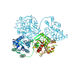
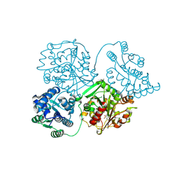
8QVF
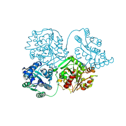 | | Comparison of room-temperature and cryogenic structures of soluble Epoxide Hydrolase with ligands bound. | | 分子名称: | 1-(1-adamantyl)-3-(1-methylsulfonylpiperidin-4-yl)urea, Bifunctional epoxide hydrolase 2 | | 著者 | Dunge, A, Uwangue, O, Phan, C, Bjelcic, M, Gunnarsson, J, Wehlander, G, Kack, H, Branden, G. | | 登録日 | 2023-10-18 | | 公開日 | 2024-08-14 | | 最終更新日 | 2024-09-11 | | 実験手法 | X-RAY DIFFRACTION (2.4 Å) | | 主引用文献 | Exploring serial crystallography for drug discovery.
Iucrj, 11, 2024
|
|
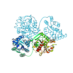
8QVH
 | | Comparison of room-temperature and cryogenic structures of soluble Epoxide Hydrolase with ligands bound. | | 分子名称: | 4-[(trans-4-{[(3s,5s,7s)-tricyclo[3.3.1.1~3,7~]dec-1-ylcarbamoyl]amino}cyclohexyl)oxy]benzoic acid, Bifunctional epoxide hydrolase 2 | | 著者 | Dunge, A, Uwangue, O, Phan, C, Bjelcic, M, Gunnarsson, J, Wehlander, G, Kack, H, Branden, G. | | 登録日 | 2023-10-18 | | 公開日 | 2024-08-14 | | 最終更新日 | 2024-09-11 | | 実験手法 | X-RAY DIFFRACTION (2.24 Å) | | 主引用文献 | Exploring serial crystallography for drug discovery.
Iucrj, 11, 2024
|
|
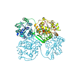
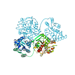
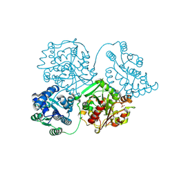
8QWG
 | | Comparison of room-temperature and cryogenic structures of soluble Epoxide Hydrolase with ligands bound. | | 分子名称: | Bifunctional epoxide hydrolase 2, TRIETHYLENE GLYCOL | | 著者 | Dunge, A, Uwangue, O, Phan, C, Bjelcic, M, Gunnarsson, J, Wehlander, G, Kack, H, Branden, G. | | 登録日 | 2023-10-19 | | 公開日 | 2024-08-14 | | 最終更新日 | 2024-09-11 | | 実験手法 | X-RAY DIFFRACTION (2.2 Å) | | 主引用文献 | Exploring serial crystallography for drug discovery.
Iucrj, 11, 2024
|
|
8QVL
 | | Comparison of room-temperature and cryogenic structures of soluble Epoxide Hydrolase with ligands bound. | | 分子名称: | 2-[(5-BROMO-2-PYRIDYL)-METHYL-AMINO]ETHANOL, Bifunctional epoxide hydrolase 2, DIMETHYL SULFOXIDE, ... | | 著者 | Dunge, A, Uwangue, O, Phan, C, Bjelcic, M, Gunnarsson, J, Wehlander, G, Kack, H, Branden, G. | | 登録日 | 2023-10-18 | | 公開日 | 2024-08-14 | | 最終更新日 | 2024-09-11 | | 実験手法 | X-RAY DIFFRACTION (2.14 Å) | | 主引用文献 | Exploring serial crystallography for drug discovery.
Iucrj, 11, 2024
|
|
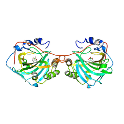
8Q8I
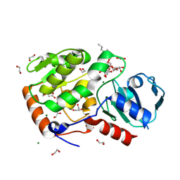 | | AO75L in complex with a synthetic trisaccharide acceptor. | | 分子名称: | (2~{R},3~{S},4~{S},5~{S},6~{R})-2-[(2~{S},3~{R},4~{R},5~{S},6~{R})-5-[(2~{R},3~{R},4~{S},5~{R},6~{R})-6-(hydroxymethyl)-3,4,5-tris(oxidanyl)oxan-2-yl]oxy-2-methyl-6-octoxy-3-oxidanyl-oxan-4-yl]oxy-6-methyl-oxane-3,4,5-triol, 1,2-ETHANEDIOL, BICINE, ... | | 著者 | Laugueri, M.E, Speciale, I, Gimeno, A, Sicheng, L, Poveda, A, Lowary, T, Van Etten J, L, Barbero, J, De Castro, C, Tonetti, M, Rojas A, L. | | 登録日 | 2023-08-18 | | 公開日 | 2024-08-21 | | 実験手法 | X-RAY DIFFRACTION (1.77 Å) | | 主引用文献 | AO75L in complex with a synthetic trisaccharide acceptor.
To Be Published
|
|
8QVG
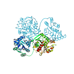 | | Comparison of room-temperature and cryogenic structures of soluble Epoxide Hydrolase with ligands bound. | | 分子名称: | Bifunctional epoxide hydrolase 2, N-(3,3-DIPHENYLPROPYL)PYRROLIDINE-1-CARBOXAMIDE | | 著者 | Dunge, A, Uwangue, O, Phan, C, Bjelcic, M, Gunnarsson, J, Wehlander, G, Kack, H, Branden, G. | | 登録日 | 2023-10-18 | | 公開日 | 2024-08-14 | | 最終更新日 | 2024-09-11 | | 実験手法 | X-RAY DIFFRACTION (2.2 Å) | | 主引用文献 | Exploring serial crystallography for drug discovery.
Iucrj, 11, 2024
|
|
8QVK
 | | Comparison of room-temperature and cryogenic structures of soluble Epoxide Hydrolase with ligands bound. | | 分子名称: | Bifunctional epoxide hydrolase 2, N-(5,5-dioxodibenzothiophen-2-yl)-4,4-difluoro-piperidine-1-carboxamide | | 著者 | Dunge, A, Uwangue, O, Phan, C, Bjelcic, M, Gunnarsson, J, Wehlander, G, Kack, H, Branden, G. | | 登録日 | 2023-10-18 | | 公開日 | 2024-08-14 | | 最終更新日 | 2024-09-11 | | 実験手法 | X-RAY DIFFRACTION (2.1 Å) | | 主引用文献 | Exploring serial crystallography for drug discovery.
Iucrj, 11, 2024
|
|
8QVM
 | | Comparison of room-temperature and cryogenic structures of soluble Epoxide Hydrolase with ligands bound. | | 分子名称: | Bifunctional epoxide hydrolase 2, TRIETHYLENE GLYCOL | | 著者 | Dunge, A, Uwangue, O, Phan, C, Bjelcic, M, Gunnarsson, J, Wehlander, G, Kack, H, Branden, G. | | 登録日 | 2023-10-18 | | 公開日 | 2024-08-14 | | 最終更新日 | 2024-09-11 | | 実験手法 | X-RAY DIFFRACTION (2 Å) | | 主引用文献 | Exploring serial crystallography for drug discovery.
Iucrj, 11, 2024
|
|
8QWI
 | | Comparison of room-temperature and cryogenic structures of soluble Epoxide Hydrolase with ligands bound. | | 分子名称: | 2-(1H-BENZIMIDAZOL-2-YLSULFANYL)ETHANOL, Bifunctional epoxide hydrolase 2, SULFATE ION | | 著者 | Dunge, A, Uwangue, O, Phan, C, Bjelcic, M, Gunnarsson, J, Wehlander, G, Kack, H, Branden, G. | | 登録日 | 2023-10-19 | | 公開日 | 2024-08-14 | | 最終更新日 | 2024-09-11 | | 実験手法 | X-RAY DIFFRACTION (2.12 Å) | | 主引用文献 | Exploring serial crystallography for drug discovery.
Iucrj, 11, 2024
|
|
8Q6L
 | | human Carbonic Anhydrase I in complex with 3,4-dihydro-1H-benzo[c][1,2]oxaborinin-1-ol | | 分子名称: | 1,1-bis(oxidanyl)-3,4-dihydro-2,1$l^{4}-benzoxaborinine, Carbonic anhydrase 1, SODIUM ION, ... | | 著者 | Angeli, A, Ferraroni, M. | | 登録日 | 2023-08-13 | | 公開日 | 2024-08-21 | | 実験手法 | X-RAY DIFFRACTION (1.72 Å) | | 主引用文献 | human Carbonic Anhydrase I in complex with 3,4-dihydro-1H-benzo[c][1,2]oxaborinin-1-ol
To Be Published
|
|


