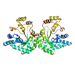
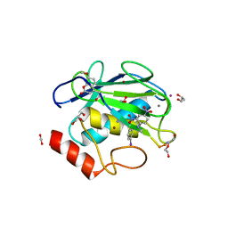
4NO6
 
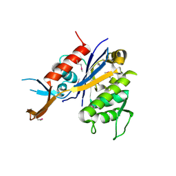
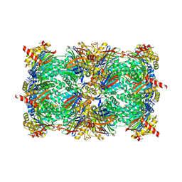 | | yCP in complex with Z-Leu-Leu-Leu-vinylsulfone | | 分子名称: | MAGNESIUM ION, N-[(benzyloxy)carbonyl]-L-leucyl-N-[(3S)-5-methyl-1-(methylsulfonyl)hexan-3-yl]-L-leucinamide, Probable proteasome subunit alpha type-7, ... | | 著者 | Stein, M.L, Cui, H, Beck, P, Dubiella, C, Voss, C, Krueger, A, Schmidt, B, Groll, M. | | 登録日 | 2013-11-19 | | 公開日 | 2014-02-12 | | 最終更新日 | 2024-03-13 | | 実験手法 | X-RAY DIFFRACTION (3 Å) | | 主引用文献 | Systematic Comparison of Peptidic Proteasome Inhibitors Highlights the alpha-Ketoamide Electrophile as an Auspicious Reversible Lead Motif.
Angew.Chem.Int.Ed.Engl., 53, 2014
|
|
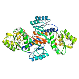
1JH7
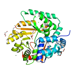
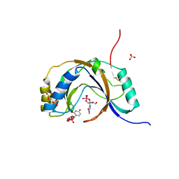 | | Semi-reduced Inhibitor-bound Cyclic Nucleotide Phosphodiesterase from Arabidopsis thaliana | | 分子名称: | SULFATE ION, URIDINE-2',3'-VANADATE, cyclic phosphodiesterase | | 著者 | Hofmann, A, Grella, M, Botos, I, Filipowicz, W, Wlodawer, A. | | 登録日 | 2001-06-27 | | 公開日 | 2002-02-06 | | 最終更新日 | 2024-04-03 | | 実験手法 | X-RAY DIFFRACTION (2.4 Å) | | 主引用文献 | Crystal structures of the semireduced and inhibitor-bound forms of cyclic nucleotide phosphodiesterase from Arabidopsis thaliana.
J.Biol.Chem., 277, 2002
|
|
3D2Q
 | |
3OUU
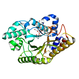
 | | Crystal Structure of Biotin Carboxylase-beta-gamma-ATP Complex from Campylobacter jejuni | | 分子名称: | Biotin carboxylase, CACODYLATE ION, CALCIUM ION, ... | | 著者 | Maltseva, N, Kim, Y, Makowska-Grzyska, M, Mulligan, R, Papazisi, L, Anderson, W.F, Joachimiak, A, Center for Structural Genomics of Infectious Diseases (CSGID) | | 登録日 | 2010-09-15 | | 公開日 | 2010-10-20 | | 最終更新日 | 2024-04-03 | | 実験手法 | X-RAY DIFFRACTION (2.252 Å) | | 主引用文献 | Crystal Structure of Biotin Carboxylase-beta-gamma-ATP Complex from Campylobacter jejuni
TO BE PUBLISHED
|
|
6R8H
 | | Triosephosphate isomerase from liver fluke (Fasciola hepatica). | | 分子名称: | SULFATE ION, Triosephosphate isomerase | | 著者 | Ferraro, F, Corvo, I, Bergalli, L, Ilarraz, A, Cabrera, M, Gil, J, Susuki, B, Caffrey, C, Timson, D.J, Robert, X, Guillon, C, Alvarez, G. | | 登録日 | 2019-04-01 | | 公開日 | 2020-02-12 | | 最終更新日 | 2024-01-24 | | 実験手法 | X-RAY DIFFRACTION (1.9 Å) | | 主引用文献 | Novel and selective inactivators of Triosephosphate isomerase with anti-trematode activity.
Sci Rep, 10, 2020
|
|
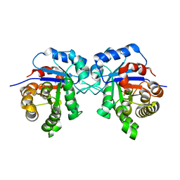
1JEZ
 | | THE STRUCTURE OF XYLOSE REDUCTASE, A DIMERIC ALDO-KETO REDUCTASE FROM CANDIDA TENUIS | | 分子名称: | XYLOSE REDUCTASE | | 著者 | Kavanagh, K.L, Klimacek, M, Nidetzky, B, Wilson, D.K. | | 登録日 | 2001-06-19 | | 公開日 | 2002-07-05 | | 最終更新日 | 2023-08-16 | | 実験手法 | X-RAY DIFFRACTION (2.2 Å) | | 主引用文献 | The structure of apo and holo forms of xylose reductase, a dimeric aldo-keto reductase from Candida tenuis.
Biochemistry, 41, 2002
|
|
4NEH
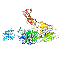 | | An internal ligand-bound, metastable state of a leukocyte integrin, aXb2 | | 分子名称: | 2-acetamido-2-deoxy-beta-D-glucopyranose, 2-acetamido-2-deoxy-beta-D-glucopyranose-(1-4)-2-acetamido-2-deoxy-beta-D-glucopyranose, CALCIUM ION, ... | | 著者 | Sen, M, Yuki, K, Springer, T.A. | | 登録日 | 2013-10-29 | | 公開日 | 2014-01-15 | | 最終更新日 | 2023-09-20 | | 実験手法 | X-RAY DIFFRACTION (2.7505 Å) | | 主引用文献 | An internal ligand-bound, metastable state of a leukocyte integrin, alpha X beta 2.
J.Cell Biol., 203, 2013
|
|
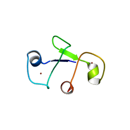
6JPT
 | | Crystal structure of human PAC3 homodimer (trigonal form) | | 分子名称: | POTASSIUM ION, Proteasome assembly chaperone 3, THIOCYANATE ION | | 著者 | Satoh, T, Yagi-Utsumi, M, Okamoto, K, Kurimoto, E, Tanaka, K, Kato, K. | | 登録日 | 2019-03-27 | | 公開日 | 2019-05-29 | | 最終更新日 | 2023-11-22 | | 実験手法 | X-RAY DIFFRACTION (0.96 Å) | | 主引用文献 | Molecular and Structural Basis of the Proteasome alpha Subunit Assembly Mechanism Mediated by the Proteasome-Assembling Chaperone PAC3-PAC4 Heterodimer.
Int J Mol Sci, 20, 2019
|
|
6GE7
 | |
3OWQ
 | | X-Ray Structure of Lin1025 protein from Listeria innocua, Northeast Structural Genomics Consortium Target LkR164 | | 分子名称: | DI(HYDROXYETHYL)ETHER, Lin1025 protein | | 著者 | Kuzin, A, Su, M, Lew, S, Seetharaman, J, Patel, P, Xiao, R, Ciccosanti, C, Lee, D, Everett, J.K, Nair, R, Acton, T.B, Rost, B, Montelione, G.T, Hunt, J.F, Tong, L, Northeast Structural Genomics Consortium (NESG) | | 登録日 | 2010-09-20 | | 公開日 | 2010-10-20 | | 最終更新日 | 2012-02-22 | | 実験手法 | X-RAY DIFFRACTION (2.606 Å) | | 主引用文献 | Northeast Structural Genomics Consortium Target LkR164
To be published
|
|
3OS4
 | | The Crystal Structure of Nicotinate Phosphoribosyltransferase from Yersinia pestis | | 分子名称: | ACETIC ACID, CHLORIDE ION, DI(HYDROXYETHYL)ETHER, ... | | 著者 | Maltseva, N, Kim, Y, Gu, M, Anderson, W.F, Joachimiak, A, Center for Structural Genomics of Infectious Diseases (CSGID) | | 登録日 | 2010-09-08 | | 公開日 | 2010-09-22 | | 最終更新日 | 2017-11-08 | | 実験手法 | X-RAY DIFFRACTION (1.601 Å) | | 主引用文献 | The Crystal Structure of Nicotinate Phosphoribosyltransferase from Yersinia pestis
TO BE PUBLISHED
|
|
3CUH
 | |
4JNK
 | | Lactate Dehydrogenase A in complex with inhibitor compound 22 | | 分子名称: | (2R)-2-{[5-cyano-4-(3,4-dichlorophenyl)-6-oxo-1,6-dihydropyrimidin-2-yl]sulfanyl}-N-(4-sulfamoylphenyl)propanamide, 1,4-DIHYDRONICOTINAMIDE ADENINE DINUCLEOTIDE, 4-(2-HYDROXYETHYL)-1-PIPERAZINE ETHANESULFONIC ACID, ... | | 著者 | Eigenbrot, C, Ultsch, M. | | 登録日 | 2013-03-15 | | 公開日 | 2013-05-22 | | 最終更新日 | 2023-11-15 | | 実験手法 | X-RAY DIFFRACTION (1.896 Å) | | 主引用文献 | Identification of substituted 2-thio-6-oxo-1,6-dihydropyrimidines as inhibitors of human lactate dehydrogenase.
Bioorg.Med.Chem.Lett., 23, 2013
|
|
4JIJ
 | | Crystal structure of an inactive mutant of MMP-9 catalytic domain in complex with a fluorogenic synthetic peptidic substrate | | 分子名称: | 1,2-ETHANEDIOL, AZIDE ION, CALCIUM ION, ... | | 著者 | Stura, E.A, Vera, L, Cassar-Lajeunesse, E, Tranchant, I, Amoura, M, Dive, V. | | 登録日 | 2013-03-06 | | 公開日 | 2014-03-12 | | 最終更新日 | 2023-12-06 | | 実験手法 | X-RAY DIFFRACTION (1.698 Å) | | 主引用文献 | Halogen Bonding Controls Selectivity of FRET Substrate Probes for MMP-9.
Chem.Biol., 21, 2014
|
|
4JOQ
 | | Putative ribose ABC transporter, periplasmic solute-binding protein from Rhodobacter sphaeroides | | 分子名称: | 1,2-ETHANEDIOL, ABC ribose transporter, periplasmic solute-binding protein, ... | | 著者 | Osipiuk, J, Tesar, C, Endres, M, Joachimiak, A, Midwest Center for Structural Genomics (MCSG) | | 登録日 | 2013-03-18 | | 公開日 | 2013-04-10 | | 最終更新日 | 2017-11-15 | | 実験手法 | X-RAY DIFFRACTION (1.9 Å) | | 主引用文献 | Putative ribose ABC transporter, periplasmic solute-binding protein from Rhodobacter sphaeroides.
To be Published
|
|
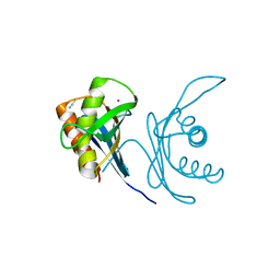
5OXW
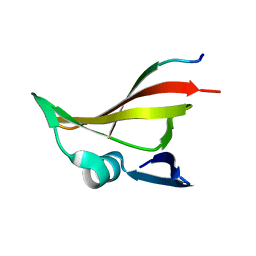 | | Structure of NeqN from Nanoarchaeum equitans | | 分子名称: | ALA-SER-GLY-SER-PHE-LYS-VAL-ILE-TYR-GLY-ASP, NEQ068 | | 著者 | Aparicio, D, Perez-Luque, R, Ribo, M, Fita, I. | | 登録日 | 2017-09-07 | | 公開日 | 2018-10-10 | | 最終更新日 | 2024-05-08 | | 実験手法 | X-RAY DIFFRACTION (2.61 Å) | | 主引用文献 | Structural Insights into Subunits Assembly and the Oxyester Splicing Mechanism of Neq pol Split Intein.
Cell Chem Biol, 25, 2018
|
|
4NEL
 | | Crystal structure of a putative transcriptional regulator from Saccharomonospora viridis in complex with N,N-dimethylmethanamine | | 分子名称: | N,N-dimethylmethanamine, Transcriptional regulator | | 著者 | Halavaty, A.S, Filippova, E.V, Minasov, G, Kiryukhina, O, Shuvalova, L, Endres, M, Joachimiak, A, Anderson, W.F, Midwest Center for Structural Genomics (MCSG) | | 登録日 | 2013-10-29 | | 公開日 | 2013-12-04 | | 最終更新日 | 2023-12-06 | | 実験手法 | X-RAY DIFFRACTION (2.05 Å) | | 主引用文献 | Crystal structure of a putative transcriptional regulator from Saccharomonospora viridis in complex with N,N-dimethylmethanamine
To be Published
|
|
4JIU
 | | Crystal structure of the metallopeptidase zymogen of Pyrococcus abyssi abylysin | | 分子名称: | GLYCEROL, Proabylysin, ZINC ION | | 著者 | Lopez-Pelegrin, M, Cerda-Costa, N, Martinez-Jimenez, F, Cintas-Pedrola, A, Canals, A, Peinado, J.R, Marti-Renom, M.A, Lopez-Otin, C, Arolas, J.L, Gomis-Ruth, F.X. | | 登録日 | 2013-03-07 | | 公開日 | 2013-06-12 | | 最終更新日 | 2024-03-20 | | 実験手法 | X-RAY DIFFRACTION (1.15 Å) | | 主引用文献 | A novel family of soluble minimal scaffolds provides structural insight into the catalytic domains of integral membrane metallopeptidases
J.Biol.Chem., 288, 2013
|
|
3OZD
 | | Crystal Structure of human 5'-deoxy-5'-methyladenosine phosphorylase in complex with pCl-phenylthioDADMeImmA | | 分子名称: | (3R,4S)-1-[(4-amino-5H-pyrrolo[3,2-d]pyrimidin-7-yl)methyl]-4-{[(4-chlorophenyl)sulfanyl]methyl}pyrrolidin-3-ol, S-methyl-5'-thioadenosine phosphorylase | | 著者 | Ho, M, Guan, R, Almo, S.C, Schramm, V.L. | | 登録日 | 2010-09-24 | | 公開日 | 2011-09-28 | | 最終更新日 | 2017-11-08 | | 実験手法 | X-RAY DIFFRACTION (2.1 Å) | | 主引用文献 | Crystal Structure of human 5'-deoxy-5'-methyladenosine phosphorylase
to be published
|
|
6G10
 | |
6RBR
 | |
4JQI
 | | Structure of active beta-arrestin1 bound to a G protein-coupled receptor phosphopeptide | | 分子名称: | 1,2-ETHANEDIOL, Beta-arrestin-1, CHLORIDE ION, ... | | 著者 | Shukla, A.K, Manglik, A, Kruse, A.C, Xiao, K, Reis, R.I, Tseng, W.C, Staus, D.P, Hilger, D, Uysal, S, Huang, L.H, Paduch, M, Shukla, P.T, Koide, A, Koide, S, Weis, W.I, Kossiakoff, A.A, Kobilka, B.K, Lefkowitz, R.J. | | 登録日 | 2013-03-20 | | 公開日 | 2013-04-17 | | 最終更新日 | 2023-09-20 | | 実験手法 | X-RAY DIFFRACTION (2.6 Å) | | 主引用文献 | Structure of active beta-arrestin-1 bound to a G-protein-coupled receptor phosphopeptide.
Nature, 497, 2013
|
|
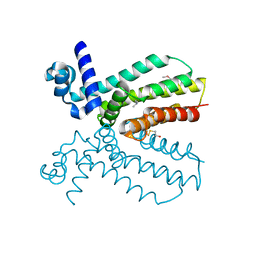
6JHD
 | | Solution structure of IFN alpha8 | | 分子名称: | Interferon alpha-8 | | 著者 | Ken-ichi, A, Shigeyuki, M. | | 登録日 | 2019-02-18 | | 公開日 | 2020-02-19 | | 最終更新日 | 2023-06-14 | | 実験手法 | SOLUTION NMR | | 主引用文献 | Determination of solution structure of interferon alpha8:
Novel insights into the preferred interaction with IFNAR2 among its subtypes
To Be Published
|
|
6RC2
 | |
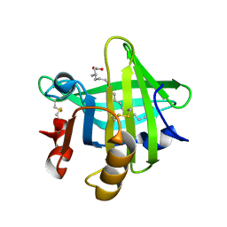
3F8D
 | | Structure of Sulfolobus solfataricus Thioredoxin reductase Mutant C147A | | 分子名称: | ACETIC ACID, FLAVIN-ADENINE DINUCLEOTIDE, SULFATE ION, ... | | 著者 | Ruggiero, A, Masullo, M, Ruocco, M.R, Arcari, P, Zagari, A, Vitagliano, L. | | 登録日 | 2008-11-12 | | 公開日 | 2009-01-13 | | 最終更新日 | 2023-11-01 | | 実験手法 | X-RAY DIFFRACTION (1.4 Å) | | 主引用文献 | Structure and stability of a thioredoxin reductase from Sulfolobus solfataricus: a thermostable protein with two functions
Biochim.Biophys.Acta, 1794, 2009
|
|


