Loading
PDBj
メニューPDBj@FacebookPDBj@TwitterPDBj@YouTubewwPDB FoundationwwPDB
RCSB PDBPDBeBMRBAdv. SearchSearch help
PDB: 135 件

7YHL
DownloadVisualize
BU of 7yhl by Molmil
Crystal Structure of the ring nuclease Sso2081 from Saccharolobus solfataricus in complex with free phosphate
分子名称: CRISPR system ring nuclease SSO2081, PHOSPHATE ION
著者Lin, Z, Du, L, Luo, Z.
登録日2022-07-13
公開日2023-02-15
最終更新日2023-03-29
実験手法X-RAY DIFFRACTION (2.7 Å)
主引用文献Molecular basis of stepwise cyclic tetra-adenylate cleavage by the type III CRISPR ring nuclease Crn1/Sso2081.
Nucleic Acids Res., 51, 2023
7YGH
DownloadVisualize
BU of 7ygh by Molmil
Crystal Structure of the ring nuclease Sso2081 from Saccharolobus solfataricus in complex with cyclic-tetraadenylate (cA4)
分子名称: CRISPR system ring nuclease SSO2081, RNA (5'-R(P*AP*AP*AP*A)-3')
著者Lin, Z, Du, L, Luo, Z.
登録日2022-07-11
公開日2023-02-15
最終更新日2023-11-29
実験手法X-RAY DIFFRACTION (3.11 Å)
主引用文献Molecular basis of stepwise cyclic tetra-adenylate cleavage by the type III CRISPR ring nuclease Crn1/Sso2081.
Nucleic Acids Res., 51, 2023
7YGL
DownloadVisualize
BU of 7ygl by Molmil
Crystal Structure of the ring nuclease Sso2081 from Saccharolobus solfataricus in complex with A4>p cleavage intermediate
分子名称: CRISPR system ring nuclease SSO2081, RNA (5'-R(*AP*AP*AP*(A23))-3')
著者Lin, Z, Du, L, Luo, Z.
登録日2022-07-11
公開日2023-02-15
最終更新日2023-11-29
実験手法X-RAY DIFFRACTION (2.5 Å)
主引用文献Molecular basis of stepwise cyclic tetra-adenylate cleavage by the type III CRISPR ring nuclease Crn1/Sso2081.
Nucleic Acids Res., 51, 2023
7ECF
DownloadVisualize
BU of 7ecf by Molmil
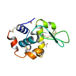
Crystal Structure of d(G4C2)2-Ba in C2221 space group
分子名称: BARIUM ION, DNA (5'-D(*GP*GP*GP*GP*CP*CP*GP*GP*GP*GP*CP*C)-3')
著者Geng, Y, Liu, C, Cai, Q, Luo, Z, Zhu, G.
登録日2021-03-12
公開日2021-06-09
最終更新日2024-03-27
実験手法X-RAY DIFFRACTION (1.6 Å)
主引用文献Crystal structure of parallel G-quadruplex formed by the two-repeat ALS- and FTD-related GGGGCC sequence.
Nucleic Acids Res., 49, 2021
6LAW
DownloadVisualize
BU of 6law by Molmil
MicroED structure of proteinase K at 1.50A determained using crystal lamellas prepared by focused ion beam milling
分子名称: Proteinase K, SULFATE ION
著者Zhou, H, Luo, Z, Li, X.
登録日2019-11-13
公開日2019-12-04
実験手法ELECTRON CRYSTALLOGRAPHY (1.5 Å)
主引用文献Using focus ion beam to prepare crystal lamella for electron diffraction.
J. Struct. Biol., 205, 2019
6LAV
DownloadVisualize
BU of 6lav by Molmil
MicroED structure of lysozyme at 1.73A determained using crystal lamellas prepared by focused ion beam milling
分子名称: ACETATE ION, Lysozyme C
著者Zhou, H, Luo, Z, Li, X.
登録日2019-11-13
公開日2019-11-27
実験手法ELECTRON CRYSTALLOGRAPHY (1.73 Å)
主引用文献Using focus ion beam to prepare crystal lamella for electron diffraction.
J. Struct. Biol., 205, 2019
7WOK
DownloadVisualize
BU of 7wok by Molmil
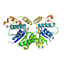
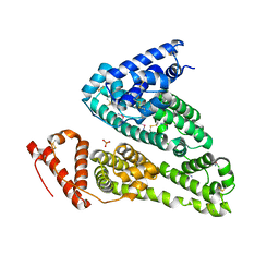
Crystal structure of HSA soaked with cisplatin for one week
分子名称: Albumin, Cisplatin, PHOSPHATE ION
著者Chen, S.L, Yuan, C, Jiang, L.G, Luo, Z.P, Huang, M.D.
登録日2022-01-21
公開日2022-07-20
最終更新日2023-11-29
実験手法X-RAY DIFFRACTION (2.9 Å)
主引用文献Crystallographic analysis of interaction between cisplatin and human serum albumin: Effect of fatty acid.
Int.J.Biol.Macromol., 216, 2022
7WOJ
DownloadVisualize
BU of 7woj by Molmil
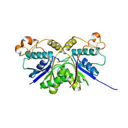
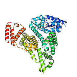
Crystal structure of HSA-Myr complex soaked with cisplatin for one week
分子名称: Albumin, Cisplatin, MYRISTIC ACID
著者Chen, S.L, Yuan, C, Jiang, L.G, Luo, Z.P, Huang, M.D.
登録日2022-01-21
公開日2022-07-20
最終更新日2023-11-29
実験手法X-RAY DIFFRACTION (2.89 Å)
主引用文献Crystallographic analysis of interaction between cisplatin and human serum albumin: Effect of fatty acid.
Int.J.Biol.Macromol., 216, 2022
7XLT
DownloadVisualize
BU of 7xlt by Molmil
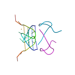
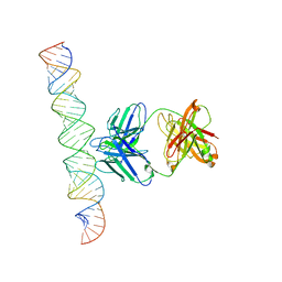
Cryo-EM Structure of R-loop monoclonal antibody S9.6 in recognizing RNA:DNA hybrids
分子名称: DNA, RNA, S9.6 Fab HC, ...
著者Li, Q, Lin, C, Luo, Z, Li, H, Li, X, Sun, Q.
登録日2022-04-22
公開日2022-05-25
最終更新日2022-08-03
実験手法ELECTRON MICROSCOPY (4.4 Å)
主引用文献Cryo-EM structure of R-loop monoclonal antibody S9.6 in recognizing RNA:DNA hybrids.
J Genet Genomics, 49, 2022
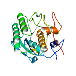
4I8B
DownloadVisualize
BU of 4i8b by Molmil
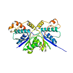
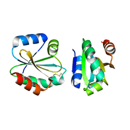
Crystal Structure of Thioredoxin from Schistosoma Japonicum
分子名称: Thioredoxin
著者Wu, Q, Peng, Y, Zhao, J, Li, X, Fan, X, Zhou, X, Chen, J, Luo, Z, Shi, D.
登録日2012-12-03
公開日2013-12-04
最終更新日2015-06-17
実験手法X-RAY DIFFRACTION (2 Å)
主引用文献Expression, characterization and crystal structure of thioredoxin from Schistosoma japonicum.
Parasitology, 142, 2015

220472

件を2024-05-29に公開中

PDB statisticsPDBj update infoContact PDBjnumon