Loading
PDBj
MenuPDBj@FacebookPDBj@TwitterPDBj@YouTubewwPDB FoundationwwPDB
RCSB PDBPDBeBMRBAdv. SearchSearch help
PDB: 104 results

8HQ2
DownloadVisualize
BU of 8hq2 by Molmil
Crystal structure of human ADAM22 in complex with human LGI1 mutant
Descriptor: 2-acetamido-2-deoxy-beta-D-glucopyranose, CALCIUM ION, Disintegrin and metalloproteinase domain-containing protein 22, ...
Authors:Liu, H, Lin, Z, Xu, F.
Deposit date:2022-12-13
Release date:2023-12-13
Method:X-RAY DIFFRACTION (2.93 Å)
Cite:Crystal structure of human ADAM22 in complex with human LGI1 mutant
To Be Published
6LW3
DownloadVisualize
BU of 6lw3 by Molmil
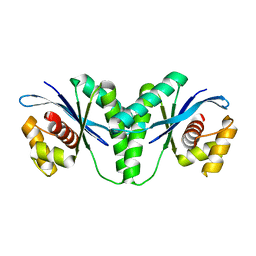
Crystal structure of RuvC from Pseudomonas aeruginosa
Descriptor: Crossover junction endodeoxyribonuclease RuvC
Authors:Hu, Y, He, Y, Lin, Z.
Deposit date:2020-02-07
Release date:2020-02-26
Last modified:2023-11-29
Method:X-RAY DIFFRACTION (2.38 Å)
Cite:Biochemical and structural characterization of the Holliday junction resolvase RuvC from Pseudomonas aeruginosa.
Biochem.Biophys.Res.Commun., 525, 2020
2H25
DownloadVisualize
BU of 2h25 by Molmil
Solution Structure of Maltose Binding Protein complexed with beta-cyclodextrin
Descriptor: Maltose-binding periplasmic protein
Authors:Xu, Y, Lin, Z, Zheng, Y, Yang, D.
Deposit date:2006-05-18
Release date:2006-10-31
Last modified:2024-05-29
Method:SOLUTION NMR
Cite:A new strategy for structure determination of large proteins in solution without deuteration
Nat.Methods, 3, 2006
7XSC
DownloadVisualize
BU of 7xsc by Molmil
Crystal structure of SARS-CoV-2 spike receptor binding domain bound with P5S-2B10
Descriptor: P5S-2B10 Heavy chain, P5S-2B10 Light chain, Spike protein S1
Authors:Wang, X, Wang, Z, Lin, Z.
Deposit date:2022-05-13
Release date:2023-05-24
Last modified:2023-11-29
Method:X-RAY DIFFRACTION (2.88 Å)
Cite:Infection with wild-type SARS-CoV-2 elicits broadly neutralizing and protective antibodies against omicron subvariants.
Nat.Immunol., 24, 2023

220472

PDB entries from 2024-05-29

PDB statisticsPDBj update infoContact PDBjnumon