6JKP
 
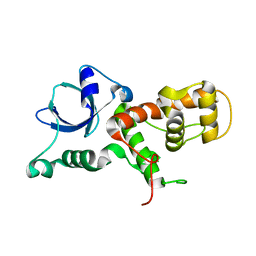
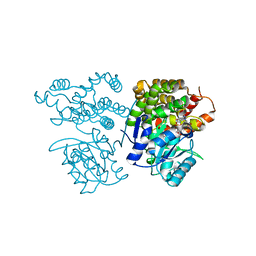 | | Crystal structure of sulfoacetaldehyde reductase from Bifidobacterium kashiwanohense in complex with NAD+ | | 分子名称: | Methanol dehydrogenase, NICOTINAMIDE-ADENINE-DINUCLEOTIDE, ZINC ION | | 著者 | Zhou, Y, Xu, T, Lin, L, Zhang, Y, Yuchi, Z. | | 登録日 | 2019-03-01 | | 公開日 | 2019-06-12 | | 最終更新日 | 2023-11-22 | | 実験手法 | X-RAY DIFFRACTION (3.008 Å) | | 主引用文献 | Identification and characterization of a new sulfoacetaldehyde reductase from the human gut bacteriumBifidobacterium kashiwanohense.
Biosci.Rep., 39, 2019
|
|
6JKO
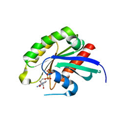
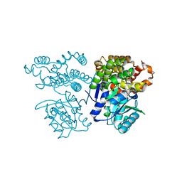 | | Crystal structure of sulfoacetaldehyde reductase from Bifidobacterium kashiwanohense | | 分子名称: | Methanol dehydrogenase, ZINC ION | | 著者 | Zhou, Y, Xu, T, Lin, L, Zhang, Y, Yuchi, Z. | | 登録日 | 2019-03-01 | | 公開日 | 2019-06-12 | | 最終更新日 | 2023-11-22 | | 実験手法 | X-RAY DIFFRACTION (1.9 Å) | | 主引用文献 | Identification and characterization of a new sulfoacetaldehyde reductase from the human gut bacteriumBifidobacterium kashiwanohense.
Biosci.Rep., 39, 2019
|
|
6JMU
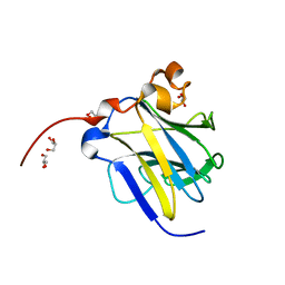
 | | Crystal structure of GIT1/Paxillin complex | | 分子名称: | ARF GTPase-activating protein GIT1, Paxillin | | 著者 | Zhu, J, Lin, L, Xia, Y, Zhang, R, Zhang, M. | | 登録日 | 2019-03-13 | | 公開日 | 2020-05-20 | | 最終更新日 | 2023-11-22 | | 実験手法 | X-RAY DIFFRACTION (2 Å) | | 主引用文献 | GIT/PIX Condensates Are Modular and Ideal for Distinct Compartmentalized Cell Signaling.
Mol.Cell, 79, 2020
|
|
6KIM
 | |
6JMT
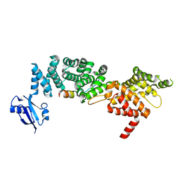
 | | Crystal structure of GIT/PIX complex | | 分子名称: | ARF GTPase-activating protein GIT2, ZINC ION, beta PIX | | 著者 | Zhu, J, Lin, L, Xia, Y, Zhang, R, Zhang, M. | | 登録日 | 2019-03-13 | | 公開日 | 2020-05-20 | | 最終更新日 | 2023-11-22 | | 実験手法 | X-RAY DIFFRACTION (2.8 Å) | | 主引用文献 | GIT/PIX Condensates Are Modular and Ideal for Distinct Compartmentalized Cell Signaling.
Mol.Cell, 79, 2020
|
|
6IDX
 | | Crystal Structure of BAI1/ELMO2 complex | | 分子名称: | Adhesion G protein-coupled receptor B1, Engulfment and cell motility protein 2 | | 著者 | Weng, Z.F, Lin, L, Zhu, J.W, Zhang, R.G. | | 登録日 | 2018-09-11 | | 公開日 | 2019-01-23 | | 最終更新日 | 2023-11-22 | | 実験手法 | X-RAY DIFFRACTION (1.699 Å) | | 主引用文献 | Structure of BAI1/ELMO2 complex reveals an action mechanism of adhesion GPCRs via ELMO family scaffolds
Nat Commun, 10, 2019
|
|
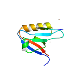
8I98
 | | Crystal structure of TePixD Y8F | | 分子名称: | FLAVIN MONONUCLEOTIDE, Tll0078 protein | | 著者 | Hu, R, Lin, L, Lu, Q. | | 登録日 | 2023-02-06 | | 公開日 | 2023-09-20 | | 実験手法 | X-RAY DIFFRACTION (2.54 Å) | | 主引用文献 | Structure of the BLUF Protein TePixD Y8F Mutant
Progress in Biochemistry and Biophysics, 2023
|
|
7WI8
 | | Cryo-EM structure of inactive mGlu3 bound to LY341495 | | 分子名称: | 2-[(1S,2S)-2-carboxycyclopropyl]-3-(9H-xanthen-9-yl)-D-alanine, 2-acetamido-2-deoxy-beta-D-glucopyranose, Metabotropic glutamate receptor 3 | | 著者 | Fang, W, Yang, F, Xu, C.J, Ling, S.L, Lin, L, Zhou, Y.X, Sun, W.J, Wang, X.M, Liu, P, Rondard, P, Pan, S, Pin, J.P, Tian, C.L, Liu, J.F. | | 登録日 | 2022-01-03 | | 公開日 | 2022-03-16 | | 最終更新日 | 2022-07-20 | | 実験手法 | ELECTRON MICROSCOPY (4.17 Å) | | 主引用文献 | Structural basis of the activation of metabotropic glutamate receptor 3.
Cell Res., 32, 2022
|
|
7WI6
 | | Cryo-EM structure of LY341495/NAM-bound mGlu3 | | 分子名称: | 2-[(1S,2S)-2-carboxycyclopropyl]-3-(9H-xanthen-9-yl)-D-alanine, 2-acetamido-2-deoxy-beta-D-glucopyranose, Metabotropic glutamate receptor 3 | | 著者 | Fang, W, Yang, F, Xu, C.J, Ling, S.L, Lin, L, Zhou, Y.X, Sun, W.J, Wang, X.M, Liu, P, Rondard, P, Pan, S, Pin, J.P, Tian, C.L, Liu, J.F. | | 登録日 | 2022-01-03 | | 公開日 | 2022-03-16 | | 最終更新日 | 2022-07-20 | | 実験手法 | ELECTRON MICROSCOPY (3.71 Å) | | 主引用文献 | Structural basis of the activation of metabotropic glutamate receptor 3.
Cell Res., 32, 2022
|
|
7WIH
 | | Cryo-EM structure of LY2794193-bound mGlu3 | | 分子名称: | (1S,2S,4S,5R,6S)-2-amino-4-[(3-methoxybenzene-1-carbonyl)amino]bicyclo[3.1.0]hexane-2,6-dicarboxylic acid, 2-acetamido-2-deoxy-beta-D-glucopyranose, Metabotropic glutamate receptor 3 | | 著者 | Fang, W, Yang, F, Xu, C.J, Ling, S.L, Lin, L, Zhou, Y.X, Sun, W.J, Wang, X.M, Liu, P, Rondard, P, Pan, S, Pin, J.P, Tian, C.L, Liu, J.F. | | 登録日 | 2022-01-03 | | 公開日 | 2022-03-16 | | 最終更新日 | 2022-07-20 | | 実験手法 | ELECTRON MICROSCOPY (3.68 Å) | | 主引用文献 | Structural basis of the activation of metabotropic glutamate receptor 3.
Cell Res., 32, 2022
|
|
6CW0
 | | Crystal structure of Cryptosporidium parvum bromodomain cgd2_2690 | | 分子名称: | 4-{[(7R)-8-cyclopentyl-7-ethyl-5-methyl-6-oxo-5,6,7,8-tetrahydropteridin-2-yl]amino}-3-methoxy-N-(1-methylpiperidin-4-yl)benzamide, Cgd2_2690 protein, GLYCEROL, ... | | 著者 | Dong, A, Lin, L, Bountra, C, Arrowsmith, C.H, Edwards, A.M, Hui, R, Structural Genomics Consortium (SGC) | | 登録日 | 2018-03-29 | | 公開日 | 2018-04-18 | | 最終更新日 | 2023-10-04 | | 実験手法 | X-RAY DIFFRACTION (1.38 Å) | | 主引用文献 | Crystal structure of Cryptosporidium parvum bromodomain cgd2_2690
to be published
|
|
3G7N
 | | Crystal Structure of a Triacylglycerol Lipase from Penicillium Expansum at 1.3 | | 分子名称: | DI(HYDROXYETHYL)ETHER, Lipase, PENTAETHYLENE GLYCOL, ... | | 著者 | Bian, C.B, Yuan, C, Chen, L.Q, Edward, J.M, Lin, L, Jiang, L.G, Huang, Z.X, Huang, M.D. | | 登録日 | 2009-02-10 | | 公開日 | 2010-02-23 | | 最終更新日 | 2017-11-01 | | 実験手法 | X-RAY DIFFRACTION (1.3 Å) | | 主引用文献 | Crystal structure of a triacylglycerol lipase from Penicillium expansum at 1.3 A determined by sulfur SAD
Proteins, 78, 2010
|
|
4J12
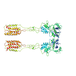
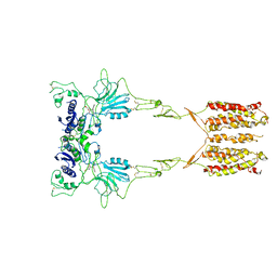
 | | monomeric Fc | | 分子名称: | 2-acetamido-2-deoxy-beta-D-glucopyranose, 2-acetamido-2-deoxy-beta-D-glucopyranose-(1-2)-alpha-D-mannopyranose-(1-3)-[2-acetamido-2-deoxy-beta-D-glucopyranose-(1-2)-alpha-D-mannopyranose-(1-6)]beta-D-mannopyranose-(1-4)-2-acetamido-2-deoxy-beta-D-glucopyranose-(1-4)-2-acetamido-2-deoxy-beta-D-glucopyranose, human Fc fragment | | 著者 | Ishino, T, Wang, M, Mosyak, L, Tam, A, Duan, W, Svenson, K, Joyce, A, O'Hara, D, Lin, L, Somers, W, Kriz, R. | | 登録日 | 2013-01-31 | | 公開日 | 2013-05-01 | | 最終更新日 | 2020-07-29 | | 実験手法 | X-RAY DIFFRACTION (1.9 Å) | | 主引用文献 | Engineering a Monomeric Fc Domain Modality by N-Glycosylation for the Half-life Extension of Biotherapeutics.
J.Biol.Chem., 288, 2013
|
|
3ES5
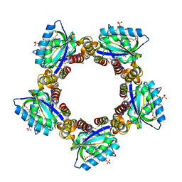
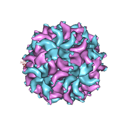 | | Crystal Structure of Partitivirus (PsV-F) | | 分子名称: | Putative capsid protein | | 著者 | Pan, J, Dong, L, Lin, L, Ochoa, W.F, Sinkovits, R.S, Havens, W.M, Nibert, M.L, Baker, T.S, Ghabrial, S.A, Tao, Y.J. | | 登録日 | 2008-10-03 | | 公開日 | 2009-03-10 | | 最終更新日 | 2024-04-03 | | 実験手法 | X-RAY DIFFRACTION (3.3 Å) | | 主引用文献 | Atomic structure reveals the unique capsid organization of a dsRNA virus.
Proc.Natl.Acad.Sci.USA, 106, 2009
|
|
7V8N
 | |
7SFA
 | |
7SF9
 | |
3BFK
 | | Crystal structure of Plasmodium falciparum Rab11a in complex with GDP | | 分子名称: | GLYCEROL, GUANOSINE-5'-DIPHOSPHATE, Small GTPase Rab11 | | 著者 | Pizarro, J.C, Sukumar, D, Hassanali, A, Lin, L, Wernimont, A.K, Lew, J, Kozieradzki, I, Edwards, A.M, Arrowsmith, C.H, Weigelt, J, Sundstrom, M, Bochkarev, A, Hui, R, Structural Genomics Consortium (SGC) | | 登録日 | 2007-11-21 | | 公開日 | 2007-12-04 | | 最終更新日 | 2023-08-30 | | 実験手法 | X-RAY DIFFRACTION (1.8 Å) | | 主引用文献 | Crystal structure of Plasmodium falciparum Rab11a in complex with GDP.
To be Published
|
|
7WEG
 | | Complex structure of PDZD7 and FCHSD2 | | 分子名称: | FCHSD2, PDZ domain-containing protein 7, ZINC ION | | 著者 | Wang, H, Lin, L, Lu, Q. | | 登録日 | 2021-12-23 | | 公開日 | 2022-11-16 | | 最終更新日 | 2023-11-29 | | 実験手法 | X-RAY DIFFRACTION (2 Å) | | 主引用文献 | Deafness-related protein PDZD7 forms complex with the C-terminal tail of FCHSD2.
Biochem.J., 479, 2022
|
|
1S1E
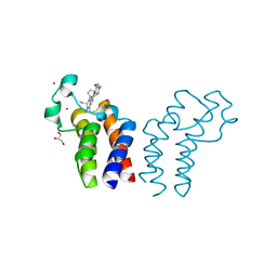
 | | Crystal Structure of Kv Channel-interacting protein 1 (KChIP-1) | | 分子名称: | CALCIUM ION, Kv channel interacting protein 1 | | 著者 | Scannevin, R.H, Wang, K.-W, Jow, F, Megules, J, Kopsco, D.C, Edris, W, Carroll, K.C, Lu, Q, Xu, W.-X, Xu, Z.-B, Katz, A.H, Olland, S, Lin, L, Taylor, M, Stahl, M, Malakian, K, Somers, W, Mosyak, L, Bowlby, M.R, Chanda, P, Rhodes, K.J. | | 登録日 | 2004-01-06 | | 公開日 | 2005-01-11 | | 最終更新日 | 2023-08-23 | | 実験手法 | X-RAY DIFFRACTION (2.3 Å) | | 主引用文献 | Two N-terminal domains of Kv4 K(+) channels regulate binding to and modulation by KChIP1.
Neuron, 41, 2004
|
|
1S1G
 | | Crystal Structure of Kv4.3 T1 Domain | | 分子名称: | Potassium voltage-gated channel subfamily D member 3, ZINC ION | | 著者 | Scannevin, R.H, Wang, K.W, Jow, F, Megules, J, Kopsco, D.C, Edris, W, Carroll, K.C, Lu, Q, Xu, W.X, Xu, Z.B, Katz, A.H, Olland, S, Lin, L, Taylor, M, Stahl, M, Malakian, K, Somers, W, Mosyak, L, Bowlby, M.R, Chanda, P, Rhodes, K.J. | | 登録日 | 2004-01-06 | | 公開日 | 2004-03-23 | | 最終更新日 | 2023-08-23 | | 実験手法 | X-RAY DIFFRACTION (2.6 Å) | | 主引用文献 | Two N-terminal domains of Kv4 K(+) channels regulate binding to and modulation by KChIP1.
Neuron, 41, 2004
|
|
7YKF
 | | Crystal structure of MAGI2 PDZ0-GK/pEphexin4 complex | | 分子名称: | Ephexin4, Membrane-associated guanylate kinase, WW and PDZ domain-containing protein 2 | | 著者 | Zhang, M, Lin, L, Zhu, J. | | 登録日 | 2022-07-22 | | 公開日 | 2023-08-02 | | 最終更新日 | 2024-02-28 | | 実験手法 | X-RAY DIFFRACTION (2.28 Å) | | 主引用文献 | Phosphorylation-dependent recognition of diverse protein targets by the cryptic GK domain of MAGI MAGUKs.
Sci Adv, 9, 2023
|
|
7YKI
 | | Crystal structure of MAGI2 PDZ0-GK domain in complex with phospho-SAPAP1 GBR3 peptide | | 分子名称: | GLYCEROL, Membrane-associated guanylate kinase, WW and PDZ domain-containing protein 2, ... | | 著者 | Zhang, M, Lin, L, Zhu, J. | | 登録日 | 2022-07-22 | | 公開日 | 2023-08-02 | | 最終更新日 | 2024-02-28 | | 実験手法 | X-RAY DIFFRACTION (2 Å) | | 主引用文献 | Phosphorylation-dependent recognition of diverse protein targets by the cryptic GK domain of MAGI MAGUKs.
Sci Adv, 9, 2023
|
|
7YKG
 | | Crystal structure of MAGI2 PDZ0-GK/pSGEF complex | | 分子名称: | Membrane-associated guanylate kinase, WW and PDZ domain-containing protein 2, SGEF | | 著者 | Zhang, M, Lin, L, Zhu, J. | | 登録日 | 2022-07-22 | | 公開日 | 2023-08-02 | | 最終更新日 | 2024-02-28 | | 実験手法 | X-RAY DIFFRACTION (2.16 Å) | | 主引用文献 | Phosphorylation-dependent recognition of diverse protein targets by the cryptic GK domain of MAGI MAGUKs.
Sci Adv, 9, 2023
|
|
7YKH
 | | Crystal structure of MAGI2 PDZ0-GK domain in complex with phospho-SAPAP1 GBR2 peptide | | 分子名称: | GLYCEROL, Membrane-associated guanylate kinase, WW and PDZ domain-containing protein 2, ... | | 著者 | Zhang, M, Lin, L, Zhu, J. | | 登録日 | 2022-07-22 | | 公開日 | 2023-08-16 | | 最終更新日 | 2024-02-28 | | 実験手法 | X-RAY DIFFRACTION (2.5 Å) | | 主引用文献 | Phosphorylation-dependent recognition of diverse protein targets by the cryptic GK domain of MAGI MAGUKs.
Sci Adv, 9, 2023
|
|


