5MK1
 
 | |
5MJZ
 | |
5MK3
 | |
5MK0
 | |
5LM1
 | |
5MJY
 | |
5MK2
 | |
6Y2V
 | |
6Y2W
 | |
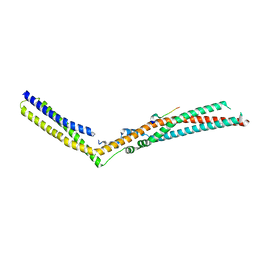
7NDS
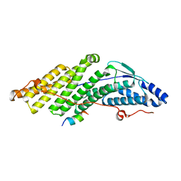
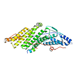
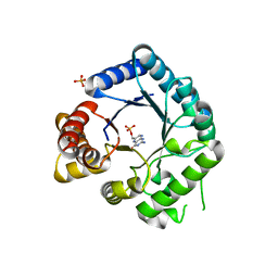
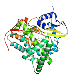
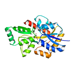 | | Crystal structure of TphC in a closed conformation | | 分子名称: | Tripartite tricarboxylate transporter substrate binding protein, terephthalic acid | | 著者 | Levy, C. | | 登録日 | 2021-02-02 | | 公開日 | 2021-11-03 | | 最終更新日 | 2024-01-31 | | 実験手法 | X-RAY DIFFRACTION (2.4 Å) | | 主引用文献 | Structural basis of terephthalate recognition by solute binding protein TphC.
Nat Commun, 12, 2021
|
|
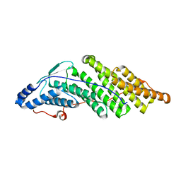
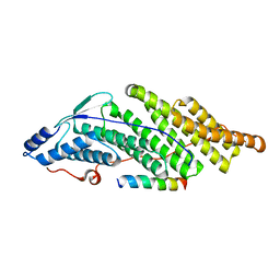
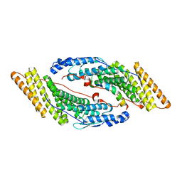
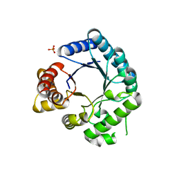
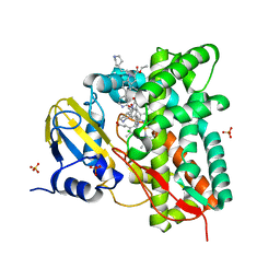
7NDR
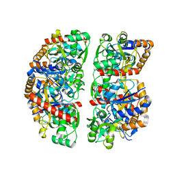 | | Crystal structure of TphC in an open conformation | | 分子名称: | 1,2-ETHANEDIOL, Tripartite tricarboxylate transporter substrate binding protein | | 著者 | Levy, C. | | 登録日 | 2021-02-02 | | 公開日 | 2021-11-03 | | 最終更新日 | 2024-01-31 | | 実験手法 | X-RAY DIFFRACTION (1.97 Å) | | 主引用文献 | Structural basis of terephthalate recognition by solute binding protein TphC.
Nat Commun, 12, 2021
|
|
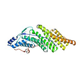
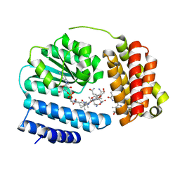
7YXF
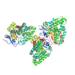
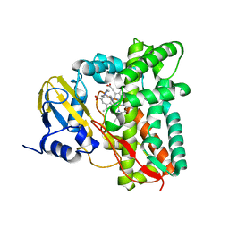 | | Crystal structure of CYP125 from Mycobacterium tuberculosis in complex with an inhibitor | | 分子名称: | 1-(2-piperazin-1-ylethyl)-5-pyridin-4-yl-indole, PROTOPORPHYRIN IX CONTAINING FE, SULFATE ION, ... | | 著者 | Snee, M, Katariya, M, Levy, C, Leys, D. | | 登録日 | 2022-02-16 | | 公開日 | 2023-03-01 | | 最終更新日 | 2024-02-07 | | 実験手法 | X-RAY DIFFRACTION (1.85 Å) | | 主引用文献 | Structure Based Discovery of Inhibitors of CYP125 and CYP142 from Mycobacterium tuberculosis.
Chemistry, 29, 2023
|
|
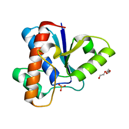
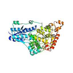
4NSH
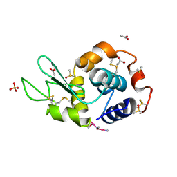 | | Carboplatin binding to HEWL in 0.2M NH4SO4, 0.1M NaAc in 25% PEG 4000 at pH 4.6 | | 分子名称: | ACETATE ION, DIMETHYL SULFOXIDE, Lysozyme C, ... | | 著者 | Tanley, S.W.M, Diederichs, K, Kroon-Batenburg, L.M.J, Levy, C, Schreurs, A.M.M, Helliwell, J.R. | | 登録日 | 2013-11-28 | | 公開日 | 2014-10-15 | | 最終更新日 | 2023-09-20 | | 実験手法 | X-RAY DIFFRACTION (2.1 Å) | | 主引用文献 | Carboplatin binding to histidine.
Acta Crystallogr F Struct Biol Commun, 70, 2014
|
|
6RQ5
 | | CYP121 in complex with 3,5-dimethyl dicyclotyrosine | | 分子名称: | (3~{S},6~{S})-3-[(3,5-dimethyl-4-oxidanyl-phenyl)methyl]-6-[(4-hydroxyphenyl)methyl]piperazine-2,5-dione, 2-(N-MORPHOLINO)-ETHANESULFONIC ACID, Mycocyclosin synthase, ... | | 著者 | Poddar, H, Levy, C. | | 登録日 | 2019-05-15 | | 公開日 | 2020-04-22 | | 最終更新日 | 2024-01-24 | | 実験手法 | X-RAY DIFFRACTION (1.55 Å) | | 主引用文献 | Structure-Activity Relationships of cyclo (l-Tyrosyl-l-tyrosine) Derivatives Binding to Mycobacterium tuberculosis CYP121: Iodinated Analogues Promote Shift to High-Spin Adduct.
J.Med.Chem., 62, 2019
|
|
8J2X
 | | Saccharothrix syringae photocobilins protein, light state | | 分子名称: | BILIVERDINE IX ALPHA, COBALAMIN, Cobalamin-binding protein, ... | | 著者 | Zhang, S, Poddar, H, Levy, C, Leys, D. | | 登録日 | 2023-04-15 | | 公開日 | 2024-04-10 | | 実験手法 | X-RAY DIFFRACTION (1.98 Å) | | 主引用文献 | Photocobilins integrate B12 and bilin photochemistry for enzyme control.
Nat Commun, 15, 2024
|
|
2VEG
 | |
2VEF
 | |
4GU5
 | | Structure of Full-length Drosophila Cryptochrome | | 分子名称: | Cryptochrome-1, FLAVIN-ADENINE DINUCLEOTIDE, MAGNESIUM ION | | 著者 | Zoltowski, B.D, Vaidya, A.T, Top, D, Widom, J, Young, M.W, Levy, C, Jones, A.R, Scrutton, N.S, Leys, D, Crane, B.R. | | 登録日 | 2012-08-29 | | 公開日 | 2012-09-26 | | 最終更新日 | 2023-09-13 | | 実験手法 | X-RAY DIFFRACTION (2.3 Å) | | 主引用文献 | Updated structure of Drosophila cryptochrome.
Nature, 495, 2013
|
|
2CG8
 | | The bifunctional dihydroneopterin aldolase 6-hydroxymethyl-7,8- dihydropterin synthase from Streptococcus pneumoniae | | 分子名称: | DIHYDRONEOPTERIN ALDOLASE 6-HYDROXYMETHYL-7,8-DIHYDROPTERIN SYNTHASE | | 著者 | Garcon, A, Levy, C, Derrick, J.P. | | 登録日 | 2006-02-28 | | 公開日 | 2006-06-21 | | 最終更新日 | 2023-12-13 | | 実験手法 | X-RAY DIFFRACTION (2.9 Å) | | 主引用文献 | Crystal Structure of the Bifunctional Dihydroneopterin Aldolase/6-Hydroxymethyl-7,8-Dihydropterin Pyrophosphokinase from Streptococcus Pneumoniae.
J.Mol.Biol., 360, 2006
|
|
8C3W
 | | Crystal structure of a computationally designed heme binding protein, dnHEM1 | | 分子名称: | (4S)-2-METHYL-2,4-PENTANEDIOL, 1,2-ETHANEDIOL, DI(HYDROXYETHYL)ETHER, ... | | 著者 | Ortmayer, M, Levy, C. | | 登録日 | 2022-12-29 | | 公開日 | 2023-07-05 | | 最終更新日 | 2024-02-07 | | 実験手法 | X-RAY DIFFRACTION (1.6 Å) | | 主引用文献 | Design of Heme Enzymes with a Tunable Substrate Binding Pocket Adjacent to an Open Metal Coordination Site.
J.Am.Chem.Soc., 145, 2023
|
|
7QKE
 | | Crystal structure of CYP125 from Mycobacterium tuberculosis in complex with inhibitor (surface entropy reduction mutant) | | 分子名称: | PROTOPORPHYRIN IX CONTAINING FE, Steroid C26-monooxygenase, ethyl 1-(cyclohexylmethyl)-5-pyridin-4-yl-indole-2-carboxylate | | 著者 | Snee, M, Tunnicliffe, R, Leys, D, Levy, C, Katariya, M. | | 登録日 | 2021-12-17 | | 公開日 | 2022-12-28 | | 最終更新日 | 2024-02-07 | | 実験手法 | X-RAY DIFFRACTION (2.3 Å) | | 主引用文献 | Structure Based Discovery of Inhibitors of CYP125 and CYP142 from Mycobacterium tuberculosis.
Chemistry, 29, 2023
|
|
7QNN
 | | Crystal structure of CYP125 from Mycobacterium tuberculosis in complex with inhibitor (surface entropy reduction mutant) | | 分子名称: | PROTOPORPHYRIN IX CONTAINING FE, Steroid C26-monooxygenase, ethyl 1-(cyclopentylmethyl)-5-pyridin-4-yl-indole-2-carboxylate | | 著者 | Snee, M, Tunnicliffe, R, Leys, D, Levy, C, Katariya, M. | | 登録日 | 2021-12-21 | | 公開日 | 2022-12-28 | | 最終更新日 | 2024-02-07 | | 実験手法 | X-RAY DIFFRACTION (2.47 Å) | | 主引用文献 | Structure Based Discovery of Inhibitors of CYP125 and CYP142 from Mycobacterium tuberculosis.
Chemistry, 29, 2023
|
|
7QJL
 | | Crystal structure of CYP142 from Mycobacterium tuberculosis in complex with an inhibitor | | 分子名称: | ACETATE ION, BROMIDE ION, PROTOPORPHYRIN IX CONTAINING FE, ... | | 著者 | Snee, M, Levy, C, Katariya, M. | | 登録日 | 2021-12-16 | | 公開日 | 2022-12-28 | | 最終更新日 | 2024-02-07 | | 実験手法 | X-RAY DIFFRACTION (1.38 Å) | | 主引用文献 | Structure Based Discovery of Inhibitors of CYP125 and CYP142 from Mycobacterium tuberculosis.
Chemistry, 29, 2023
|
|
7QWN
 | | Crystal structure of CYP125 from Mycobacterium tuberculosis in complex with an inhibitor | | 分子名称: | CHLORIDE ION, PROTOPORPHYRIN IX CONTAINING FE, SULFATE ION, ... | | 著者 | Snee, M, Katariya, M, Levy, C, Leys, D. | | 登録日 | 2022-01-25 | | 公開日 | 2023-02-01 | | 最終更新日 | 2024-02-07 | | 実験手法 | X-RAY DIFFRACTION (1.93 Å) | | 主引用文献 | Structure Based Discovery of Inhibitors of CYP125 and CYP142 from Mycobacterium tuberculosis.
Chemistry, 29, 2023
|
|
7R1I
 | | Crystal structure of CYP125 from Mycobacterium tuberculosis in complex with an inhibitor | | 分子名称: | CHLORIDE ION, PROTOPORPHYRIN IX CONTAINING FE, SULFATE ION, ... | | 著者 | Snee, M, Katariya, M, Levy, C, Leys, D. | | 登録日 | 2022-02-03 | | 公開日 | 2023-02-15 | | 最終更新日 | 2024-02-07 | | 実験手法 | X-RAY DIFFRACTION (2.24 Å) | | 主引用文献 | Structure Based Discovery of Inhibitors of CYP125 and CYP142 from Mycobacterium tuberculosis.
Chemistry, 29, 2023
|
|


