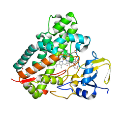
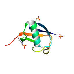
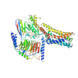
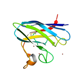
7FUP
 
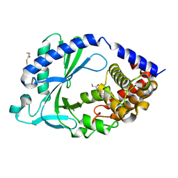 | | Crystal Structure of human cyclic GMP-AMP synthase in complex with methyl 1-benzoyl-3-methyl-5-(1,2-oxazol-5-yl)pyrazole-4-carboxylate | | 分子名称: | Cyclic GMP-AMP synthase, PROPANOIC ACID, ZINC ION | | 著者 | Leibrock, L, Benz, J, Groebke-Zbinden, K, Rudolph, M.G. | | 登録日 | 2023-02-08 | | 公開日 | 2024-02-21 | | 実験手法 | X-RAY DIFFRACTION (2.411 Å) | | 主引用文献 | Crystal Structure of a human cyclic GMP-AMP synthase complex
To be published
|
|
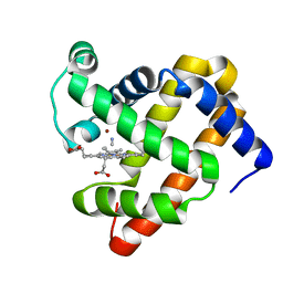
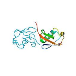
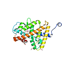
7FU1
 | |
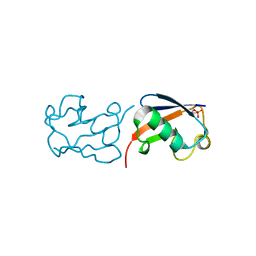
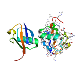
4Y8W
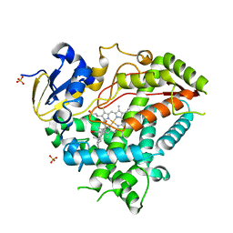 | | Crystal Structure of Human Cytochrome P450 21A2 Progesterone Complex | | 分子名称: | Cytochrome P450 21-hydroxylase, PROGESTERONE, PROTOPORPHYRIN IX CONTAINING FE, ... | | 著者 | Pallan, P.S, Lei, L, Egli, M. | | 登録日 | 2015-02-16 | | 公開日 | 2015-04-15 | | 最終更新日 | 2023-09-27 | | 実験手法 | X-RAY DIFFRACTION (2.64 Å) | | 主引用文献 | Human Cytochrome P450 21A2, the Major Steroid 21-Hydroxylase: STRUCTURE OF THE ENZYMEPROGESTERONE SUBSTRATE COMPLEX AND RATE-LIMITING C-H BOND CLEAVAGE.
J.Biol.Chem., 290, 2015
|
|
1SE6
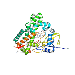 | | Crystal Structure of Streptomyces Coelicolor A3(2) CYP158A2 from antibiotic biosynthetic pathways | | 分子名称: | 2-(N-MORPHOLINO)-ETHANESULFONIC ACID, PROTOPORPHYRIN IX CONTAINING FE, SPERMINE (FULLY PROTONATED FORM), ... | | 著者 | Zhao, B, Lamb, D.C, Lei, L, Sundaramoorthy, M, Podust, L.M, Waterman, M.R. | | 登録日 | 2004-02-16 | | 公開日 | 2005-01-25 | | 最終更新日 | 2023-08-23 | | 実験手法 | X-RAY DIFFRACTION (1.75 Å) | | 主引用文献 | Binding of Two Flaviolin Substrate Molecules, Oxidative Coupling, and Crystal Structure of Streptomyces coelicolor A3(2) Cytochrome P450 158A2.
J.Biol.Chem., 280, 2005
|
|
1S1F
 | | Crystal Structure of Streptomyces Coelicolor A3(2) CYP158A2 from antibiotic biosynthetic pathways | | 分子名称: | 4-PHENYL-1H-IMIDAZOLE, GLYCEROL, MALONIC ACID, ... | | 著者 | Zhao, B, Lamb, D.C, Lei, L, Sundaramoorthy, M, Podust, L.M, Waterman, M.R. | | 登録日 | 2004-01-06 | | 公開日 | 2005-01-11 | | 最終更新日 | 2024-02-14 | | 実験手法 | X-RAY DIFFRACTION (1.5 Å) | | 主引用文献 | Binding of Two Flaviolin Substrate Molecules, Oxidative Coupling, and Crystal Structure of Streptomyces coelicolor A3(2) Cytochrome P450 158A2
J.Biol.Chem., 280, 2005
|
|
3MN0
 | | Introducing a 2-His-1-Glu Non-Heme Iron Center into Myoglobin confers Nitric Oxide Reductase activity: Cu(II)-CN-FeBMb(-His) form | | 分子名称: | COPPER (II) ION, CYANIDE ION, Myoglobin, ... | | 著者 | Lin, Y.-W, Yeung, N, Gao, Y.-G, Miner, K.D, Lei, L, Robinson, H, Lu, Y. | | 登録日 | 2010-04-20 | | 公開日 | 2010-08-11 | | 最終更新日 | 2024-02-21 | | 実験手法 | X-RAY DIFFRACTION (1.65 Å) | | 主引用文献 | Introducing a 2-his-1-glu nonheme iron center into myoglobin confers nitric oxide reductase activity.
J.Am.Chem.Soc., 132, 2010
|
|
3K9Z
 | | Rational Design of a Structural and Functional Nitric Oxide Reductase | | 分子名称: | FE (II) ION, Myoglobin, PROTOPORPHYRIN IX CONTAINING FE | | 著者 | Yeung, N, Lin, Y.-W, Gao, Y.-G, Zhao, X, Russell, B.S, Lei, L, Miner, K.D, Robinson, H, Lu, Y. | | 登録日 | 2009-10-16 | | 公開日 | 2009-12-01 | | 最終更新日 | 2024-02-21 | | 実験手法 | X-RAY DIFFRACTION (1.72 Å) | | 主引用文献 | Rational design of a structural and functional nitric oxide reductase.
Nature, 462, 2009
|
|
5X3O
 | |
5X3N
 | |
5X3M
 | |
5XPK
 | |
5YV7
 | |
8GDA
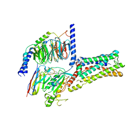 | | Cryo-EM Structure of the Prostaglandin E2 Receptor 4 Coupled to G Protein | | 分子名称: | (2S,5R)-5-[(1E,3S)-4,4-difluoro-3-hydroxy-4-phenylbut-1-en-1-yl]-1-[6-(1H-tetrazol-5-yl)hexyl]pyrrolidin-2-ol, Guanine nucleotide-binding protein G(I)/G(S)/G(T) subunit beta-1, Guanine nucleotide-binding protein G(s) subunit alpha isoforms short, ... | | 著者 | Shenming, H, Mengyao, X, Lei, L, Jiangqian, M, Sheng, C, Jinpeng, S. | | 登録日 | 2023-03-03 | | 公開日 | 2024-01-10 | | 実験手法 | ELECTRON MICROSCOPY (3.3 Å) | | 主引用文献 | Single hormone or synthetic agonist induces G s /G i coupling selectivity of EP receptors via distinct binding modes and propagating paths.
Proc.Natl.Acad.Sci.USA, 120, 2023
|
|
8GDB
 | | Cryo-EM Structure of the Prostaglandin E2 Receptor 4 Coupled to G Protein | | 分子名称: | (Z)-7-[(1R,2R,3R)-3-hydroxy-2-[(E,3S)-3-hydroxyoct-1-enyl]-5-oxo-cyclopentyl]hept-5-enoic acid, Guanine nucleotide-binding protein G(I)/G(S)/G(T) subunit beta-1, Guanine nucleotide-binding protein G(s) subunit alpha isoforms short, ... | | 著者 | Shenming, H, Mengyao, X, Lei, L, Jianqiang, M, Jinpeng, S. | | 登録日 | 2023-03-03 | | 公開日 | 2024-01-10 | | 実験手法 | ELECTRON MICROSCOPY (3.1 Å) | | 主引用文献 | Single hormone or synthetic agonist induces G s /G i coupling selectivity of EP receptors via distinct binding modes and propagating paths.
Proc.Natl.Acad.Sci.USA, 120, 2023
|
|
8GDC
 | | Cryo-EM Structure of the Prostaglandin E2 Receptor 3 Coupled to G Protein | | 分子名称: | (Z)-7-[(1R,2R,3R)-3-hydroxy-2-[(E,3S)-3-hydroxyoct-1-enyl]-5-oxo-cyclopentyl]hept-5-enoic acid, Guanine nucleotide-binding protein G(I)/G(S)/G(T) subunit beta-1, Guanine nucleotide-binding protein G(i) subunit alpha-1, ... | | 著者 | Shenming, H, Mengyao, X, Lei, L, Yang, D, Shiyi, G, Jinpeng, S. | | 登録日 | 2023-03-03 | | 公開日 | 2024-01-10 | | 実験手法 | ELECTRON MICROSCOPY (3.5 Å) | | 主引用文献 | Single hormone or synthetic agonist induces G s /G i coupling selectivity of EP receptors via distinct binding modes and propagating paths.
Proc.Natl.Acad.Sci.USA, 120, 2023
|
|
8GD9
 | | Cryo-EM Structure of the Prostaglandin E2 Receptor 4 Coupled to G Protein | | 分子名称: | 4-({2-[(1R,2R,3R,5S)-3,5-dihydroxy-2-{(3S)-3-hydroxy-4-[3-(methoxymethyl)phenyl]butyl}cyclopentyl]ethyl}sulfanyl)butanoic acid, Guanine nucleotide-binding protein G(I)/G(S)/G(T) subunit beta-1, Guanine nucleotide-binding protein G(s) subunit alpha isoforms short, ... | | 著者 | Shenming, H, Mengyao, X, Lei, L, Yang, D, Sheng, C, Jinpeng, S. | | 登録日 | 2023-03-03 | | 公開日 | 2024-01-10 | | 実験手法 | ELECTRON MICROSCOPY (3.2 Å) | | 主引用文献 | Single hormone or synthetic agonist induces G s /G i coupling selectivity of EP receptors via distinct binding modes and propagating paths.
Proc.Natl.Acad.Sci.USA, 120, 2023
|
|
3HDL
 | | Crystal Structure of Highly Glycosylated Peroxidase from Royal Palm Tree | | 分子名称: | 1,2-ETHANEDIOL, 2-(N-MORPHOLINO)-ETHANESULFONIC ACID, 2-acetamido-2-deoxy-beta-D-glucopyranose, ... | | 著者 | Watanabe, L, Moura, P.R, Bleicher, L, Nascimento, A.S, Zamorano, L.S, Calvete, J.J, Bursakov, S, Roig, M.G, Shnyrov, V.L, Polikarpov, I. | | 登録日 | 2009-05-07 | | 公開日 | 2009-11-24 | | 最終更新日 | 2020-07-29 | | 実験手法 | X-RAY DIFFRACTION (1.85 Å) | | 主引用文献 | Crystal structure and statistical coupling analysis of highly glycosylated peroxidase from royal palm tree (Roystonea regia).
J.Struct.Biol., 169, 2010
|
|
3G9V
 | | Crystal structure of a soluble decoy receptor IL-22BP bound to interleukin-22 | | 分子名称: | Interleukin 22 receptor, alpha 2, Interleukin-22 | | 著者 | de Moura, P.R, Watanabe, L, Bleicher, L, Colau, D, Renauld, J.-C, Polikarpov, I. | | 登録日 | 2009-02-14 | | 公開日 | 2009-04-14 | | 最終更新日 | 2023-11-01 | | 実験手法 | X-RAY DIFFRACTION (2.756 Å) | | 主引用文献 | Crystal structure of a soluble decoy receptor IL-22BP bound to interleukin-22
FEBS Lett., 583, 2009
|
|
3ILZ
 | | Structure of TR-alfa bound to selective thyromimetic GC-1 in P212121 space group | | 分子名称: | Thyroid hormone receptor, alpha isoform 1 variant, {4-[4-hydroxy-3-(1-methylethyl)benzyl]-3,5-dimethylphenoxy}acetic acid | | 著者 | Aparicio, R, Polikarpov, L, Bleicher, L. | | 登録日 | 2009-08-07 | | 公開日 | 2010-04-28 | | 最終更新日 | 2023-09-06 | | 実験手法 | X-RAY DIFFRACTION (1.85 Å) | | 主引用文献 | Structural basis of GC-1 selectivity for thyroid hormone receptor isoforms.
Bmc Struct.Biol., 8, 2008
|
|
2XJL
 | | Monomeric Human Cu,Zn Superoxide dismutase without Cu ligands | | 分子名称: | ACETATE ION, DI(HYDROXYETHYL)ETHER, SODIUM ION, ... | | 著者 | Saraboji, K, Leinartaite, L, Nordlund, A, Oliveberg, M, Logan, D.T. | | 登録日 | 2010-07-07 | | 公開日 | 2010-09-01 | | 最終更新日 | 2024-05-01 | | 実験手法 | X-RAY DIFFRACTION (1.55 Å) | | 主引用文献 | Folding Catalysis by Transient Coordination of Zn2+ to the Cu Ligands of the Als-Associated Enzyme Cu/Zn Superoxide Dismutase 1.
J.Am.Chem.Soc., 132, 2010
|
|
2XJK
 | | Monomeric Human Cu,Zn Superoxide dismutase | | 分子名称: | COPPER (II) ION, SUPEROXIDE DISMUTASE [CU-ZN], ZINC ION | | 著者 | Saraboji, K, Leinartaite, L, Nordlund, A, Oliveberg, M, Logan, D.T. | | 登録日 | 2010-07-07 | | 公開日 | 2010-09-01 | | 最終更新日 | 2023-12-20 | | 実験手法 | X-RAY DIFFRACTION (1.45 Å) | | 主引用文献 | Folding Catalysis by Transient Coordination of Zn2+ to the Cu Ligands of the Als-Associated Enzyme Cu/Zn Superoxide Dismutase 1.
J.Am.Chem.Soc., 132, 2010
|
|
3HZF
 | | Structure of TR-alfa bound to selective thyromimetic GC-1 in C2 space group | | 分子名称: | Thyroid hormone receptor, alpha isoform 1 variant, {4-[4-hydroxy-3-(1-methylethyl)benzyl]-3,5-dimethylphenoxy}acetic acid | | 著者 | Aparicio, R, Bleicher, L, Polikarpov, I. | | 登録日 | 2009-06-23 | | 公開日 | 2009-07-21 | | 最終更新日 | 2023-09-06 | | 実験手法 | X-RAY DIFFRACTION (2.5 Å) | | 主引用文献 | Structural basis of GC-1 selectivity for thyroid hormone receptor isoforms.
Bmc Struct.Biol., 8, 2008
|
|
3HFF
 | | Monomeric human Cu,Zn Superoxide dismutase without Zn ligands | | 分子名称: | Superoxide dismutase [Cu-Zn], ZINC ION | | 著者 | Saraboji, K, Nordlund, A, Leinartait, L, Oliveberg, M, Logan, D.T. | | 登録日 | 2009-05-11 | | 公開日 | 2009-06-16 | | 最終更新日 | 2024-05-29 | | 実験手法 | X-RAY DIFFRACTION (2.2 Å) | | 主引用文献 | Functional features cause misfolding of the ALS-provoking enzyme SOD1.
Proc.Natl.Acad.Sci.USA, 106, 2009
|
|
2XZG
 | | Clathrin Terminal Domain Complexed with Pitstop 1 | | 分子名称: | 2-(4-AMINOBENZYL)-1,3-DIOXO-2,3-DIHYDRO-1H-BENZO[DE]ISOQUINOLINE-5-SULFONATE, ACETATE ION, CLATHRIN HEAVY CHAIN 1, ... | | 著者 | Bulut, H, Von Kleist, L, Saenger, W, Haucke, V. | | 登録日 | 2010-11-25 | | 公開日 | 2011-08-17 | | 最終更新日 | 2023-12-20 | | 実験手法 | X-RAY DIFFRACTION (1.7 Å) | | 主引用文献 | Role of the Clathrin Terminal Domain in Regulating Coated Pit Dynamics Revealed by Small Molecule Inhibition.
Cell(Cambridge,Mass.), 146, 2011
|
|
2X64
 | | GLUTATHIONE-S-TRANSFERASE FROM XYLELLA FASTIDIOSA | | 分子名称: | CHLORIDE ION, GLUTATHIONE, GLUTATHIONE-S-TRANSFERASE | | 著者 | Muniz, J.R.C, Rodrigues, N.C, Bleicher, L, Travensolo, R.F, Garcia, W. | | 登録日 | 2010-02-14 | | 公開日 | 2010-03-02 | | 最終更新日 | 2024-05-08 | | 実験手法 | X-RAY DIFFRACTION (2.3 Å) | | 主引用文献 | Structural and Biophysical Characterization of the Recombinant Glutathione-S-Transferase from Xylella
To be Published
|
|


