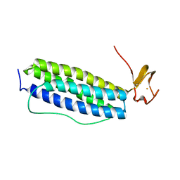
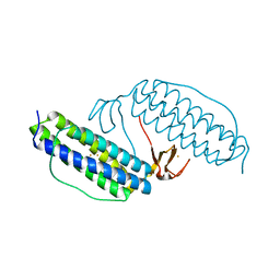
3WHN
 
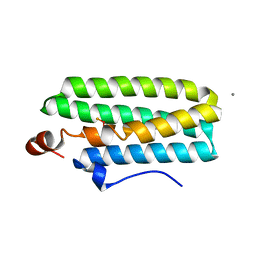 | | Hemerythrin-like domain of DcrH I119H mutant (met) | | 分子名称: | CALCIUM ION, CHLORO DIIRON-OXO MOIETY, Hemerythrin-like domain protein DcrH | | 著者 | Okamoto, Y, Onoda, A, Sugimoto, H, Takano, Y, Hirota, S, Kurtz Jr, D.M, Shiro, Y, Hayashi, T. | | 登録日 | 2013-08-29 | | 公開日 | 2014-02-26 | | 最終更新日 | 2023-11-08 | | 実験手法 | X-RAY DIFFRACTION (1.9 Å) | | 主引用文献 | H2O2-dependent substrate oxidation by an engineered diiron site in a bacterial hemerythrin.
Chem.Commun.(Camb.), 50, 2014
|
|
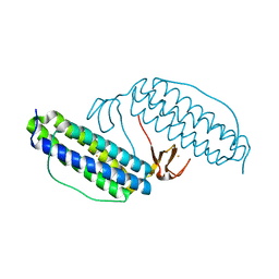
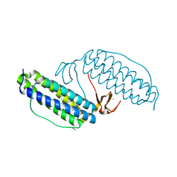
3WAQ
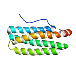 | | Hemerythrin-like domain of DcrH I119E mutant (met) | | 分子名称: | Hemerythrin-like domain protein DcrH, MU-OXO-DIIRON | | 著者 | Okamoto, Y, Onoda, A, Sugimoto, H, Takano, Y, Hirota, S, Kurtz Jr, D.M, Shiro, Y, Hayashi, T. | | 登録日 | 2013-05-07 | | 公開日 | 2014-03-19 | | 最終更新日 | 2023-11-08 | | 実験手法 | X-RAY DIFFRACTION (1.8 Å) | | 主引用文献 | Crystal structure, exogenous ligand binding, and redox properties of an engineered diiron active site in a bacterial hemerythrin
Inorg.Chem., 52, 2013
|
|
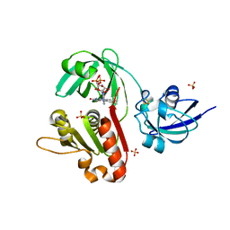
5V8S
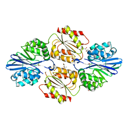 | | Flavo di-iron protein H90D mutant from Thermotoga maritima | | 分子名称: | (4R)-2-METHYLPENTANE-2,4-DIOL, ACETATE ION, CHLORIDE ION, ... | | 著者 | Taylor, A.B, Becker, A, Giri, N, Hart, P.J, Kurtz Jr, D.M. | | 登録日 | 2017-03-22 | | 公開日 | 2017-04-19 | | 最終更新日 | 2023-10-04 | | 実験手法 | X-RAY DIFFRACTION (1.41 Å) | | 主引用文献 | Flavo Di-iron protein H90D Mutant from Thermotoga Maritima
To Be Published
|
|
4DIK
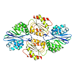 | | Flavo Di-iron protein H90A mutant from Thermotoga maritima | | 分子名称: | CHLORIDE ION, FLAVOPROTEIN, MU-OXO-DIIRON | | 著者 | Fang, H, Caranto, J.D, Taylor, A.B, Hart, P.J, Kurtz Jr, D.M. | | 登録日 | 2012-01-31 | | 公開日 | 2012-10-17 | | 最終更新日 | 2023-09-13 | | 実験手法 | X-RAY DIFFRACTION (1.75 Å) | | 主引用文献 | Histidine ligand variants of a flavo-diiron protein: effects on structure and activities.
J.Biol.Inorg.Chem., 17, 2012
|
|
1S30
 | | X-ray crystal structure of Desulfovibrio vulgaris Rubrerythrin with displacement of iron by zinc at the diiron Site | | 分子名称: | FE (III) ION, Rubrerythrin, ZINC ION | | 著者 | Jin, S, Kurtz Jr, D.M, Liu, Z.-J, Rose, J, Wang, B.-C. | | 登録日 | 2004-01-12 | | 公開日 | 2004-06-22 | | 最終更新日 | 2023-08-23 | | 実験手法 | X-RAY DIFFRACTION (2.05 Å) | | 主引用文献 | Displacement of iron by zinc at the diiron site of Desulfovibrio vulgaris rubrerythrin: X-ray crystal structure and anomalous scattering analysis
J.Inorg.Biochem., 98, 2004
|
|
1S2Z
 | | X-ray crystal structure of Desulfovibrio vulgaris Rubrerythrin with displacement of iron by zinc at the diiron Site | | 分子名称: | FE (III) ION, Rubrerythrin, ZINC ION | | 著者 | Jin, S, Kurtz Jr, D.M, Liu, Z.-J, Rose, J, Wang, B.-C. | | 登録日 | 2004-01-12 | | 公開日 | 2004-06-22 | | 最終更新日 | 2023-08-23 | | 実験手法 | X-RAY DIFFRACTION (1.75 Å) | | 主引用文献 | Displacement of iron by zinc at the diiron site of Desulfovibrio vulgaris rubrerythrin: X-ray crystal structure and anomalous scattering analysis
J.Inorg.Biochem., 98, 2004
|
|
1I4Z
 | | THE CRYSTAL STRUCTURE OF PHASCOLOPSIS GOULDII L98Y METHEMERYTHRIN | | 分子名称: | METHEMERYTHRIN, MU-OXO-DIIRON | | 著者 | Farmer, C.S, Kurtz Jr, D.M, Liu, Z.-J, Wang, B.C, Rose, J. | | 登録日 | 2001-02-23 | | 公開日 | 2001-03-21 | | 最終更新日 | 2024-04-03 | | 実験手法 | X-RAY DIFFRACTION (2.1 Å) | | 主引用文献 | The crystal structures of Phascolopsis gouldii wild type and L98Y methemerythrins: structural and functional alterations of the O2 binding pocket.
J.Biol.Inorg.Chem., 6, 2001
|
|
1I4Y
 | | THE CRYSTAL STRUCTURE OF PHASCOLOPSIS GOULDII WILD TYPE METHEMERYTHRIN | | 分子名称: | CHLORIDE ION, METHEMERYTHRIN, MU-OXO-DIIRON | | 著者 | Farmer, C.S, Kurtz Jr, D.M, Liu, Z.-J, Wang, B.C, Rose, J. | | 登録日 | 2001-02-23 | | 公開日 | 2001-03-21 | | 最終更新日 | 2023-08-09 | | 実験手法 | X-RAY DIFFRACTION (1.8 Å) | | 主引用文献 | The crystal structures of Phascolopsis gouldii wild type and L98Y methemerythrins: structural and functional alterations of the O2 binding pocket.
J.Biol.Inorg.Chem., 6, 2001
|
|
1LKM
 | | Crystal structure of Desulfovibrio vulgaris rubrerythrin all-iron(III) form | | 分子名称: | FE (III) ION, Rubrerythrin all-iron(III) form | | 著者 | Jin, S, Kurtz Jr, D.M, Liu, Z.J, Rose, J, Wang, B.C. | | 登録日 | 2002-04-25 | | 公開日 | 2002-09-18 | | 最終更新日 | 2023-08-16 | | 実験手法 | X-RAY DIFFRACTION (1.69 Å) | | 主引用文献 | X-ray Crystal Structures of Reduced Rubrerythrin and its Azide Adduct: A Structure-Based Mechanism for a Non-Heme DiIron Peroxidase
J.Am.Chem.Soc., 124, 2002
|
|
1LKO
 | | Crystal structure of Desulfovibrio vulgaris rubrerythrin all-iron(II) form | | 分子名称: | FE (II) ION, Rubrerythrin all-iron(II) form | | 著者 | Jin, S, Kurtz Jr, D.M, Liu, Z.J, Rose, J, Wang, B.C. | | 登録日 | 2002-04-25 | | 公開日 | 2002-09-18 | | 最終更新日 | 2024-02-14 | | 実験手法 | X-RAY DIFFRACTION (1.63 Å) | | 主引用文献 | X-ray Crystal Structures of Reduced Rubrerythrin and its Azide Adduct: A Structure-Based Mechanism for a Non-Heme DiIron Peroxidase
J.Am.Chem.Soc., 124, 2002
|
|
1KRH
 | | X-ray Structure of Benzoate Dioxygenase Reductase | | 分子名称: | Benzoate 1,2-Dioxygenase Reductase, FE2/S2 (INORGANIC) CLUSTER, FLAVIN-ADENINE DINUCLEOTIDE, ... | | 著者 | Karlsson, A, Beharry, Z.M, Eby, D.M, Coulter, E.D, Niedle, E.L, Kurtz Jr, D.M, Eklund, H, Ramaswamy, S. | | 登録日 | 2002-01-09 | | 公開日 | 2002-05-15 | | 最終更新日 | 2024-02-14 | | 実験手法 | X-RAY DIFFRACTION (1.5 Å) | | 主引用文献 | X-ray crystal structure of benzoate 1,2-dioxygenase reductase from Acinetobacter sp. strain ADP1.
J.Mol.Biol., 318, 2002
|
|
1LKP
 | | Crystal structure of Desulfovibrio vulgaris rubrerythrin all-iron(II) form, azide adduct | | 分子名称: | AZIDE ION, FE (II) ION, Rubrerythrin | | 著者 | Jin, S, Kurtz Jr, D.M, Liu, Z.J, Rose, J, Wang, B.C. | | 登録日 | 2002-04-25 | | 公開日 | 2002-09-18 | | 最終更新日 | 2024-04-03 | | 実験手法 | X-RAY DIFFRACTION (1.64 Å) | | 主引用文献 | X-ray Crystal Structures of Reduced Rubrerythrin and its Azide Adduct: A Structure-Based Mechanism for a Non-Heme Diiron Peroxidase
J.Am.Chem.Soc., 124, 2002
|
|


