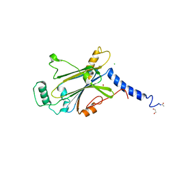
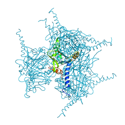
8X8S
 
 | | Crystal structure of Cypovirus Polyhedra mutant fused with c-Myc fragment | | 分子名称: | Polyhedrin,Myc proto-oncogene protein | | 著者 | Kojima, M, Ueno, T, Abe, S, Hirata, K. | | 登録日 | 2023-11-28 | | 公開日 | 2024-06-05 | | 最終更新日 | 2024-07-03 | | 実験手法 | X-RAY DIFFRACTION (2.04 Å) | | 主引用文献 | High-throughput structure determination of an intrinsically disordered protein using cell-free protein crystallization.
Proc.Natl.Acad.Sci.USA, 121, 2024
|
|
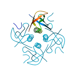
8WLG
 | | Crystal structure of Cypovirus Polyhedra mutant fused with c-Myc fragment | | 分子名称: | Polyhedrin,Myc proto-oncogene protein | | 著者 | Kojima, M, Ueno, T, Abe, S, Hirata, K. | | 登録日 | 2023-09-29 | | 公開日 | 2024-06-05 | | 最終更新日 | 2024-07-03 | | 実験手法 | X-RAY DIFFRACTION (2.55 Å) | | 主引用文献 | High-throughput structure determination of an intrinsically disordered protein using cell-free protein crystallization.
Proc.Natl.Acad.Sci.USA, 121, 2024
|
|
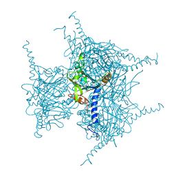
8X8V
 | | Crystal structure of Cypovirus Polyhedra mutant fused with c-Myc fragment | | 分子名称: | Polyhedrin,Myc proto-oncogene protein | | 著者 | Kojima, M, Ueno, T, Abe, S, Hirata, K. | | 登録日 | 2023-11-29 | | 公開日 | 2024-06-05 | | 最終更新日 | 2024-07-03 | | 実験手法 | X-RAY DIFFRACTION (2 Å) | | 主引用文献 | High-throughput structure determination of an intrinsically disordered protein using cell-free protein crystallization.
Proc.Natl.Acad.Sci.USA, 121, 2024
|
|
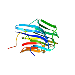
8J2Q
 | | Crystal structure of Cypovirus Polyhedra mutant fused with c-Myc fragment | | 分子名称: | Polyhedrin,Myc proto-oncogene protein | | 著者 | Kojima, M, Ueno, T, Abe, S, Hirata, K. | | 登録日 | 2023-04-15 | | 公開日 | 2024-04-17 | | 最終更新日 | 2024-07-03 | | 実験手法 | X-RAY DIFFRACTION (1.92 Å) | | 主引用文献 | High-throughput structure determination of an intrinsically disordered protein using cell-free protein crystallization.
Proc.Natl.Acad.Sci.USA, 121, 2024
|
|
8JAE
 | |
7WYR
 | | Crystal structure of Cypovirus Polyhedra mutant fused with CLN025 | | 分子名称: | 1,2-ETHANEDIOL, CHLORIDE ION, Polyhedrin fused with CLN025 | | 著者 | Kojima, M, Abe, S, Hirata, K, Yamashita, K, Ueno, T. | | 登録日 | 2022-02-16 | | 公開日 | 2023-02-22 | | 最終更新日 | 2023-11-29 | | 実験手法 | X-RAY DIFFRACTION (1.75 Å) | | 主引用文献 | Engineering of an in-cell protein crystal for fastening a metastable conformation of a target miniprotein.
Biomater Sci, 11, 2023
|
|
8IJU
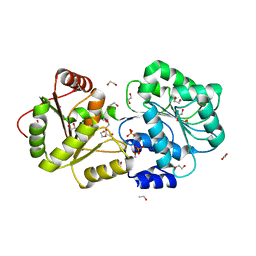 | | ATP-dependent RNA helicase DDX39A (URH49delta41) | | 分子名称: | 1,2-ETHANEDIOL, ATP-dependent RNA helicase DDX39A, PHOSPHATE ION, ... | | 著者 | Mikami, B, Fujita, K, Masuda, S, Kojima, M. | | 登録日 | 2023-02-28 | | 公開日 | 2024-01-24 | | 実験手法 | X-RAY DIFFRACTION (1.82 Å) | | 主引用文献 | Structural differences between the closely related RNA helicases, UAP56 and URH49, fashion distinct functional apo-complexes.
Nat Commun, 15, 2024
|
|
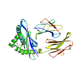
1Y43
 | | crystal structure of aspergilloglutamic peptidase from Aspergillus niger | | 分子名称: | Aspergillopepsin II heavy chain, Aspergillopepsin II light chain, SULFATE ION | | 著者 | Sasaki, H, Nakagawa, A, Iwata, S, Muramatsu, T, Suganuma, M, Sawano, Y, Kojima, M, Kubota, K, Takahashi, K. | | 登録日 | 2004-11-30 | | 公開日 | 2005-12-13 | | 最終更新日 | 2013-02-27 | | 実験手法 | X-RAY DIFFRACTION (1.4 Å) | | 主引用文献 | The three-dimensional structure of aspergilloglutamic peptidase from Aspergillus niger
Proc.Jpn.Acad.,Ser.B, 80, 2004
|
|
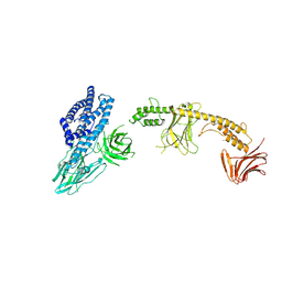
2CII
 | | The crystal structure of H-2Db complexed with a partial peptide epitope suggests an MHC Class I assembly-intermediate | | 分子名称: | BETA-2-MICROGLOBULIN, GLYCEROL, H-2 CLASS I HISTOCOMPATIBILITY ANTIGEN D-B ALPHA CHAIN, ... | | 著者 | Glithero, A, Tormo, J, Doering, K, Kojima, M, Jones, E.Y, Elliott, T. | | 登録日 | 2006-03-21 | | 公開日 | 2006-03-29 | | 最終更新日 | 2023-12-13 | | 実験手法 | X-RAY DIFFRACTION (2.55 Å) | | 主引用文献 | The crystal structure of H-2D(b) complexed with a partial peptide epitope suggests a major histocompatibility complex class I assembly intermediate.
J. Biol. Chem., 281, 2006
|
|
1IYY
 | | NMR STRUCTURE OF Gln25-RIBONUCLEASE T1, 24 STRUCTURES | | 分子名称: | RIBONUCLEASE T1 | | 著者 | Hatano, K, Kojima, M, Suzuki, E, Tanokura, M, Takahashi, K. | | 登録日 | 2002-09-12 | | 公開日 | 2003-10-07 | | 最終更新日 | 2023-12-27 | | 実験手法 | SOLUTION NMR | | 主引用文献 | Determination of the NMR structure of Gln25-ribonuclease T1.
Biol. Chem., 384, 2003
|
|
8W7N
 | | Crystal structure of the in-cell Cry1Aa purified from Bacillus thuringiensis | | 分子名称: | Pesticidal crystal protein Cry1Aa, UNKNOWN ATOM OR ION | | 著者 | Tanaka, J, Abe, S, Hayakawa, T, Kojima, M, Yamashita, K, Hirata, K, Ueno, T. | | 登録日 | 2023-08-31 | | 公開日 | 2024-03-06 | | 実験手法 | X-RAY DIFFRACTION (3.6 Å) | | 主引用文献 | Crystal structure of the in-cell Cry1Aa purified from Bacillus thuringiensis.
Biochem.Biophys.Res.Commun., 685, 2023
|
|
6KS2
 | | Structure of anti-Ghrelin receptor antibody | | 分子名称: | Fab7881 Heavy Chain (FabH), Fab7881 Light Chain (FabL) | | 著者 | Shiimura, Y, Horita, S, Asada, H, Hirata, K, Iwata, S, Kojima, M. | | 登録日 | 2019-08-23 | | 公開日 | 2020-08-12 | | 最終更新日 | 2023-11-22 | | 実験手法 | X-RAY DIFFRACTION (1.753 Å) | | 主引用文献 | Structure of an antagonist-bound ghrelin receptor reveals possible ghrelin recognition mode.
Nat Commun, 11, 2020
|
|
6KO5
 | | Complex structure of Ghrelin receptor with Fab | | 分子名称: | 6-(4-bromanyl-2-fluoranyl-phenoxy)-2-methyl-3-[[(3~{S})-1-propan-2-ylpiperidin-3-yl]methyl]pyrido[3,2-d]pyrimidin-4-one, Chimera of Soluble cytochrome b562 and Growth hormone secretagogue receptor type 1, Fab7881 Heavy Chain, ... | | 著者 | Shiimura, Y, Horita, S, Asada, H, Hirata, K, Iwata, S, Kojima, M. | | 登録日 | 2019-08-08 | | 公開日 | 2020-08-12 | | 最終更新日 | 2023-11-22 | | 実験手法 | X-RAY DIFFRACTION (3.3 Å) | | 主引用文献 | Structure of an antagonist-bound ghrelin receptor reveals possible ghrelin recognition mode.
Nat Commun, 11, 2020
|
|
7XWS
 | | Crystal structure of Wild Type Cypovirus Polyhedra produced by cell-free protein synthesis with small volume | | 分子名称: | ACETYL GROUP, CHLORIDE ION, Polyhedrin | | 著者 | Abe, S, Tanaka, J, Kojima, M, Hirata, K, Yamashita, K, Ueno, T. | | 登録日 | 2022-05-27 | | 公開日 | 2023-02-01 | | 最終更新日 | 2023-11-29 | | 実験手法 | X-RAY DIFFRACTION (1.95 Å) | | 主引用文献 | Cell-free protein crystallization for nanocrystal structure determination.
Sci Rep, 12, 2022
|
|
7XHS
 | | Crystal structure of CipA crystal produced by cell-free protein synthesis | | 分子名称: | Cro/Cl family transcriptional regulator | | 著者 | Abe, S, Tanaka, J, Kojima, M, Kanamaru, S, Yamashita, K, Hirata, K, Ueno, T. | | 登録日 | 2022-04-10 | | 公開日 | 2023-02-01 | | 最終更新日 | 2024-05-29 | | 実験手法 | X-RAY DIFFRACTION (2.11 Å) | | 主引用文献 | Cell-free protein crystallization for nanocrystal structure determination.
Sci Rep, 12, 2022
|
|
7XHR
 | | Crystal structure of Wild Type Cypovirus Polyhedra produced by cell-free protein synthesis | | 分子名称: | ACETYL GROUP, CHLORIDE ION, Polyhedrin | | 著者 | Abe, S, Tanaka, J, Kojima, M, Hirata, K, Yamashita, K, Ueno, T. | | 登録日 | 2022-04-10 | | 公開日 | 2023-02-01 | | 最終更新日 | 2023-11-29 | | 実験手法 | X-RAY DIFFRACTION (1.801 Å) | | 主引用文献 | Cell-free protein crystallization for nanocrystal structure determination.
Sci Rep, 12, 2022
|
|
3TRS
 | | The crystal structure of aspergilloglutamic peptidase from Aspergillus niger | | 分子名称: | Aspergillopepsin-2 heavy chain, Aspergillopepsin-2 light chain, DIMETHYL SULFOXIDE | | 著者 | Sasaki, H, Kubota, K, Lee, W.C, Ohtsuka, J, Kojima, M, Takahashi, K, Tanokura, M. | | 登録日 | 2011-09-10 | | 公開日 | 2012-08-22 | | 最終更新日 | 2023-11-01 | | 実験手法 | X-RAY DIFFRACTION (1.6 Å) | | 主引用文献 | The crystal structure of an intermediate dimer of aspergilloglutamic peptidase that mimics the enzyme-activation product complex produced upon autoproteolysis.
J.Biochem., 152, 2012
|
|


