2CZV
 
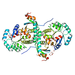 | | Crystal structure of archeal RNase P protein ph1481p in complex with ph1877p | | 分子名称: | ACETIC ACID, Ribonuclease P protein component 2, Ribonuclease P protein component 3, ... | | 著者 | Kawano, S, Kakuta, Y, Nakashima, T, Tanaka, I, Kimura, M. | | 登録日 | 2005-07-19 | | 公開日 | 2006-06-27 | | 最終更新日 | 2024-05-29 | | 実験手法 | X-RAY DIFFRACTION (2 Å) | | 主引用文献 | Crystal structure of protein Ph1481p in complex with protein Ph1877p of archaeal RNase P from Pyrococcus horikoshii OT3: implication of dimer formation of the holoenzyme
J.Mol.Biol., 357, 2006
|
|
5ETA
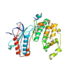 | | Structure of MAPK14 with bound the KIM domain of the Toxoplasma protein GRA24 | | 分子名称: | Mitogen-activated protein kinase 14, Putative transmembrane protein | | 著者 | Pellegrini, E, Palencia, A, Braun, L, Kapp, U, Bougdour, A, Belrhali, H, Bowler, M.W, Hakimi, M. | | 登録日 | 2015-11-17 | | 公開日 | 2016-10-26 | | 最終更新日 | 2019-02-20 | | 実験手法 | X-RAY DIFFRACTION (2.8 Å) | | 主引用文献 | Structural Basis for the Subversion of MAP Kinase Signaling by an Intrinsically Disordered Parasite Secreted Agonist.
Structure, 25, 2017
|
|
5ETF
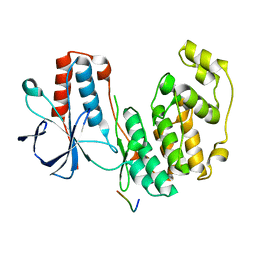 | | Structure of dead kinase MAPK14 with bound the KIM domain of MKK6 | | 分子名称: | Dual specificity mitogen-activated protein kinase kinase 6, Mitogen-activated protein kinase 14 | | 著者 | Pellegrini, E, Palencia, A, Braun, L, Kapp, U, Bougdour, A, Belrhali, H, Bowler, M.W, Hakimi, M. | | 登録日 | 2015-11-17 | | 公開日 | 2016-10-26 | | 最終更新日 | 2024-05-08 | | 実験手法 | X-RAY DIFFRACTION (2.4 Å) | | 主引用文献 | Structural Basis for the Subversion of MAP Kinase Signaling by an Intrinsically Disordered Parasite Secreted Agonist.
Structure, 25, 2017
|
|
3VU2
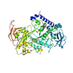 | | Structure of the Starch Branching Enzyme I (BEI) complexed with maltopentaose from Oryza sativa L | | 分子名称: | 1,4-alpha-glucan-branching enzyme, chloroplastic/amyloplastic, GLYCEROL, ... | | 著者 | Chaen, K, Kakuta, Y, Kimura, M. | | 登録日 | 2012-06-14 | | 公開日 | 2013-05-08 | | 最終更新日 | 2024-03-20 | | 実験手法 | X-RAY DIFFRACTION (2.23 Å) | | 主引用文献 | Crystal structure of the rice branching enzyme I (BEI) in complex with maltopentaose.
Biochem.Biophys.Res.Commun., 424, 2012
|
|
2E2R
 | | Crystal structure of human estrogen-related receptor gamma ligand binding domain complex with bisphenol A | | 分子名称: | 4,4'-PROPANE-2,2-DIYLDIPHENOL, Estrogen-related receptor gamma, GLYCEROL | | 著者 | Matsushima, A, Kakuta, Y, Teramoto, T, Koshiba, T, Kimura, M, Shimohigashi, Y. | | 登録日 | 2006-11-16 | | 公開日 | 2007-09-11 | | 最終更新日 | 2023-10-25 | | 実験手法 | X-RAY DIFFRACTION (1.6 Å) | | 主引用文献 | Structural Evidence for Endocrine Disruptor Bisphenol A Binding to Human Nuclear Receptor ERR{gamma}
J.Biochem.(Tokyo), 142, 2007
|
|


